. 2024 Jul 8;19(7):e0306323.
doi: 10.1371/journal.pone.0306323. eCollection 2024.

Left atrial reservoir strain is a marker of atrial fibrotic remodeling in patients undergoing cardiovascular surgery: Analysis of gene expression

Affiliations

Left atrial reservoir strain is a marker of atrial fibrotic remodeling in patients undergoing cardiovascular surgery: Analysis of gene expression

Toshiaki Nakajima et al. PLoS One. .

Abstract

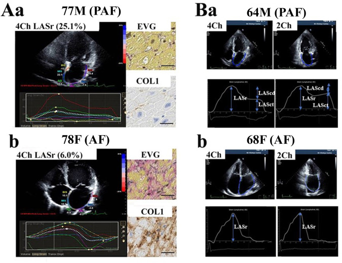
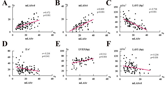
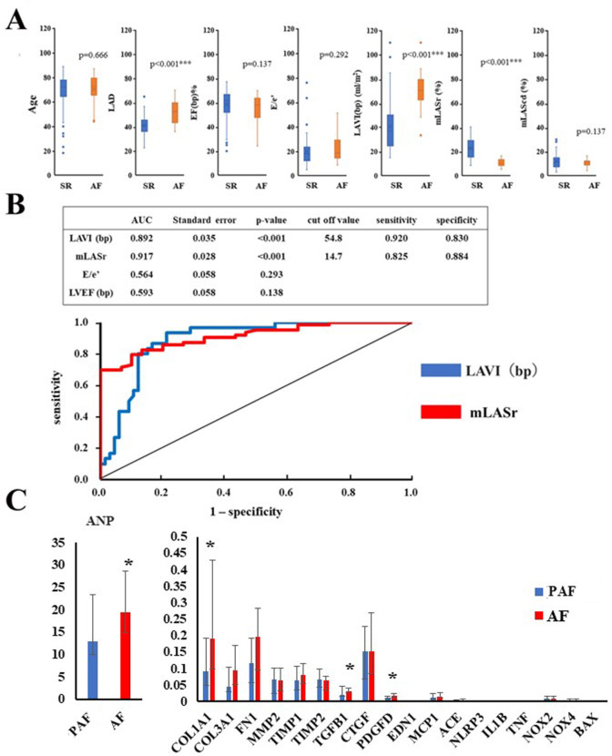
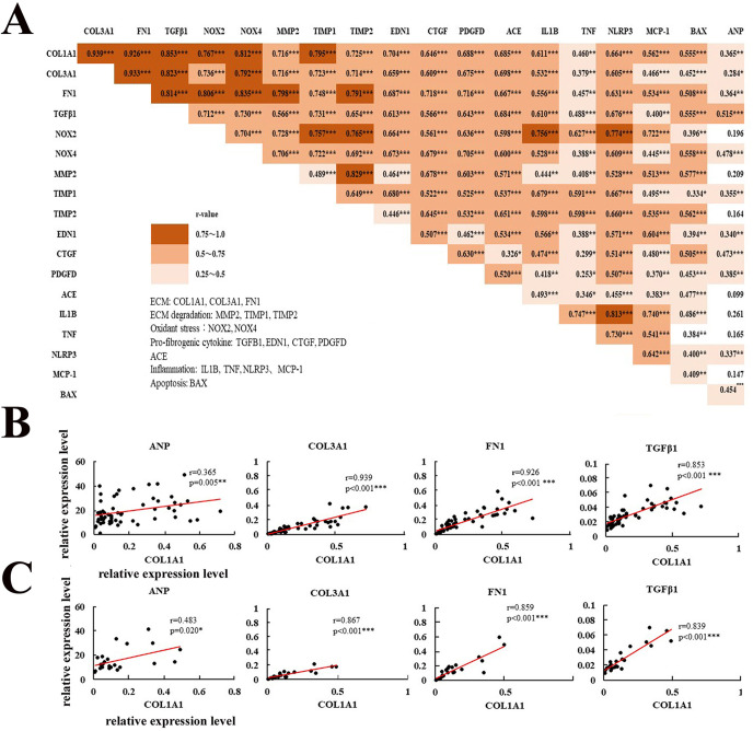
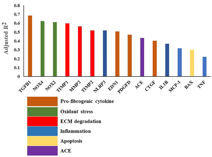
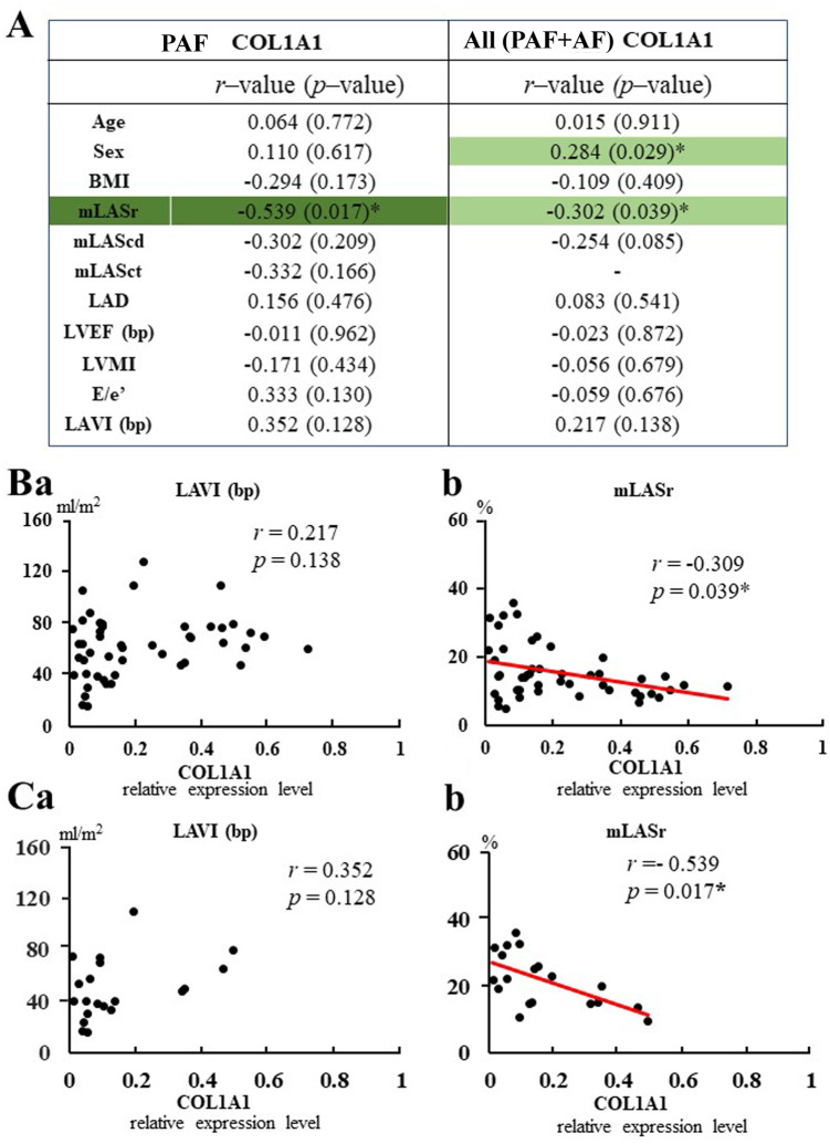
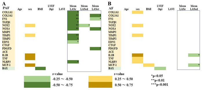
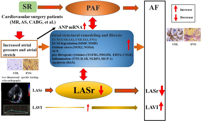
Left atrial strain (LAS) measured by two-dimensional speckle tracking echocardiography (2DSTE) is considered to be a marker of LA structural remodeling, but it remains unsettled. We investigated the potential usefulness and clinical relevance of LAS to detect atrial remodeling including fibrosis by analyzing gene expression in cardiovascular surgery patients. Preoperative 2DSTE was performed in 131 patients (92 patients with sinus rhythm [SR] patients including paroxysmal AF [PAF], 39 atrial fibrillation [AF]) undergoing cardiovascular surgery. Atrial samples were obtained from the left atrial appendages, and mRNA expression level was analyzed by real-time reverse transcription polymerase chain reaction (RT-PCR) in 59 cases (24 PAF, 35 AF). Mean value of left atrial reservoir strain (mLASr) correlated with left atrial volume index (LAVI), and left atrial conduit strain (mLAScd). mLASr also correlated with left atrial contractile strain (mLASct) in SR patients including PAF. mLASr was significantly lower, and LAVI was higher, in the AF group, compared with SR patients including PAF. The expression of COL1A1 mRNA encoding collagen type I α1 significantly increased in AF patients (p = 0.031). mLASr negatively correlated with COL1A1 expression level, and multivariate regression analysis showed that mLASr was an independent predictor of atrial COL1A1 expression level, even after adjusting for age, sex, and BMI. But, neither mLAScd / mLASct nor LAVI (bp) correlated with COL1A1 gene expression. The expression level of COL1A1 mRNA strongly correlated with ECM-related genes (COL3A1, FN1). It also correlated ECM degradation-related genes (MMP2, TIMP1, and TIMP2), pro-fibrogenic cytokines (TGFB1 encoding TGFβ1, END1, PDGFD, CTGF), oxidant stress-related genes (NOX2, NOX4), ACE, inflammation-related genes (NLRP, IL1B, MCP-1), and apoptosis (BAX). Among the fibrosis-related genes examined, univariable regression analysis showed that log (COL1A1) was associated with log (TGFB1) (adjusted R2 = 0.685, p<0.001), log (NOX4) (adjusted R2 = 0.622, p<0.001), log (NOX2) (adjusted R2 = 0.611, p<0.001), suggesting that TGFB1 and NOX4 was the potent independent determinants of COL1A1 expression level. mLASr negatively correlated with the ECM-related genes, and fibrosis-related gene expression level including TGFB1, NOX2, and NLRP3 in PAF patients. PAF patients with low mLASr had higher expression of the fibrosis-related gene expression, compared with those with high mLASr. These results suggest that LASr correlates with atrial COL1A1 gene expression associated with fibrosis-related gene expression. Patients with low LASr exhibit increased atrial fibrosis-related gene expression, even those with PAF, highlighting the utility of LAS as a marker for LA fibrosis in cardiovascular surgery patients.


Conflict of interest statement

The authors have declared that no competing interests exist.

Figures

Fig 1
Fig 1
Typical left atrial strain (LAS) and histological findings in a patient with PAF (Aa, Ba) and a patient with atrial fibrillation (AF) (Ab, Bb). Aa and Ab: Quad view of longitudinal left atrial (LA) strain by 2DSTE. Elastica van Gieson stain (EVG), and immunohistochemical staining of collagen type 1a are also shown for each patient. Ba and Bb: Two typical LA strain images from 4-chamber (4CH) and 2-chamber (2CH) apical views obtained from a patient with PAF (Ba) and a patient with AF (Bb). The mean values were averaged for 6 segments in the 4CH view (left part) and 6 segments in the 2CH view (right part), as shown in the lower part. LASr, LA reservoir strain; LASct, LA contractile strain; LAScd, LA conduit strain.
Fig 2
Fig 2. Relationships between left atrial strain (LAS) and echocardiographic findings (LAVI (bp), E/e’, LVEF (bp).
The data of mLASr and mLAScd were obtained from 118 patients including PAF and AF. The data of mLASct were obtained from 84 patients with SR patients including PAF. mean LASr value (mLASr), LAScd (mLAScd), LASct (mLASct)). R- and P-value are shown. *P<0.05, **P<0.01, ***P<0.001.
Fig 3
Fig 3. Comparison of echocardiographic parameters and gene expression level between patients with SR including PAF and those with AF.
A: Comparison of echocardiographic parameters. SR including PAF (blue, n = 84), AF (red, n = 34). B: ROC to identify the optimal cut-off level of echocardiographic findings to detect AF (n = 118). C: Comparison of ANP, and fibrosis-related gene expression level in between patients with PAF (n = 23–24) and those with AF (n = 33–35). PAF (blue), AF (red). *P<0.05, ***P<0.001.
Fig 4
Fig 4. Correlations among atrial gene expression level including ANP, and fibrosis-related gene expression in total patients.
A: Correlations among atrial gene expression level including ANP, and fibrosis-related gene expression in total patients receiving LA resection (n = 57–59). The R-value and P-value among genes are indicated. *P<0.05, **P<0.01, ***P<0.001. The orange area shows statistical significance. B: Correlations between COL1A1 and fibrosis-related gene expression level (ANP, COL3A1, FN1, TGFB1) in total patients receiving LA resection (n = 57–59). C: Correlations between COL1A1 and fibrosis-related gene expression level (ANP, COL3A1, FN1, TGFB1) in PAF patients (n = 23–24). *P<0.05, **P<0.01, ***P<0.001.
Fig 5
Fig 5. The univariate regression analysis with log (COL1A1) expression level as the dependent variable and fibrosis-related gene expression as the independent variable.
The adjusted R-squared value (R2) is shown. The data were obtained in 57–59 patients receiving LA resection. Pro-fibrogenic cytokine genes (TGFB1, EDN1, PDGFD, CTGF), oxidant stress-related genes (NOX2, NOX4), ECM degradation-related genes (MMP2, TIMP1, TIMP2), inflammation-related genes (NLRP3, IL1B, MCP-1, TNF), angiotensin-related gene (ACE), apoptosis-related gene (BAX). All data are P<0.001.
Fig 6
Fig 6. Relationships between COL1A1 mRNA expression level and echocardiographic parameters.
A: Relationships between COL1A1 mRNA expression and clinical data (age, sex, BMI) and echocardiographic parameters in PAF patients (n = 20) and total patients (n = 52) receiving LA resection. R- and P–values are shown. *P<0.05 B and C: Relationships between COL1A1 mRNA expression level and LAVI (bp) or mLASr in total patients (B, n = 52) and PAF patients (C, n = 20) receiving LA resection. *P<0.05.
Fig 7
Fig 7
Correlation matrix between clinical data (age, sex, BMI), echocardiographic findings and fibrosis-related gene expression level in PAF (A) and AF (B) patients. The data were obtained from PAF patients (n = 19–20) and AF patients (n = 30–32) receiving LA resection. R–value and P–value in mLASr, mLAScd, and mLASct are shown. *P<0.05, **P<0.01, ***P<0.001. Significant negative relationships (R = –0.25 ~ –0.5, R = –0.5 ~ –0.75) are shown in green area, and positive relationships (R = 0.25~ 0.5, R = 0.5 ~ 0.75) are shown in orange area.
Fig 8
Fig 8. Graphic abstract of the study.
SR, sinus rhythm; PAF, paroxysmal atrial fibrillation; AF, atrial fibrillation; ANP, atrial natriuretic peptide; LASr, left atrial reservoir strain; LAVI, left atrial volume index; COL collagen; EVG, Elastica van Gieson staining.

Similar articles

Cited by

References

    1. Barbier P, Solomon SB, Schiller NB, Glantz SA. Left atrial relaxation and left ventricular systolic function determine left atrial reservoir function. Circulation. 1999; 100(4): 427–436. doi: 10.1161/01.cir.100.4.427 . - DOI - PubMed
    1. Nattel S, Burstein B, Dobrev D. Atrial remodeling and atrial fibrillation: mechanisms and implications. Circ Arrhythm Electrophysiol. 2008; 1(1): 62–73. doi: 10.1161/CIRCEP.107.754564 . - DOI - PubMed
    1. Casaclang-Verzosa G, Gersh BJ, Tsang TS. Structural and functional remodeling of the left atrium: clinical and therapeutic implications for atrial fibrillation. J Am Coll Cardiol. 2008; 51(1): 1–11. doi: 10.1016/j.jacc.2007.09.026 . - DOI - PubMed
    1. Hoit BD. Left atrial size and function: role in prognosis. J Am Coll Cardiol. 2014; 63(6): 493–505. doi: 10.1016/j.jacc.2013.10.055 . - DOI - PubMed
    1. Vieira MJ, Teixeira R, Gonçalves L, Gersh BJ. Left atrial mechanics: echocardiographic assessment and clinical implications. J Am Soc Echocardiogr. 2014; 27(5): 463–478. doi: 10.1016/j.echo.2014.01.021 . - DOI - PubMed

MeSH terms