. 2024 Jul 8;19(7):e0304074.
doi: 10.1371/journal.pone.0304074. eCollection 2024.

The expression of MIR125B transcripts and bone phenotypes in Mir125b2-deficient mice

Affiliations

The expression of MIR125B transcripts and bone phenotypes in Mir125b2-deficient mice

Tomohiro Ogasawara et al. PLoS One. .

Abstract

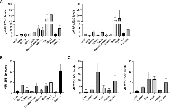
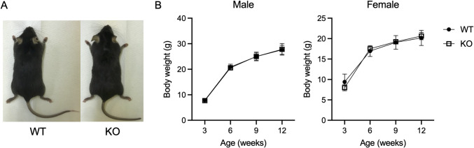
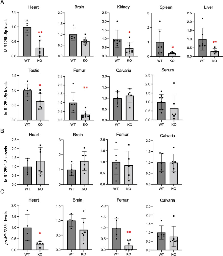
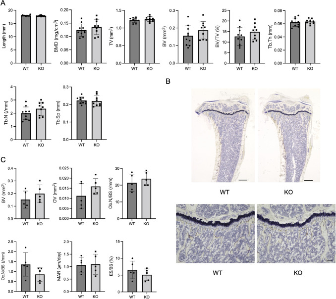
MIR125B, particularly its 5p strand, is apparently involved in multiple cellular processes, including osteoblastogenesis and osteoclastogenesis. Given that MIR125B is transcribed from the loci Mir125b1 and Mir125b2, three mature transcripts (MIR125B-5p, MIR125B1-3p, and MIR125B2-3p) are generated (MIR125B-5p is common to both); however, their expression profiles and roles in the bones remain poorly understood. Both primary and mature MIR125B transcripts were differentially expressed in various organs, tissues, and cells, and their expression patterns did not necessarily correlate in wild-type (WT) mice. We generated Mir125b2 knockout (KO) mice to examine the contribution of Mir125b2 to MIR125B expression profiles and bone phenotypes. Mir125b2 KO mice were born and grew normally without any changes in bone parameters. Interestingly, in WT and Mir125b2 KO, MIR125B-5p was abundant in the calvaria and bone marrow stromal cells. These results indicate that the genetic ablation of Mir125b2 does not impinge on the bones of mice, attracting greater attention to MIR125B-5p derived from Mir125b1. Future studies should investigate the conditional deletion of Mir125b1 and both Mir125b1 and Mir125b2 in mice.


Conflict of interest statement

The authors have declared that no competing interests exist.

Figures

Fig 1
Fig 1. Transcriptional profiling of Mir125b genes in wild-type (WT) mice.
Relative levels of pri-Mir125b1 and pri-Mir125b2 (A), MIR125B-5p (B), MIR125B1-3p, and MIR125B2-3p (C) in various organs/tissues of 12-week-old male mice are shown. Actb (A) and Rnu6 (B, C) were used as internal controls. n = 4−6.
Fig 2
Fig 2. The gross appearance of WT and Mir125b2 KO mice and their growth curves.
(A) Representative images of 12-week-old male mice. (B) Body weight changes in mice from 3–12 weeks of age. n = 15−18 (males) and 7−9 (females).
Fig 3
Fig 3. Levels of MIR125B-5p and MIR125B1 transcripts in WT and Mir125b2 KO mice.
Relative levels of MIR125B-5p (A), MIR125B1-3p (B), and pri-Mir125b1 (C) in various organs/tissues of 12-week-old male mice are shown. Mean values of the WT group were set at 1.0. Rnu6 (A, B) and Actb (C) were used as internal controls. n = 4−6. *, p < 0.05 and **, p < 0.01 versus WT mice.
Fig 4
Fig 4. μCT and histomorphometric analyses of WT and Mir125b2 KO mouse tibiae.
(A) Trabecular bone parameters in 12-week-old male mice were assessed using μCT. BMD, bone mineral density; TV, tissue volume; BV, bone volume; BV/TV, bone volume/tissue volume; Tb.N, trabecular number; Tb.Th, trabecular thickness; Tb.Sp, trabecular separation. n = 9−10. (B) Representative images of longitudinal sections with Villanueva staining are shown. The scales in the upper and lower panels represent 500 μm and 100 μm, respectively. (C) Trabecular bone parameters were assessed by bone histomorphometry. BV, bone volume; OV, osteoid volume; Ob. N/BS, number of osteoblasts per bone surface; Oc. N/BS, number of osteoclasts per bone surface; MAR, mineral apposition rate; ES/BS, eroded surface per bone surface. n = 5.
Fig 5
Fig 5. Levels of mature MIR125B transcripts in BMCs, BMSCs, and BMMs of WT and Mir125b2 KO mice.
Bone marrow stroma cells (BMSCs) and bone marrow macrophage (BMMs) were obtained from bone marrow cells (BMCs) of 10−15-week-old male mouse femurs and tibiae. Relative levels of MIR125B-5p, MIR125B1-3p, and MIR125B2-3p in BMCs, BMSCs, and BMMs are shown. Femurs of 12-week-old WT male mice were used for comparison. Rnu6 was used as internal control.

Similar articles

Cited by

References

    1. Ambros V. The functions of animal microRNAs. Nature. 2004;431: 350–355. doi: 10.1038/nature02871 - DOI - PubMed
    1. Valadi H, Ekström K, Bossios A, Sjöstrand M, Lee JJ, Lötvall JO. Exosome-mediated transfer of mRNAs and microRNAs is a novel mechanism of genetic exchange between cells. Nat Cell Biol. 2007;9: 654–659. doi: 10.1038/ncb1596 - DOI - PubMed
    1. Kelch S, Balmayor ER, Seeliger C, Vester H, Kirschke JS, Van Griensven M. MiRNAs in bone tissue correlate to bone mineral density and circulating miRNAs are gender independent in osteoporotic patients. Sci Rep. 2017;7: 15861. doi: 10.1038/s41598-017-16113-x - DOI - PMC - PubMed
    1. Seeliger C, Karpinski K, Haug AT, Vester H, Schmitt A, Bauer JS, et al.. Five freely circulating miRNAs and bone tissue miRNAs are associated with osteoporotic fractures. J Bone Miner Res. 2014;29: 1718–1728. doi: 10.1002/jbmr.2175 - DOI - PubMed
    1. Panach L, Mifsut D, Tarín JJ, Cano A, García-Pérez MÁ. Serum circulating microRNAs as biomarkers of osteoporotic fracture. Calcif Tissue Int. 2015;97: 495–505. doi: 10.1007/s00223-015-0036-z - DOI - PubMed

LinkOut - more resources