. 2024 Jul 16;121(29):e2322864121.
doi: 10.1073/pnas.2322864121. Epub 2024 Jul 8.

A wound-induced differentiation trajectory for neurons

Affiliations

A wound-induced differentiation trajectory for neurons

Ryan E Hulett et al. Proc Natl Acad Sci U S A. .

Abstract

Animals capable of whole-body regeneration can replace any missing cell type and regenerate fully functional new organs, including new brains, de novo. The regeneration of a new brain requires the formation of diverse neural cell types and their assembly into an organized structure with correctly wired circuits. Recent work in various regenerative animals has revealed transcriptional programs required for the differentiation of distinct neural subpopulations, however, how these transcriptional programs are initiated in response to injury remains unknown. Here, we focused on the highly regenerative acoel worm, Hofstenia miamia, to study wound-induced transcriptional regulatory events that lead to the production of neurons and subsequently a functional brain. Footprinting analysis using chromatin accessibility data on a chromosome-scale genome assembly revealed that binding sites for the Nuclear Factor Y (NFY) transcription factor complex were significantly bound during regeneration, showing a dynamic increase in binding within one hour upon amputation specifically in tail fragments, which will regenerate a new brain. Strikingly, NFY targets were highly enriched for genes with neuronal function. Single-cell transcriptome analysis combined with functional studies identified soxC+ stem cells as a putative progenitor population for multiple neural subtypes. Further, we found that wound-induced soxC expression is likely under direct transcriptional control by NFY, uncovering a mechanism for the initiation of a neural differentiation pathway by early wound-induced binding of a transcriptional regulator.

Keywords: functional genomics; neural differentiation; regeneration; stem cells; wound response.


Conflict of interest statement

Competing interests statement:The authors declare no competing interest.

Figures

Fig. 1.
Fig. 1.
Nuclear Factor Y (NFY) is the most significantly bound motif in the H. miamia genome during regeneration. (A) Schematic phylogeny of select metazoan lineages based on published literature (52) identifying animals capable of WBR and extensive adult neural regeneration. The origin of the Bilateria ~550 million years ago (mya) is indicated on the lineage leading to animals with bilateral symmetry. Silhouettes of animals were modified from PhyloPic. Credit to B. Duygu Özpolot (tunicate and annelid) and Michelle Site (placozoan). (B) Heatmap showing the mean binding score calculated at the binding sites associated with the curated list of 78 H. miamia TFs. In head and tail fragments sampled during regeneration, NFY is the most differentially bound TF binding site in the H. miamia genome. (C) Volcano plot showing that NFIC is the most bound motif at 0 hpa and NFYA is the most bound motif at 1 hpa in the regenerating tail fragment in the H. miamia genome (relative to 0 hpa). Volcano plot showing that NFIC is the most bound motif at 0 hpa and NEUROD1 is the most bound motif at 1 hpa in the regenerating head fragment in the H. miamia genome (relative to 0 hpa). The comparison here is relative measure, not an absolute measure of binding at 1 hpa vs. 0 hpa. (D) Dynamically bound NFY sites at 1 hpa compared to 0 hpa are linked to genes associated with neural function. Gene ratio is the number of genes in a particular biological process category over the total list of genes.
Fig. 2.
Fig. 2.
NFY RNAi drastically impacts nervous system regeneration, with some other anterior structures also impacted. (A) NFY RNAi (consisting of NFYA, NFYB1, NFYB2, and NFYC) a majority of animals make an atypical blastema lacking a mouth (SI Appendix, Fig. S2E). Regenerating head and tail fragments are shown 7 d postamputation (7 dpa). The white arrowhead indicates proper blastema in control RNAi. The yellow arrowhead indicates an atypical blastema lacking a mouth. (Scale bars, 200 µm.) (B) Plot showing the average velocities of 7 dpa control RNAi regenerating tail fragments (n = 3) and 7dpa NFY RNAi regenerating tail fragments (n = 3). Comparison of average velocities from control RNAi and NFY RNAi fragments using a Welch’s T test (P = 0.0003). (C) NFY RNAi regenerating tail fragments lack concentrated expression of neural markers gad-1 and TrpC-1 in the anterior but do show expression of anterior markers sFRP-1 and FoxD as well the pharynx marker, phar. Regenerating head and tail fragments are shown 7 dpa. These anterior and pharyngeal markers do seem impacted by NFY RNAi but not as drastically as the nervous system. White arrowheads indicate normal gene expression in control RNAi. Yellow arrowheads indicate lack of anterior neural expression. White arrowheads in the NFY RNAi condition for sfrp-1, foxD, and phar denote expression, albeit at lower levels than in controls. (Scale bars, 200 µm.) Associated head fragments are in SI Appendix, Fig. S2H.
Fig. 3.
Fig. 3.
Identification of major neural subpopulations during regeneration. (A) Uniform Manifold Approximation and Projection (UMAP) representation of neural cells from a merged regeneration scRNA-seq dataset (55) that were reclustered to identify neural subpopulations. There are 20 putative neural subtypes identified. UMAP colored by cluster in SI Appendix, Fig. S3A. (B) A dot plot showing neural subpopulations on the y axis from the UMAP with the dot size indicating the number of cells expressing a given gene and dot color indicating the average expression of a given gene in a given cluster. The genes displayed along the x axis represent the neoblast marker piwi-1 and a top enriched marker gene from each neural subpopulation. There are four major neural types, falling into groups 1 to 4. A major transcriptional distinction between the populations is the presence of relatively high piwi-1 expression (dotted black box on the Left) and relatively high pc2 expression (dotted black box on the Right). pc2 is also supported as a panneural marker because of its presence across groups 2 to 4. Group 1 likely represents a neural progenitor population and is the only group that expresses relatively high levels of soxC (dotted black box, second from the Left). (C) FISH corroboration of selected markers corresponding to each of neural groups. (Scale bars, 200 µm.) Additional FISH in SI Appendix, Fig. S3B.
Fig. 4.
Fig. 4.
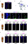
soxC is wound-induced in a subset of stem cells and is required for the expression of neural genes associated with distinct neural subpopulations (A) soxC expression is wound-induced in regenerating tail fragments at 12 hpa relative to a 0 hpa. The white arrowhead indicates wound-induced gene expression. (Scale bars, 200 µm.) (B) UMAP representation of neural subpopulations depicting soxC expression. (C) Double FISH of the putative neural progenitor marker soxC and neoblast marker piwi-1 in regenerating tail fragments at 12 hpa. The dotted black box indicates the region of interest where high-magnification imaging was performed. Coexpression of soxC and piwi-1 (denoted by white arrowheads) was detected in a subset of piwi-1+ cells. (Scale bars, 10 µm.) (D) RNAi of soxC impacts TF gene expression associated with neural groups 2 to 4. Regenerating tail fragments are shown 3 dpa. White arrowheads indicate normal gene expression in control RNAi. Yellow arrowheads indicate impacted neural gene expression. (Scale bars, 200 µm.) (E) RNAi of TFs associated with and expressed within different neural groups. Regenerating tail fragments are shown 3 dpa. For each group, we performed RNAi of a TF associated with that group and assessed expression of a putative differentiated gene associated with the same group. White arrowheads indicate normal gene expression in control RNAi. Yellow arrowheads indicate impacted neural gene expression. (Scale bars, 200 µm.)
Fig. 5.
Fig. 5.
NFY wound-induced TF binding regulates soxC expression during regeneration. (A) Schematic depicting the ~12.1 kb region that encompasses the soxC gene locus showing ATAC-seq data for 0, 1, and 6 hpa regenerating tail fragments along with sites that are putatively bound by the NFY TF (at the NFYA site). The NFY TF binding summary of the NFY site on the right, which is initially unbound at 0 hpa and becomes statistically significantly bound at 1 hpa, is depicted as a heatmap where the light gray color indicates unbound and dark gray indicates bound. The black arrowhead on the gene model denotes the transcription start site. (B) RNAi of NFY leads to a lack of soxC upregulation in the anterior of regenerating tail fragments 12 hpa and 1 dpa. The white arrowhead indicates soxC upregulation in control RNAi. The yellow arrowhead indicates a failure of soxC upregulation. The soxC Inset shows a zoom-in of the anterior region. (Scale bars, 200 µm and Inset scale bars 200 µm.) (C) In the NFY RNAi ATAC-seq data, dynamically bound NFY sites located proximal to genes associated with neural function are no longer accessible at 6 hpa. Gene ratio is the number of genes in a particular biological process category over the total list of genes. (D) A proposed model of NFY wound-induced TF binding which leads to the upregulation of soxC gene expression in neoblasts during regeneration which is required for the expression of genes associated with distinct neural subpopulations.

Update of

References

    1. Bely A. E., Nyberg K. G., Evolution of animal regeneration: Re-emergence of a field. Trends Ecol. Evol. 25, 161–170 (2010). - PubMed
    1. Lai A. G., Aboobaker A. A., EvoRegen in animals: Time to uncover deep conservation or convergence of adult stem cell evolution and regenerative processes. Dev. Biol. 433, 118–131 (2018). - PubMed
    1. Thor S., Andersson S. G. E., Tomlinson A., Thomas J. B., A LIM-homeodomain combinatorial code for motor-neuron pathway selection. Nature 397, 76 (1999). - PubMed
    1. Rincón-Limas D. E., et al. , Conservation of the expression and function of apterous orthologs in Drosophila and mammals. Proc. Natl. Acad. Sci. U.S.A. 96, 2165–2170 (1999). - PMC - PubMed
    1. Melkman T., Regulation of chemosensory and GABAergic motor neuron development by the C. elegans Aristaless/Arx homolog alr-1. Development 132, 1935–1949 (2005). - PubMed

LinkOut - more resources