. 2024 Oct 1;110(10):6558-6572.
doi: 10.1097/JS9.0000000000001849.

Crystalloid volume versus catecholamines for management of hemorrhagic shock during esophagectomy: assessment of microcirculatory tissue oxygenation of the gastric conduit in a porcine model using hyperspectral imaging - an experimental study

Affiliations

Crystalloid volume versus catecholamines for management of hemorrhagic shock during esophagectomy: assessment of microcirculatory tissue oxygenation of the gastric conduit in a porcine model using hyperspectral imaging - an experimental study

Alexander Studier-Fischer et al. Int J Surg. .

Abstract

Introduction: Oncologic esophagectomy is a two-cavity procedure with considerable morbidity and mortality. Complex anatomy and the proximity to major vessels constitute a risk for massive intraoperative hemorrhage. Currently, there is no conclusive consensus on the ideal anesthesiologic countermeasure in case of such immense blood loss. The objective of this work was to identify the most promising anesthesiologic management in case of intraoperative hemorrhage with regards to tissue perfusion of the gastric conduit during esophagectomy using hyperspectral imaging.

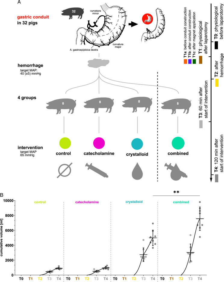
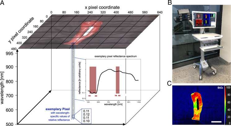
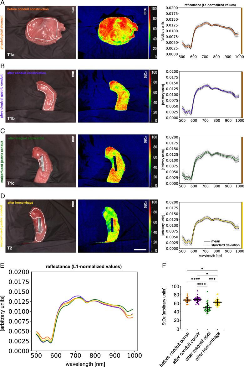
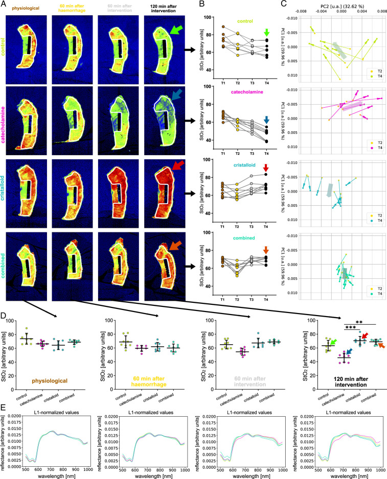
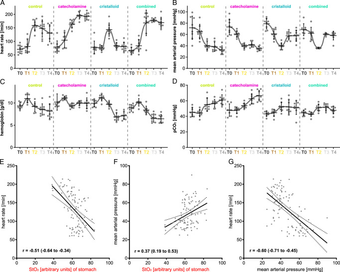
Material and methods: An established live porcine model ( n =32) for esophagectomy was used with gastric conduit formation and simulation of a linear stapled side-to-side esophagogastrostomy. After a standardized procedure of controlled blood loss of about 1 l per pig, the four experimental groups ( n =8 each) differed in anesthesiologic intervention, that is, (I) permissive hypotension, (II) catecholamine therapy using noradrenaline, (III) crystalloid volume supplementation, and (IV) combined crystalloid volume supplementation with noradrenaline therapy. Hyperspectral imaging tissue oxygenation (StO 2 ) of the gastric conduit was evaluated and correlated with systemic perfusion parameters. Measurements were conducted before (T0) and after (T1) laparotomy, after hemorrhage (T2), and 60 min (T3) and 120 min (T4) after anesthesiologic intervention.

Results: StO 2 values of the gastric conduit showed significantly different results between the four experimental groups, with 63.3% (±7.6%) after permissive hypotension (I), 45.9% (±6.4%) after catecholamine therapy (II), 70.5% (±6.1%) after crystalloid volume supplementation (III), and 69.0% (±3.7%) after combined therapy (IV). StO 2 values correlated strongly with systemic lactate values (r=-0.67; CI -0.77 to -0.54), which is an established prognostic factor.

Conclusion: Crystalloid volume supplementation (III) yields the highest StO 2 values and lowest systemic lactate values and therefore appears to be the superior primary treatment strategy after hemorrhage during esophagectomy with regards to microcirculatory tissue oxygenation of the gastric conduit.


Conflict of interest statement

Felix Nickel reports support for courses and travel from Johnson and Johnson, Medtronic, Intuitive Surgical, Cambridge Medical Robotics and KARL STORZ as well as consultancy fees from KARL STORZ. The remaining authors report no conflicts of interest.

Figures

Figure 1
Figure 1
Recording protocol. A, visualization of the recording protocol with four groups along five timepoints. B, quantification of applied intravenous fluid.
Figure 2
Figure 2
Hyperspectral Imaging technology. Visualization of HSI technology. A, HSI datacube with indicated spectral bands relevant for computing StO2. B, TIVITA Tissue camera system. C, example StO2 index image. Scale bar equals 5 cm.
Figure 3
Figure 3
Visualization of physiological baseline stomach and gastric conduit data. HSI color index pictures and respective spectra in a porcine model (n=32). A, T1a: before gastric conduit construction. B, T1b: after gastric conduit construction. C, T1c: malperfused region of gastric conduit after magnet application (separate region). D, T2: gastric conduit after hemorrhage. E, comparison of L1-normalized reflectance spectra. F, quantification of hyperspectral index values for StO2. Scale bar equals 5 cm.
Figure 4
Figure 4
Temporal course of changes in reflectance of the gastric conduit (T1, T2, T3, T4). HSI color index pictures, StO2 quantifications and respective spectra for the gastric conduit over the course of baseline (T1), after hemorrhage (T2), 60 min after intervention (T3) and 120 min after intervention (T4) stratified for interventional groups. A, HSI StO2 color index pictures. B, StO2 quantifications and respective spectra comparing groups. C, PCA visualizing the development from T2 to T4. D–E, StO2 quantifications and respective L1-normalized reflectance spectra comparing timepoints stratified for interventional groups. Scale bar equals 5.
Figure 5
Figure 5
Circulatory physiology parameter values. A, heart rate. B, mean arterial blood pressure. C, hemoglobin concentration. D, pCO2. E, regression model for StO2 values of the stomach and heart rate (R2: 0.2565; y-intercept: 299.2 (SE: 30.06); slope: −2.707 (SE: 0.4753); F: 32.44; df: 1, 94; P: <0.0001). F, regression model for StO2 values of the stomach and mean arterial blood pressure (R2: 0.1401; y-intercept: 11.52 (SE: 9.261); slope: 0.5730 (SE: 0.1465); F: 15.31; df: 1, 94; P: 0.0002). G, regression model for mean arterial blood pressure and heart rate (R2: 0.3571; y-intercept: 0.228.6 (SE: 14.22); slope: −2.086 (SE: 0.2887); F: 52.22; df: 1, 94; P: <0.0001).
Figure 6
Figure 6
Corresponding lactate values. Depiction of lactate values of the experimental groups. A, lactate values (n=156). B, regression model of arterial and venous lactate values (R2: 0.97; y-intercept: −0.1238 (SE: 0.04747); slope: 1.007 (SE: 0.01537); F: 4290; df: 1, 114; P: <0.0001). C, a regression model for gastric conduit and arterial lactate (R2: 0.4459; y-intercept: 11.25 (SE: 1.010); slope: −0.1370 (SE: 0.01601); F: 73.22; df: 1, 91; P: <0.0001. Orange markings depict the outliers of an individual with impaired circulation. Scale bar equals 5 cm.
Figure 7
Figure 7
Comparison of human and porcine gastric spectra. HSI color index pictures and respective spectra for porcine and human gastric conduit. A, porcine gastric conduit physiological (pcp) (n=32, I=32). B, porcine gastric conduit malperfused (pcm) (n=32, I=32). C, human gastric conduit physiological (hcp) (n=57, I=10). D, human gastric conduit malperfused (hcm) (n=14, I=4). E, PCA visualization with an explained variability of 64.4% on the x-axis and 20.3% on the y-axis. F, comparison of L-1 normalized reflectance spectra. G, quantification of StO2 index values. Scale bar equals 5 cm.
Figure 8
Figure 8
Examples of clinical application. Exemplary human data of single patients with useful clinical application. a, malperfused gastric conduit stump in the abdomen. b, gastric conduit before (physiological) and after (clearly malperfused) mobilization into the thoracic cavity compared to an intrathoracic physiological gastric conduit in another patient. Scale bar equals 5 cm.

Similar articles

Cited by

References

    1. Esagian SM, Ziogas IA, Skarentzos K, et al. . Robot-assisted minimally invasive esophagectomy versus open esophagectomy for esophageal cancer: a systematic review and meta-analysis. Cancers (Basel) 2022;14:3177. - PMC - PubMed
    1. Casas MA, Angeramo CA, Bras Harriott C, et al. . Surgical outcomes after totally minimally invasive Ivor Lewis esophagectomy. A systematic review and meta-analysis. Eur J Surg Oncol 2022;48:473–481. - PubMed
    1. Biere SS, Cuesta MA, van der Peet DL. Minimally invasive versus open esophagectomy for cancer: a systematic review and meta-analysis. Minerva Chir 2009;64:121–133. - PubMed
    1. Bras Harriott C, Angeramo CA, Casas MA, et al. . Open versus hybrid versus totally minimally invasive Ivor Lewis esophagectomy: systematic review and meta-analysis. J Thorac Cardiovasc Surg 2022;164:e233–e254. - PubMed
    1. Taurchini M, Cuttitta A. Minimally invasive and robotic esophagectomy: state of the art. J Vis Surg 2017;3:125. - PMC - PubMed

MeSH terms

LinkOut - more resources