

[Exogenous leptin improves cerebral ischemia-reperfusion-induced glutamate excitotoxic injury in mice by up-regulating GLT-1 and GLAST expression in astrocytes]
- PMID: 38977337
- PMCID: PMC11237293
- DOI: 10.12122/j.issn.1673-4254.2024.06.08
[Exogenous leptin improves cerebral ischemia-reperfusion-induced glutamate excitotoxic injury in mice by up-regulating GLT-1 and GLAST expression in astrocytes]
Abstract
Objective: To investigate the protective effect of exogenous leptin against focal cerebral ischemia-reperfusion (I/R) injury in mice and explore the underlying mechanism.
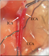
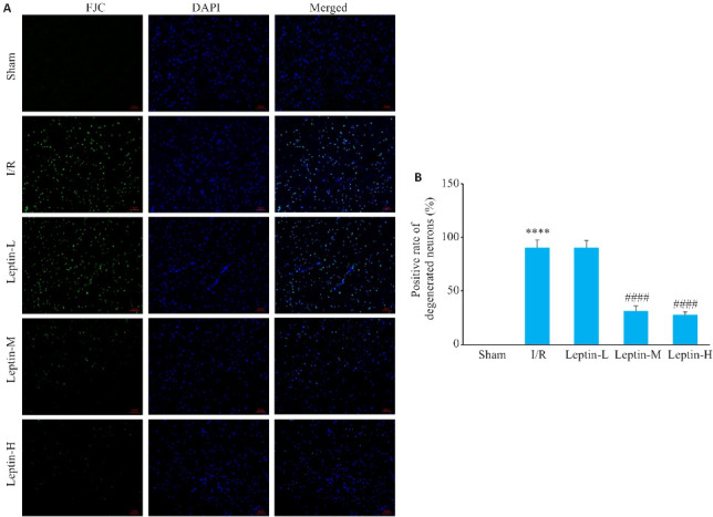
Methods: A total of 100 C57BL/6 mice were randomly divided into 5 groups, including a sham-operated group, cerebral I/R model group, and 3 leptin treatment groups with intraperitoneal injections of 0.5, 1.0 or 2.0 leptin immediately after occlusion of the internal carotid artery. At 24 h after reperfusion, neurological function scores of the mice were assessed, and TTC staining was used to determine the area of cerebral infarction. The pathological changes in the cortical brain tissue of the mice were observed using HE staining, and degenerative damage of the cortical neurons were assessed with Fluoro-Jade C staining. The expression of glial fibrillary acidic protein in cortical brain tissues was detected using immunohistochemistry and Western blotting. In another 45 C57BL/6 mice with sham operation, I/R modeling, or leptin (1 mg/kg) treatment, glutamic acid in the cortical brain tissue was detected using glutamate assay, and cortical glutamate-aspartate transporter (GLAST) and glutamate transporter-1 (GLT-1) protein expressions were detected using immunohistochemistry.
Results: Compared with the I/R model mice, the leptin-treated mice had significantly lower neurological deficit scores, smaller cerebral infarct area, milder pathologies in the cortical brain tissue, and lessened cortical neuronal damage with normal morphology and less excessive proliferation of the astrocytes. Leptin treatment significantly up-regulated the expressions of GLT-1 and GLAST and lowered the content of glutamic acid in the brain tissue of the I/R mice.
Conclusion: Exogenous leptin has obvious neuroprotective effect against cerebral I/R injury in mice, mediated probably by controlling excessive astrocyte proliferation and up-regulating cortical GLT-1 and GLAST expressions to reduce glutamate-mediated excitotoxic injury of the astrocytes.
目的: 探究外源性瘦素(Leptin)对小鼠局灶性脑缺血再灌注损伤的保护作用及其内在机制。
方法: 将100只C57BL/6小鼠随机分为5组:假手术(Sham)组、脑缺血/再灌注模型组(I/R组)、瘦素低剂量组(Leptin-L组,0.5 mg/kg)、瘦素中剂量组(Leptin-M组,1 mg/kg)和瘦素高剂量组(Leptin-H组,2 mg/kg),20只/组。采用改良线栓法复制小鼠局灶性脑缺血/再灌注模型,缺血1.5 h再灌注24 h后进行神经功能评分评估神经功能缺损程度,TTC染色测定脑梗死面积,HE染色观察皮层脑组织病理变化,退化神经元染色观察皮层神经元变性损伤程度,免疫组织化学染色测定皮层脑组织胶原酸性纤维蛋白的表达和形态变化,Western blot测定皮层脑组织胶原酸性纤维蛋白蛋白表达。在此基础上,另将45只C57BL/6小鼠随机分为Sham组、I/R组和Leptin组(1 mg/kg),15只/组。谷氨酸检测试剂盒检测皮层脑组织谷氨酸含量,免疫组织化学染色测定皮层谷氨酸-天冬氨酸转运体(GLAST)和谷氨酸转运体1(GLT-1)表达,Western blotting测定GLAST、GLT-1蛋白表达。
结果: 与I/R组比较,瘦素明显降低小鼠神经功能缺损评分(P<0.0001),缩小脑梗死面积(P<0.001),减轻皮层脑组织病理损伤程度,降低皮层神经元损伤程度(P<0.0001),维持星形胶质细胞正常形态及降低星形胶质细胞的过度增生和表达(P<0.01),使GLT-1、GLAST表达上调(P<0.01、P<0.05),脑组织谷氨酸含量降低(P<0.01)。
结论: 外源性瘦素对小鼠脑缺血再灌注损伤具有明显的神经保护作用,其机制可能与调节星形胶质细胞过度增生和上调GLT-1、GLAST的表达从而降低谷氨酸的兴奋毒性损伤有关。
Keywords: astrocytes; cerebral ischemia-reperfusion injury; excitotoxicity; glutamate transporter-1; glutamate-aspartate transporter; leptin.
Figures


References
Publication types
MeSH terms
Substances
LinkOut - more resources
Full Text Sources