. 2024 Jul 9;213(1):14.
doi: 10.1007/s00430-024-00794-z.

Exploring the potential of Lactocaseibacillus rhamnosus PMC203 in inducing autophagy to reduce the burden of Mycobacterium tuberculosis

Affiliations

Exploring the potential of Lactocaseibacillus rhamnosus PMC203 in inducing autophagy to reduce the burden of Mycobacterium tuberculosis

Md Abdur Rahim et al. Med Microbiol Immunol. .

Abstract

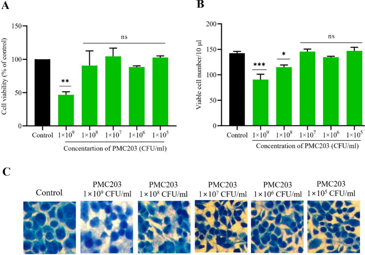
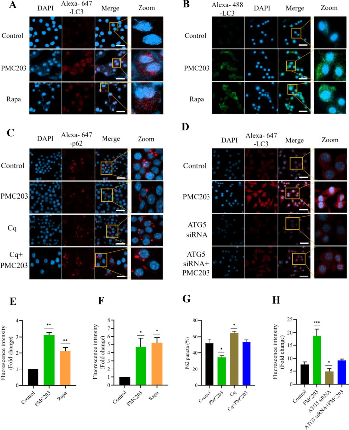
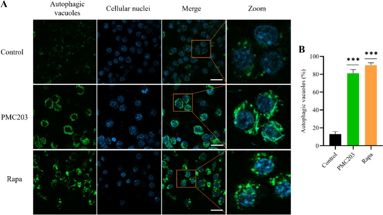
Mycobacterium tuberculosis, a lethal pathogen in human history, causes millions of deaths annually, which demands the development of new concepts of drugs. Considering this fact, earlier research has explored the anti-tuberculosis potential of a probiotic strain, Lactocaseibacillus rhamnosus PMC203, leading to a subsequent focus on the molecular mechanism involved in its effect, particularly on autophagy. In this current study, immunoblotting-based assay exhibited a remarkable expression of autophagy marker LC3-II in the PMC203 treated group compared to an untreated group. A remarkable degradation of p62 was also noticed within treated cells compared to control. Furthermore, the immunofluorescence-based assay showed significant fold change in fluorescence intensity for alexa-647-LC3 and alexa-488-LC3, whereas p62 was degraded noticeably. Moreover, lysosomal biogenesis generation was elevated significantly in terms of LAMP1 and acidic vesicular organelles. As a result, PMC203-induced autophagy played a vital role in reducing M. tuberculosis burden within the macrophages in treated groups compared to untreated group. A colony -forming unit assay also revealed a significant reduction in M. tuberculosis in the treated cells over time. Additionally, the candidate strain significantly upregulated the expression of autophagy induction and lysosomal biogenesis genes. Together, these results could enrich our current knowledge of probiotics-mediated autophagy in tuberculosis and suggest its implications for innovatively managing tuberculosis.

Keywords: Lactocaseibacillus rhamnosus PMC203; Mycobacterium tuberculosis; Anti-tuberculosis; Autophagy; Probiotics.


Conflict of interest statement

The authors declare no competing interests.

Figures

Fig. 1
Fig. 1
Evaluation of potential cytotoxicity of PMC203 to macrophages. Macrophages were incubated with a range of PMC203 concentrations and then cytotoxicity was evaluated employing (A) WST (water-soluble tetrazolium salt), and (B) trypan blue exclusion assay indicating no cytotoxicity except for higher doses. (C) Cell morphology for each concentration treatment condition is presented in light microscope images stained with methylene blue dye. The experiment was performed in triplicate, presenting the results as mean values with corresponding standard deviations. Significance was determined compared to the untreated group using one-way analysis of variance (*p < 0.05; **p < 0.01; ***p < 0.001; ns: non-significant)
Fig. 2
Fig. 2
Investigation of PMC203-induced autophagy in macrophage cells. The effect of autophagic flux in response to PMC203 was explored by immunoblotting over time. (A, B) shows the expression of LC3 and p62, in which visible differences observed in PMC203-treated cells. The obtained protein bands were analyzed by Image J software, showing the increased (C) LC3-II to β-actin ratio (D) and decreased p62 to β-actin ratio in the treated cells compared to untreated cells. (E) LC3-II or (F) p62 to β-actin ratio were compared to their corresponding 1 h values, showing a time-dependent autophagy induction by PMC203. (G, H) Furthermore, autophagy induction was confirmed by using chloroquine (Cq). The experiment was performed in triplicate, presenting the results as mean values with corresponding standard deviations. The statistical significance of the obtained data was determined using with a one-way analysis of variance (*p < 0.05; **p < 0.01; ***p < 0.001; ****p < 0.0001; ##p < 0.01). *, statistical significance compared with the control; #, denotes statistical significance of PMC203 vs. Cq + PMC203 and Cq vs. Cq + PMC203 treated group
Fig. 3
Fig. 3
Evaluation of PMC203 mediated autophagic flux based on immunofluorescence. Macrophages were treated with PMC203 or rapamycin (Rapa) and/or chloroquine (Cq) or siRNA. Results show a visual increased of LC3, (A, D) red and (B) green fluorescence, whereas (C) p62 puncta was decreased in PMC203 treated cells compared to untreated cells. In images, the scale bar indicates a length of 10 μm. (E, F, H) The fluorescence intensities of the obtained images were analyzed by image J and (G) the number of p62 puncta-positive cells was counted from 100 cells per sample. The experiment was performed in triplicate, presenting the results as mean values with corresponding standard deviations. The statistical significance of obtained data was determined compared to the untreated group using a one-way analysis of variance (*p < 0.05; **p < 0.01; ****p < 0.0001)
Fig. 4
Fig. 4
Determination of PMC203-induced autophagic vacuole formation. (A) The effects of PMC203 on the formation of LC3 and vesicle colocalization were measured, indicating an elevated visual expression of green fluorescence compared to an untreated group. The scale bar in these images indicates a length of 10 μm. (B) The number of positive cells with > 3 green puncta was calculated, with data obtained from 100 cells per sample. The experiment was performed in triplicate. Results are presented as mean values with corresponding standard deviations. The statistical significance of obtained data compared to an untreated group was determined using a one-way analysis of variance (****p < 0.0001)
Fig. 5
Fig. 5
Assessment of PMC203-induced lysosomal biogenesis formation. The generation of lysosomal biogenesis in response to PMC203 treatment was investigated, showing a visual increase of (A) AVOs and (C) LAMP1 in the treated cells compared to the control. In these images, the scale bar represents 10 µm. (B, D) Image J analysis of fluorescence intensities of the obtained images. The experiment was performed in triplicate. Results are presented as mean values with corresponding standard deviations. The statistical significance of the obtained data compared to an untreated group was determined using a one-way analysis of variance (**, p < 0.01; ****, p < 0.0001)
Fig. 6
Fig. 6
Evaluation of M. tuberculosis reduction through induction of autophagy by PMC203. (A) Macrophages were infected, treated with PMC203, and colocalization of GFP-H37Ra with LC3 puncta was observed, indicating increased colocalization in the treated group. (B) Meanwhile, the number of positive cells with > 1 colocalization was calculated using image J. (C) At the same time, the number of positive cells containing > 6 GFP-H37Ra signal was calculated, in which H37Ra signal was reduced in the treated group compared to the untreated group. In these images, the scale bar represents 10 µm. (B, D) Image J analysis of fluorescence intensities of the obtained images. (D) The conversion of LC3-I to LC3-II was examined using an immunoblot assay, showing a significant statistical difference between the groups. To observe the (E) bactericidal and (F) phagocytosis effect, CFU was determined in a time-dependent manner using M. tuberculosis-specific Middlebrook H710 agar media, showing the reduction of H37Rv in the PMC203-treated groups, whereas the reduction was disrupted in the Chloroquine (Cq)/3-methyladenine (3-MA) treated cells. (G) Additionally, the impact of PMC203 induced autophagy on M. tuberculosis survival was evaluated with siRNA using the acid-fact bacilli staining method. The images of acid-fast bacilli staining include a scale bar representing 10 µm. (B, D) Image J analysis of fluorescence intensities of the obtained images. The experiment was performed in triplicate. Results are presented as mean values with corresponding standard deviations. The statistical significance of the obtained data was determined compared to the untreated group using one-way analysis of variance (*p < 0.05; **p < 0.01; ****p < 0.0001; #p < 0.05)
Fig. 7
Fig. 7
Measurement of PMC203-induced autophagic gene expression. RT-PCR assay revealed a significant upregulation of most of the (A) autophagy induction and (B) lysosomal biogenesis genes in cells treated with H37Rv + PMC203, while H37Rv treatment alone failed to reach significant levels, both in comparison with the control. The experiment was performed in triplicate. Results are presented as mean values with corresponding standard deviations. The statistical significance of the obtained data was determined compared to the untreated group using a one-way analysis of variance (*p < 0.05; **p < 0.01)

Similar articles

Cited by

References

    1. Floyd K, Glaziou P, Zumla A, Raviglione M (2018) The global tuberculosis epidemic and progress in care, prevention, and research: an overview in year 3 of the End TB era. Lancet Respir Med 6:299–314. 10.1016/S2213-2600(18)30057-2 - PubMed
    1. WHO, GLOBAL TUBERCULOSIS REPORT 2022. Report No. 978–92–4–006172–9, 68 (2022).
    1. Raviglione M, Marais B, Floyd K, Lönnroth K, Getahun H, Migliori GB, Harries AD, Nunn P, Lienhardt C, Graham S et al (2012) Scaling up interventions to achieve global tuberculosis control: progress and new developments. The Lancet 379:1902–1913. 10.1016/S0140-6736(12)60727-2 - PubMed
    1. Dartois VA, Rubin EJ (2022) Anti-tuberculosis treatment strategies and drug development: challenges and priorities. Nat Rev Microbiol 20:685–701. 10.1038/s41579-022-00731-y - PMC - PubMed
    1. Singh V, Chibale K (2021) Strategies to combat multi-drug resistance in tuberculosis. Accounts Chem Res 54:2361–2376. 10.1021/acs.accounts.0c00878 - PMC - PubMed

MeSH terms

Substances

LinkOut - more resources