. 2024 Jul 8;15(1):5715.
doi: 10.1038/s41467-024-50041-5.

Egg multivesicular bodies elicit an LC3-associated phagocytosis-like pathway to degrade paternal mitochondria after fertilization

Affiliations

Egg multivesicular bodies elicit an LC3-associated phagocytosis-like pathway to degrade paternal mitochondria after fertilization

Sharon Ben-Hur et al. Nat Commun. .

Abstract

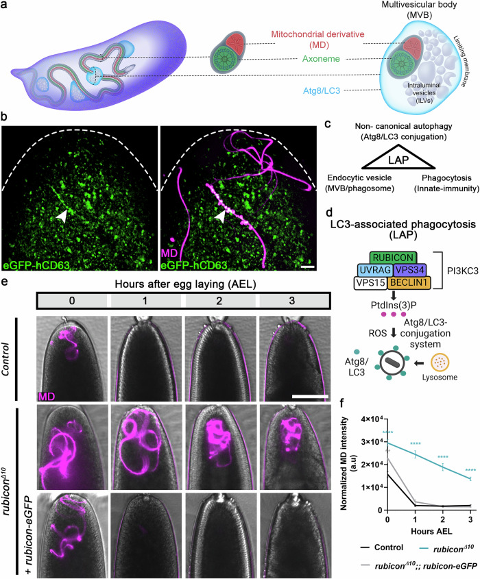
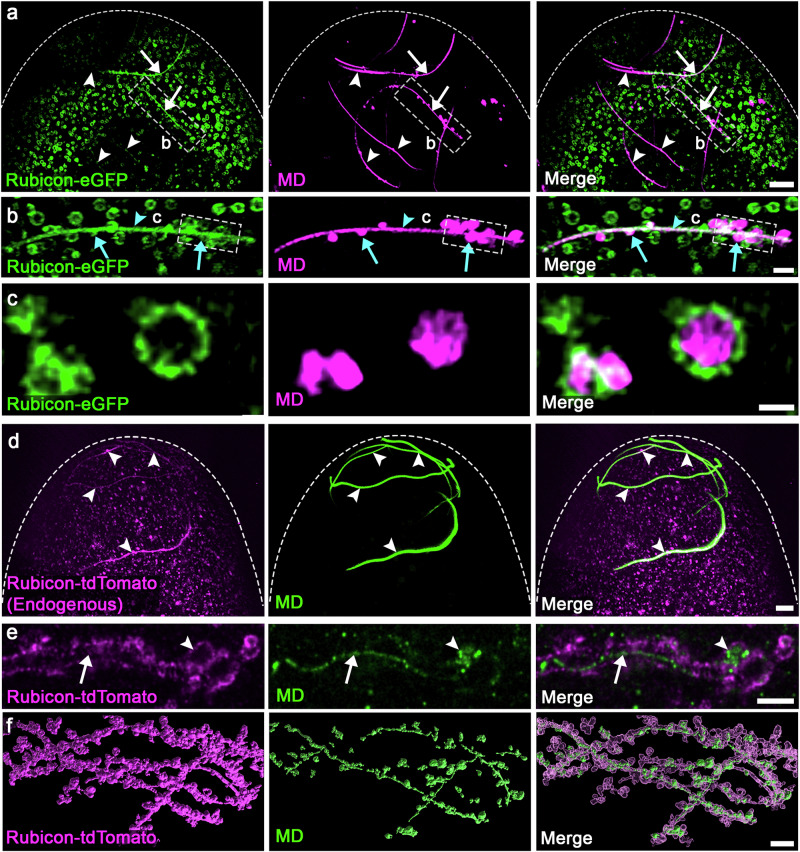
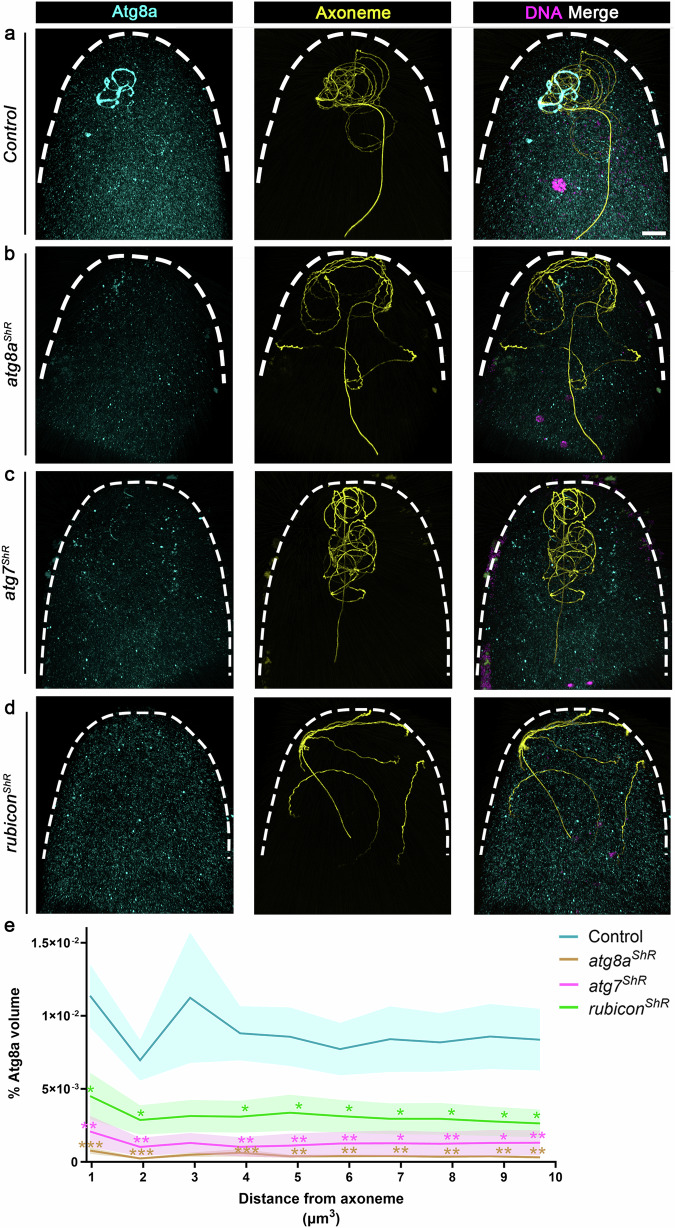
Mitochondria are maternally inherited, but the mechanisms underlying paternal mitochondrial elimination after fertilization are far less clear. Using Drosophila, we show that special egg-derived multivesicular body vesicles promote paternal mitochondrial elimination by activating an LC3-associated phagocytosis-like pathway, a cellular defense pathway commonly employed against invading microbes. Upon fertilization, these egg-derived vesicles form extended vesicular sheaths around the sperm flagellum, promoting degradation of the sperm mitochondrial derivative and plasma membrane. LC3-associated phagocytosis cascade of events, including recruitment of a Rubicon-based class III PI(3)K complex to the flagellum vesicular sheaths, its activation, and consequent recruitment of Atg8/LC3, are all required for paternal mitochondrial elimination. Finally, lysosomes fuse with strings of large vesicles derived from the flagellum vesicular sheaths and contain degrading fragments of the paternal mitochondrial derivative. Given reports showing that in some mammals, the paternal mitochondria are also decorated with Atg8/LC3 and surrounded by multivesicular bodies upon fertilization, our findings suggest that a similar pathway also mediates paternal mitochondrial elimination in other flagellated sperm-producing organisms.


Conflict of interest statement

The authors declare no competing interests.

Figures

Fig. 1
Fig. 1. Rubicon is required for PME after fertilization.
a Illustration of the onset of PME in Drosophila based on. Left, an early fertilized egg. Egg-derived MVBs associate with the 1.8 mm long sperm flagellum. Middle, a cross-section through the sperm flagellum illustrating a single cylindrical MD (red) and an intimately associated axoneme (green), both coated by a common plasma membrane (gray) and extend along the entire length of the flagellum. Right, enlargement of an MVB (shown in a cross-section displaying the ILVs and the encapsulating limiting membrane) that encapsulates the flagellum. Atg8/LC3 (cyan) is loaded on the MVB limiting membrane. b A representative confocal image of the anterior (top) region of an egg maternally expressing the MVB transgenic construct UASp-hCD63-eGFP (green), and fertilized by a coiled red-MD sperm cell (magenta; dj-(MTS)tdTomato). MVBs that coat large flagellar segments are in white (arrowhead). Scale bar, 10 μm. c Schematic view of three main features shared by the egg MVBs and the LAP pathway. d Schematic view of the LAP pathway as defined in mammalian cells (created with BioRender.com). e Live imaging of the anterior regions of early fertilized eggs laid by females of the indicated genotypes and fertilized by red-MD sperm. Early fertilized eggs carrying a single copy of the rubicon gRNA served as control. Representative time-lapse images of each fertilized egg were taken at 0, 1, 2, and 3 h AEL. Brightfield and fluorescence channel image overlay reveals the egg (gray) and the red-MD sperm flagellum (magenta), respectively. Scale bar, 100 μm. f Quantifications of normalized red-MD fluorescence intensities [arbitrary units (a.u.)] in fertilized eggs represented in e. Error bars indicate standard error of the mean (SEM). The respective numbers of scored early fertilized eggs (n) laid by females of the control, rubiconΔ10, and rubiconΔ10; rubicon-eGFP genotypes are 61, 32, and 34. **P  <   0.01 and ****P  <   0.0001. Two-way repeated measures ANOVA, followed by Dunnett’s multiple comparisons test. The P values are as follows: control vs. rubiconΔ10, P < 0.0001 (all tested hours AEL); control vs. rubiconΔ10; rubicon-eGFP, P = 0.0070 (0 h AEL). Source data are provided as a Source Data file.
Fig. 2
Fig. 2. Main members of the LAP-specific, but not the autophagy-specific PI3KC3 complex are required for PME.
a, b, d, f, h, j PME kinetic assays in control (expressing the maternal driver only) (a); and shRNA (ShR) mediated maternal knockdown in early fertilized eggs of rubicon (b), uvrag (d), pi3k (f), atg6 (h), and atg14 (j). A live imaging assay performed and presented as in Fig. 1e. Scale bar, 100 μm. c, e, g, i, k Quantifications of normalized red-MD fluorescence intensities in the fertilized eggs [magenta; arbitrary units (a.u.)] of the genotypes indicated on the left. Error bars indicate SEM. The respective numbers of scored fertilized eggs (n) laid by females of the control, rubiconShR (c), uvragShR (e), pi3kShR (g), and atg6ShR (h), are 68, 60, 24, 22, and 24. For atg14 knockdown and its control, 30 and 51 fertilized eggs were scored, respectively (k). Note that atg14 has its own control group, as this experiment was conducted at a later time. *P  <   0.05, **P  <   0.01, ***P  <   0.001, and ****P  <   0.0001. Two-way repeated measures ANOVA, followed by Šídák’s multiple comparisons test. P values in c, P < 0.0001 (1–3 h AEL); e, P < 0.0001 (1–2 h AEL), P = 0.0446 (3 h AEL); g, P = 0.0436 (0 h AEL), P < 0.0001 (1 h AEL), P = 0.0011 (2 h AEL); i, P = 0.0244 (1 h AEL); k, P = 0.0005, (0 h AEL), P = 0.0122 (1 h AEL). MD mitochondrial derivative. Source data are provided as a Source Data file.
Fig. 3
Fig. 3. Rubicon is localized to the limiting membrane of multiple egg vesicles which generate extended vesicular sheaths that enwrap the sperm flagellum.
a, d Representative confocal images of the anterior (top) region of early fertilized eggs. a An egg maternally expressing the UASz-rubicon-eGFP transgene (green) and fertilized by a red-MD sperm cell (magenta). Numerous egg vesicles display Rubicon-eGFP, many of which associate with large segments of the sperm flagellum (arrows), whereas other flagellar segments show only a few vesicles or are still free of vesicles (arrowheads), due to the asynchronous manner of the PME process along the sperm flagellum. Scale bar, 10 μm. b Magnification of the area outlined by a dashed rectangle in (a). Two types of Rubicon positive vesicles are detected: Numerous small vesicles that densely associate with flagellar segments in a rather unified order (arrowhead); Large Rubicon positive vesicles (green) that appear to bud from the flagellum, encapsulating MD pieces and debris (magenta; arrows). Scale bar, 2 μm. c Magnification of a projection of a few Z-sections in the area outlined by a dashed rectangle in (b). Rubicon (green) remains localized on the limiting membrane of the large vesicles that contain MD fragments and bud off from the flagellum. Scale bar, 1 μm. d An egg expressing tdTomato-tagged endogenous Rubicon (magenta) and fertilized by green-MD sperm (green). Rubicon is localized on multiple egg vesicles, many of which associate with large flagellar segments (arrowheads). Scale bar, 10 μm. e, f Shown are super-resolution images of flagellar regions in eggs maternally expressing the UASz-rubicon-tdTomato transgene (magenta), fertilized by green-MD sperm (green; dj-DJ-GFP), and prepared for ExM. The images feature large MD segments that are readily enwrapped within Rubicon positive FVS (arrow), as well as large Rubicon positive vesicles that contain MD fragments and bud off from the flagellum (arrowhead). Note that the image in (e) presents a single non-computed optical section, while the image in (f) presents a 3D computer rendering of segmented surfaces of this early fertilized egg. MD, mitochondrial derivative.
Fig. 4
Fig. 4. Sperm plasma membrane breakdown after fertilization occurs within the FVS at a much faster rate than that of MD degradation.
a, f Super-resolution images of flagellar regions in an egg maternally expressing the UASz-rubicon-tdTomato transgenic construct (magenta; immunostained with anti-RFP antibody), fertilized by sperm cells expressing the dj-CD8-Venus transgene that labels the plasma membrane (a) or the dj-DJ-GFP transgene that labels the MD (f, a green-MD sperm cell), both immunostained with an anti-GFP antibody (cyan), immunostained to visualize the axoneme (yellow), and prepared for ExM. The images at the top depict single optical sections before computing [scale bar (left), 1 μm; scale bar (right), 0.5 μm], while the images at the bottom depict 3D computer rendering of segmented surfaces in the early fertilized eggs (scale bar, 2 μm). Note the narrow vesicular tube within the FVS that confines the axoneme (arrowheads). b, c Representative confocal images of the anterior regions in early fertilized eggs laid by control (b) and maternal rubicon knockdown (c) mothers. The eggs were fertilized by sperm cells expressing the dj-CD8-Venus transgene (cyan; immunostained with anti-GFP antibody) and immunostained to visualize the axoneme (yellow). DAPI stained the DNA (magenta). Scale bar, 20 μm. d The graphs depict staining volumes of the sperm plasma membrane relative to the axoneme volume in each fertilized egg, corresponding to the fertilized eggs in (b,c). The respective numbers of examined fertilized eggs (n) laid by control and rubiconShR females are 22 and 18. Two-tailed unpaired Student’s t test. ****P  <  0.0001. Summary of the statistics reported in the violin plots: the center line represents the median of the frequency distribution of the data. Quartiles are represented by a dashed black line. Each dot corresponds to the relative sperm plasma membrane/Axoneme volume in a single fertilized egg. e, Live confocal imaging of the anterior region of an early fertilized egg laid by a female maternally expressing the rubicon-tdTomato transgene (magenta) and fertilized by a sperm cell expressing the CD8-Venus transgenic marker of the plasma membrane (green). Shown are representative images from Supplementary Movie 10 at the indicated times (min AEL). Scale bar, 3 μm. Yolk autofluorescence is indicated by arrowheads. MD mitochondrial derivative. Source data are provided as a Source Data file.
Fig. 5
Fig. 5. Rubicon-coated MVBs express active PI3KC3.
a, b A live imaging projection of the anterior region of an egg maternally expressing both a UASz-rubicon-eGFP (green) and the UASz-hCD63-tdTomato (magenta; MVBs) transgenic constructs, and fertilized by a WT (non-fluorescent) sperm cell. Magnification of the area outlined by a dashed rectangle in (a) is shown in (b). Rubicon positive vesicles and FVS also display hCD63-tdTomato (arrow in a). Scale bars in (a) 5 μm; (b) 0.5 μm. c Most (~85%) of the Rubicon-eGFP positive vesicles also display the MVB marker hCD63-tdTomato. Measure of center is the mean percentage. Error bar indicates standard deviation (SD). The pulled Pearson’s Correlation Coefficient (PCC) is 0.3402292 (P-value < 2.2e–16). The pulled Manders’ Colocalization Coefficient is 0.9984238. n = 4. d, e, h Representative confocal images of the anterior region of early fertilized eggs. d An egg maternally expressing both the rubicon-eGFP transgene (green) and the PtdIns(3)P reporter TagRFPt-2xFYVE (magenta), and fertilized by a WT (non-fluorescent) sperm cell. The egg contains multiple PtdIns(3)P positive vesicles (arrowhead), including large Rubicon-eGFP positive vesicles (arrows). Scale bar, 6 μm. e An egg maternally expressing the PtdIns(3)P reporter (magenta) and fertilized by green-MD sperm (green). In addition to the large PtdIns(3)P positive vesicles derived from the flagellum (arrowheads), flagellar PtdIns(3)P is also readily detected (arrows). Scale bar, 10 μm. f Representative confocal images of control (top) and maternal rubicon knockdown (bottom) early fertilized eggs at 0–15 min AEL, maternally expressing the PtdIns(3)P reporter (magenta) and fertilized by green-MD sperm (green). Arrows indicate flagellar areas. Scale bar, 2 μm. g Quantification of the number of early fertilized eggs (corresponding to the eggs in f) that display flagellar PtdIns(3)P. Two-sided Fisher’s exact test. ***P = 0.0002. The number of examined fertilized eggs (n) laid by females of both genotypes is 23. h A WT egg fertilized by red-MD sperm (magenta) and stained with the fluorescent ROS indicator H2DCFDA (green). Scale bar, 10 μm. MD mitochondrial derivative. Source data are provided as a Source Data file.
Fig. 6
Fig. 6. Atg8a recruitment to the FVS coincides with advanced MD degradation stages.
a A representative confocal image of a WT egg fertilized by green-MD sperm (magenta; stained with an anti-GFP antibody) and immunostained to visualize the axoneme (yellow) and Atg8a (cyan). Shown is a sperm flagellar region exhibiting at least three defined consecutive PME stages: Intact MD segments that are Atg8a negative and usually still reside in the vicinity of parallel axonemal segments (I); Atg8a positive FVS and derived strings of large vesicles that encapsulate degrading MD fragments (IIa) and hence result in MD-free axonemal segments (IIb); MD-free axonemal segments displaying neither MD traces nor Atg8a positive FVS, indicative of completion of the PME process (III). Scale bar, 4 μm. bc Atg8 is required for efficient MD degradation. A live imaging assay performed and presented as in Fig. 1e. Early fertilized eggs expressing the maternal driver alone served as control (b, upper panel). Scale bar, 100 μm. c Quantifications of normalized red-MD fluorescence intensities [arbitrary units (a.u.)] in early fertilized eggs represented in (b), indicating that although single maternal knockdowns of atg8a and atg8b have no effect on PME kinetics (Supplementary Fig. 6b–e), double maternal knockdown fertilized eggs exhibit significant PME attenuation. Error bars indicate SEM. The respective numbers of examined fertilized eggs (n) laid by control and atg8aShR; atg8bShR females are 51 and 76. ****P <  0.0001. Two-way repeated measures ANOVA, followed by Šídák’s multiple comparisons test. MD, mitochondrial derivative. dg Live confocal imaging of the anterior region of an early fertilized egg laid by a female maternally expressing both the UASz-rubicon-eGFP (green) and UASp-mCherry-atg8a (magenta) transgenic constructs. Shown are representative images from Supplementary Movie 7 at the indicated times (min AEL). Note the 6–12 min lag between the formation of Rubicon-eGFP positive FVS and the subsequent recruitment of mCherry-Atg8a. Scale bar, 10 μm. hk Enlargement of the areas confined by dashed rectangles in (dg), presenting the same flagellar region at different timepoints AEL. Scale bars, 4 μm. Source data are provided as a Source Data file.
Fig. 7
Fig. 7. Atg8 recruitment to FVS requires Rubicon.
ad Representative confocal images of the anterior regions in early fertilized eggs laid by control (a) and maternal atg8a (b), atg7 (c), and rubicon (d) knockdown mothers. The eggs were fertilized by WT sperm cells, and immunostained to visualize Atg8a (cyan) and the axoneme (yellow). The 0–1-hour AEL fertilized eggs were further staged by the number of nuclei, as revealed by DAPI staining of the DNA (magenta). Scale bar, 20 μm. e The graphs depict staining volumes of Atg8a in early fertilized eggs corresponding to the fertilized eggs in (ad). Calculations were performed in intervals of 1 μm in 10 μm radius area around the axoneme. The respective numbers of scored early fertilized eggs (n) laid by females of the control, atg8aShR, atg7ShR, and rubiconShR, are 20, 12, 11, and 15. *P  <  0.05, **P <  0.01, and ***P <  0.001. P values are as follows: control vs. atg8aShR, P = 0.0003, 0.0004, 0.0009, 0.0019, 0.0019, 0.0063, 0.0029, 0.0047, and 0.0035, for the distances 1, 2, 4, 5, 6, 7, 8, 9, and 10 μm3 from axoneme, respectively; control vs. atg7ShR: P = 0.0013, 0.0013, 0.0013, 0.0042, 0.0059, 0.0138, 0.0076, 0.0111, and 0.0098, for the distances 1, 2, 4, 5, 6, 7, 8, 9, and 10 μm3 from axoneme, respectively; control vs. rubiconShR: P = 0.0154, 0.0236, 0.0128, 0.0348, 0.0379, 0.0371, 0.0294, 0.025, and 0.0203, for the distances 1, 2, 4, 5, 6, 7, 8, 9, and 10 μm3 from axoneme, respectively. Two-way repeated measures ANOVA, followed by Holm-Šídák’s multiple comparisons test. Error bars indicate SEM. Source data are provided as a Source Data file.
Fig. 8
Fig. 8. Atg8 positive strings of vesicles derived from the FVS and contain MD fragments fuse with lysosomes.
a Live confocal imaging of a sperm flagellar region in an egg ubiquitously expressing a transgenic construct that marks the lysosomes (magenta; tub-GFP-LAMP1), and fertilized by a red-MD sperm cell (green). Scale bar, 2 μm. b A sperm flagellar region in an egg ubiquitously expressing the lysosomal transgenic marker, GFP-LAMP1 (magenta), fertilized by a WT (non-fluorescent) sperm cell, and immunostained to visualize Atg8a (cyan) and the axoneme (yellow). Note the budding of the string of Atg8 positive large vesicles from the FVS (i.e., Atg8a positive regions that are not associated with the axoneme), and the association of GFP-LAMP1 with these large vesicles, which correspond to the vesicles that contain degrading MD fragments (a and Fig. 6a). Scale bar, 3 μm. c Enlargement of the area confined by a dashed rectangle in (b). Scale bar, 2 μm. d An integrated model of PME by a LAP-like pathway in Drosophila. An illustration of a sperm flagellum inside the anterior part of an early fertilized egg. Relevant flagellar and LAP pathway components are indicated by different signs and colors as defined on the right-hand side. Seven PME pathway steps are indicated by Roman numerals. Egg-derived Rubicon positive MVBs engage the sperm flagellum plasma membrane in an asynchronous manner immediately after fertilization (I). The MVBs densely coat the sperm flagellum, forming extended vesicular sheaths, within which the sperm plasma membrane rapidly breaks down (II). The MD is exposed to the degradative luminal area of the FVS that express the LAP-specific Rubicon PI3KC3 (III). Active Rubicon PI3KC3 generates PtdIns(3)P (IV), which presumably with the help of ROS (produced by the egg and the MD), promotes the recruitment of Atg8/LC3 to the FVS (V). The Atg8/LC3 positive FVS bud off strings of large vesicles that contain degrading MD fragments (VI). Lysosomes fuse with the strings of large vesicles, promoting efficient degradation of the MD fragments (VII).

References

    1. Schurko AM, Logsdon JM. Using a meiosis detection toolkit to investigate ancient asexual ‘scandals’ and the evolution of sex. BioEssays. 2008;30:579–589. doi: 10.1002/bies.20764. - DOI - PubMed
    1. Bloomfield G. The molecular foundations of zygosis. Cell. Mol. Life Sci. 2020;77:323–330. doi: 10.1007/s00018-019-03187-1. - DOI - PMC - PubMed
    1. Sankaranarayanan S, Higashiyama T. Capacitation in plant and animal fertilization. Trends Plant Sci. 2018;23:129–139. doi: 10.1016/j.tplants.2017.10.006. - DOI - PubMed
    1. Lillie FB. The mechanism of fertilization. Science (1979) 1913;38:524–528. - PubMed
    1. Fuller, M. T. The Development of Drosophila melanogaster (eds. Bate, M. & Martinez-Arias, A.) 71 (Cold Spring Harbor Laboratory Press, 1993).

Substances

LinkOut - more resources