Review
. 2025 Jan;22(1):26-45.
doi: 10.1038/s41585-024-00900-z. Epub 2024 Jul 8.

Biological determinants of PSMA expression, regulation and heterogeneity in prostate cancer

Affiliations
Review

Biological determinants of PSMA expression, regulation and heterogeneity in prostate cancer

Martin K Bakht et al. Nat Rev Urol. 2025 Jan.

Abstract

Prostate-specific membrane antigen (PSMA) is an important cell-surface imaging biomarker and therapeutic target in prostate cancer. The PSMA-targeted theranostic 177Lu-PSMA-617 was approved in 2022 for men with PSMA-PET-positive metastatic castration-resistant prostate cancer. However, not all patients respond to PSMA-radioligand therapy, in part owing to the heterogeneity of PSMA expression in the tumour. The PSMA regulatory network is composed of a PSMA transcription complex, an upstream enhancer that loops to the FOLH1 (PSMA) gene promoter, intergenic enhancers and differentially methylated regions. Our understanding of the PSMA regulatory network and the mechanisms underlying PSMA suppression is evolving. Clinically, molecular imaging provides a unique window into PSMA dynamics that occur on therapy and with disease progression, although challenges arise owing to the limited resolution of PET. PSMA regulation and heterogeneity - including intertumoural and inter-patient heterogeneity, temporal changes, lineage dynamics and the tumour microenvironment - affect PSMA theranostics. PSMA response and resistance to radioligand therapy are mediated by a number of potential mechanisms, and complementary biomarkers beyond PSMA are under development. Understanding the biological determinants of cell surface target regulation and heterogeneity can inform precision medicine approaches to PSMA theranostics as well as other emerging therapies.


Conflict of interest statement

Competing interests: H.B. has served as consultant/advisory board member for Janssen, Astellas, AstraZeneca, Merck, Pfizer, Blue Earth Diagnostics, Amgen, Bayer, Daicchi Sankyo, Sanofi and Novartis, and has received research funding (to institution) from Janssen, AbbVie/Stemcentrx, Bristol Myers Squibb, Circle Pharma, Novartis and Daicchi Sankyo. M.K.B. declares no competing interests.

Figures
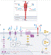
Fig. 1 |
Fig. 1 |. PSMA enzymatic active site and its crosstalk with other signalling pathways in prostate cancer.
a, Diagram showing the structure and function of the prostate-specific membrane antigen (PSMA) enzyme. The blue arrow illustrates cleavage of poly-glutamated folate and glutamate release, which most likely happens in the intestine. The green arrow illustrates cleavage of N-acetyl-L-aspartyl-L-glutamate (NAAG), which most likely happens in neuronal cells to contribute to metabotropic glutamate receptor (mGluR) signalling. b, Overview of androgen receptor (AR) signalling activated by androgens, resulting in PSA expression and secretion, and detailing the enzymatic activity of PSMA and its subsequent involvement in folate cycle and mGluR pathways. ARE, androgen response element; DHT, dihydrotestosterone; HSP, heat shock protein; NAA, N-acetylaspartate; PCFT, proton-coupled folate transporter; RFC1, reduced folate carrier. Adapted with permission from ref. , M. Bakht.
Fig. 2 |
Fig. 2 |. PSMA expression in prostate luminal epithelium.
a, Diagram depicting production of testosterone (T) in testicular Leydig cells and the anatomical structure of a prostate gland, juxtaposed (left) with a representative image of normal prostate tissue stained for prostate-specific membrane antigen (PSMA) by immunohistochemistry (right). b, t-SNE visualization of single-cell RNA sequencing data of cells obtained by radical prostatectomy from patients treated with androgen deprivation therapy (n = 4) from Karthaus et al.ʼs dataset. c, Expression of AR and FOLH1 among different epithelial subsets. AR, androgen receptor; IHC, immunohistochemistry. Part a image courtesy of Human Protein Atlas (https://www.proteinatlas.org/ENSG00000086205-FOLH1). Part a adapted with permission from ref. , M. Bakht.
Fig. 3 |
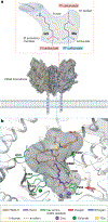
Fig. 3 |. GUL-based targeting of the PSMA enzymatic active site in prostate cancer.
Illustration of the glutamate-ureido-lysine (GUL) molecule located in the prostate-specific membrane antigen (PSMA) active site, highlighting the distinct active site regions and the probe substructure terminology (part a), as well as the predicted positioning of 68Ga-PSMA-11 within the PSMA active site (part b). The red arrow on the bottom panel shows Tyr552 residue of the PSMA active site. Adapted with permission from ref. , PNAS.
Fig. 4 |
Fig. 4 |. PSMA suppression during progression of prostate cancer.
Expression of prostate-specific membrane antigen (PSMA) gene (FOLH1) during progression of prostate cancer (PCa) towards neuroendocrine prostate cancer (NEPC) generated from the Beltran cohort,. As 22Rv1 xenograft tumours with an RNA expression of 31.3 RPKM are pathologically considered to be PSMA+ and are detectable by moderate PSMA-PET signals, we categorized samples with FOLH1 expression levels above 50 RPKM as FOLH1-high tumours and those with less than 5 RPKM as FOLH1-low tumours. This threshold-based quantification helped to provide an estimate of PSMA positivity of other models, which we confirmed using NEPC models. CRPC, castration-resistant prostate cancer.
Fig. 5 |
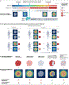
Fig. 5 |. Defining PSMA positivity is essential for selecting patients for PSMA-RLT.
a, Graphical representation of molecular-imaging prostate-specific membrane antigen (miPSMA) expression score and PSMA positivity (top) based on the reference organ uptake of PSMA-PET standardized uptake values (SUVs) (bottom). b, Inter-patient, intra-patient and temporal PSMA heterogeneity in four hypothetical patients recruited for PSMA-radioligand therapy (RLT), which two of them excluded (left) and the other ones included (right). c,d, Graphical illustration of the potential discrepancy between identified heterogeneity at the pathological (c) and radiological (d) levels owing to the inherent spatial resolution limit of clinical PET scanners. We referred to the output of the PSMA-PET signal as ‘PSMA at the radiological level’ and the intensity of PSMA identified by immunohistochemistry as ‘PSMA at the pathological level’.
Fig. 6 |
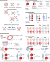
Fig. 6 |. The complexity of PSMA heterogeneity demands a more nuanced approach.
Schematic of prostate-specific membrane antigen (PSMA) heterogeneity at different levels. PSMA heterogeneity in prostate cancer is characterized by several layers of complexity, which can be delineated into specific categories. Cellular heterogeneity refers to the variability within individual tumour cells, which can evolve over time, known as temporal cellular heterogeneity. This is compounded by inter-lineage heterogeneity, indicating differences across distinct cell types within the same tumour, and is further complicated by temporal intra-patient and inter-lineage heterogeneity, reflecting changes across both time and cellular lineages. Spatial or intratumour heterogeneity describes the variability within different regions of the same tumour, subject to temporal changes or temporal intra-tumour heterogeneity. Intra-patient heterogeneity captures the variability among multiple tumours within the same patient, contrasting with inter-patient heterogeneity, which pertains to variability between tumours from different individuals. Finally, intra-lineage heterogeneity focuses on variations within a single-cell lineage over time within the same patient, whereas temporal intra-patient and inter-lineage heterogeneity track these changes over time, underscoring the dynamic nature of PSMA expression in prostate cancer and the necessity for nuanced therapeutic approaches. CRPC, castration-resistant prostate cancer; EMT, epithelial–mesenchymal transition; NEPC, neuroendocrine prostate cancer; PCa, prostate cancer.
Fig. 7 |
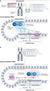
Fig. 7 |. Genomic and epigenomic features of the PSMA regulatory network.
The prostate-specific membrane antigen (PSMA) regulatory network involves four key elements including a PSMA transcription complex (PTC), an upstream enhancer that loops to the FOLH1 promoter, intergenic enhancers, and differentially methylated regions (DMRs). a, HOXB13, as part of the PTC, interacts directly with the FOLH1 upstream enhancer and promoter, leading to the expression of FOLH1. b, Either suppression of the PTC or lack of accessibility of the FOLH1 promoter can lead to FOLH1 suppression. TF, transcription factor.
Fig. 8 |
Fig. 8 |. Cellular internalization of PSMA and exposure of DNA to radiation following uptake of PSMA-radioligand.
Schematic of prostate-specific membrane antigen (PSMA) dimerization, cellular internalization by clathrin-mediated endocytosis (CME), and direct and indirect DNA damage as a result of different nuclear emissions (part a). Graphical representation of tissue range of emission (part b) and radiation-induced DNA damage (part c) by radiation exposure using 177Lu and 225Ac. Schematic of bystander and cross-fire effects of PSMA-radioligand therapy (RLT) to normal (part d) and PSMA-heterogeneous tumour (part e) tissues.

References

    1. Chen CD et al. Molecular determinants of resistance to antiandrogen therapy. Nat. Med 10, 33–39 (2004). - PubMed
    1. Polkinghorn WR et al. Androgen receptor signaling regulates DNA repair in prostate cancers. Cancer Discov 3, 1245–1253 (2013). - PMC - PubMed
    1. Chang K-H et al. Dihydrotestosterone synthesis bypasses testosterone to drive castration-resistant prostate cancer. Proc. Natl Acad. Sci. USA 108, 13728–13733 (2011). - PMC - PubMed
    1. Grasso CS et al. The mutational landscape of lethal castration-resistant prostate cancer. Nature 487, 239–243 (2012). - PMC - PubMed
    1. Scher HI et al. Increased survival with enzalutamide in prostate cancer after chemotherapy. N. Engl. J. Med 367, 1187–1197 (2012). - PubMed

Publication types

MeSH terms