Review
. 2024 Jul 8;29(1):99.
doi: 10.1186/s11658-024-00618-1.

Epigenetic control of skeletal muscle atrophy

Affiliations
Review

Epigenetic control of skeletal muscle atrophy

Wenpeng Liang et al. Cell Mol Biol Lett. .

Abstract

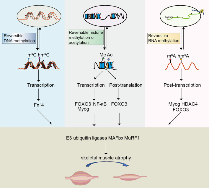
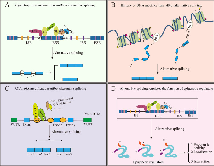
Skeletal muscular atrophy is a complex disease involving a large number of gene expression regulatory networks and various biological processes. Despite extensive research on this topic, its underlying mechanisms remain elusive, and effective therapeutic approaches are yet to be established. Recent studies have shown that epigenetics play an important role in regulating skeletal muscle atrophy, influencing the expression of numerous genes associated with this condition through the addition or removal of certain chemical modifications at the molecular level. This review article comprehensively summarizes the different types of modifications to DNA, histones, RNA, and their known regulators. We also discuss how epigenetic modifications change during the process of skeletal muscle atrophy, the molecular mechanisms by which epigenetic regulatory proteins control skeletal muscle atrophy, and assess their translational potential. The role of epigenetics on muscle stem cells is also highlighted. In addition, we propose that alternative splicing interacts with epigenetic mechanisms to regulate skeletal muscle mass, offering a novel perspective that enhances our understanding of epigenetic inheritance's role and the regulatory network governing skeletal muscle atrophy. Collectively, advancements in the understanding of epigenetic mechanisms provide invaluable insights into the study of skeletal muscle atrophy. Moreover, this knowledge paves the way for identifying new avenues for the development of more effective therapeutic strategies and pharmaceutical interventions.

Keywords: Histone modifications; Skeletal muscle atrophy; Ubiquitin–proteasome; epigenetic; m6A.


Conflict of interest statement

The author declares that no competing interests.

Figures

Fig. 1
Fig. 1
Organization of skeletal muscle. The epimysium surrounds the entire skeletal muscle. The next inner layer is the perimysium, which surrounds the fascicles and blood vessels. Below the perimysium, between the basal lamina and the endomysium, satellite cells are distributed. The endomysium surrounds individual myofibers, each of which contains myofibrils. These myofibrils are made up of sarcomeres. This figure was created with https://www.BioRender.com
Fig. 2
Fig. 2
Epigenetic control of skeletal muscle atrophy. Epigenetic modifications regulate the expression of transcription factors such as FoxO3 at the transcriptional, post-transcriptional, and post-translational levels, which in turn activate the E3 ubiquitin ligases MuRF1 and MAFbx, ultimately leading to skeletal muscle atrophy
Fig. 3
Fig. 3
Epigenetic and alternative splicing are intertwined. A Regulatory mechanism of pre-mRNA alternative splicing. Splicing regulatory elements such as exonic splicing enhancers (ESEs), exonic splicing silencers (ESSs), intronic splicing enhancers (ISEs), and intronic splicing silencers (ISSs), etc., which are located around the splice site. Splicing factor SR proteins or hnRNPs can bind to them and recruit core splicing elements such as U2AF and U1 snRNP to regulate alternative splicing. B Histone or DNA modifications affect alternative splicing. Transcription and splicing occur almost simultaneously in the same space, and various epigenetic modifications can alter the rate of Pol II elongation during transcription, which can affect recognition of the splice site by the spliceosome and, thus, alternative splicing. C RNA m6A modifications affect alternative splicing. Many m6A readers are splicing factors themselves, which can regulate numbers of alternative splicing events. m6A regulators are also able to recruit splicing factors to participate in alternative splicing regulation, e.g., YTHDC1 binds to pre-mRNAs in an m6A-dependent manner, and it can recruit the splicing factor SRSF1 to regulate alternative splicing. D Alternative splicing regulates the function of epigenetic regulators. The expression of multiple epigenetic regulators is regulated by alternative splicing, which produces full-length isoforms and N-or C-terminally reduced protein products. These truncated isoforms differ in enzymatic activity, cellular localization, and interactions

Similar articles

Cited by

References

    1. Yin L, Li N, Jia W, Wang N, Liang M, Yang X, Du G. Skeletal muscle atrophy: from mechanisms to treatments. Pharmacol Res. 2021;172:105807. doi: 10.1016/j.phrs.2021.105807. - DOI - PubMed
    1. Sartori R, Romanello V, Sandri M. Mechanisms of muscle atrophy and hypertrophy: implications in health and disease. Nat Commun. 2021;12(1):330. doi: 10.1038/s41467-020-20123-1. - DOI - PMC - PubMed
    1. Furrer R, Handschin C. Muscle wasting diseases: novel targets and treatments. Annu Rev Pharmacol Toxicol. 2019;59:315–339. doi: 10.1146/annurev-pharmtox-010818-021041. - DOI - PMC - PubMed
    1. Davegardh C, Sall J, Benrick A, Broholm C, Volkov P, Perfilyev A, Henriksen TI, Wu Y, Hjort L, Brons C, et al. VPS39-deficiency observed in type 2 diabetes impairs muscle stem cell differentiation via altered autophagy and epigenetics. Nat Commun. 2021;12(1):2431. doi: 10.1038/s41467-021-22068-5. - DOI - PMC - PubMed
    1. Bilgic SN, Domaniku A, Toledo B, Agca S, Weber BZC, Arabaci DH, Ozornek Z, Lause P, Thissen JP, Loumaye A, et al. EDA2R-NIK signalling promotes muscle atrophy linked to cancer cachexia. Nature. 2023;617(7962):827–834. doi: 10.1038/s41586-023-06047-y. - DOI - PubMed