

Smart decision support system for keratoconus severity staging using corneal curvature and thinnest pachymetry indices
- PMID: 38978067
- PMCID: PMC11229244
- DOI: 10.1186/s40662-024-00394-1
Smart decision support system for keratoconus severity staging using corneal curvature and thinnest pachymetry indices
Abstract
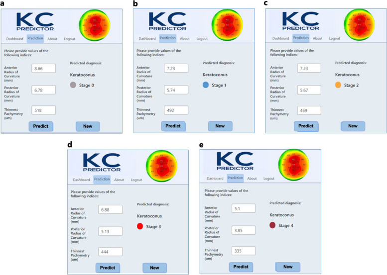
Background: This study proposes a decision support system created in collaboration with machine learning experts and ophthalmologists for detecting keratoconus (KC) severity. The system employs an ensemble machine model and minimal corneal measurements.
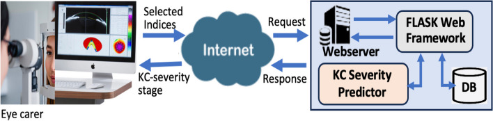
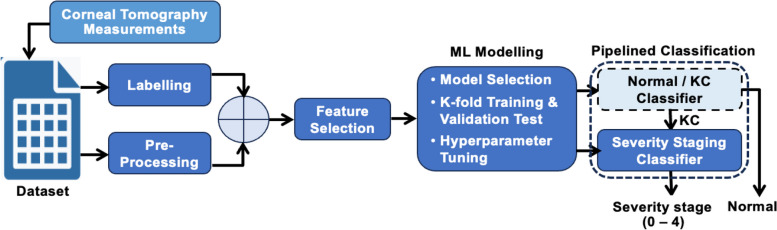
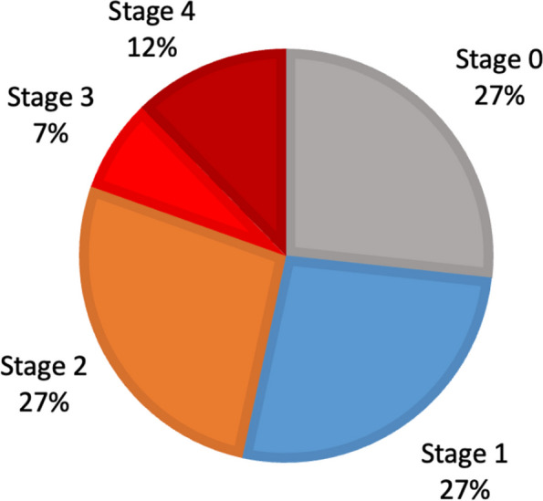
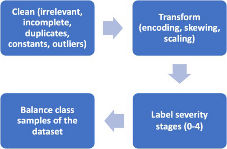
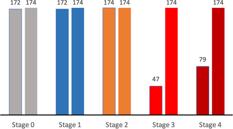
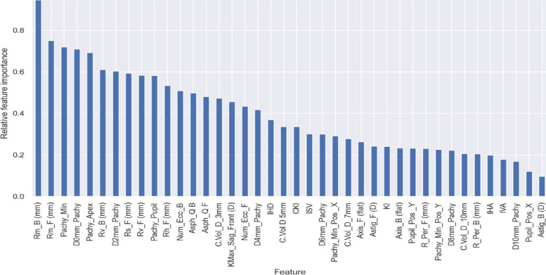
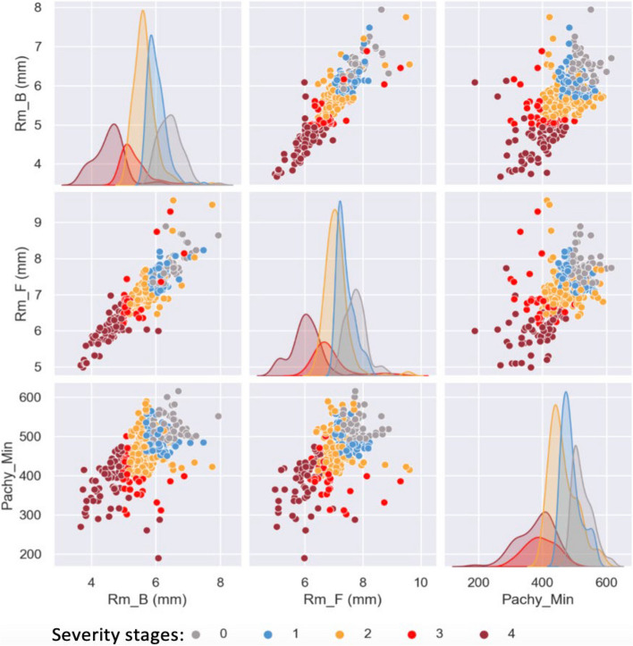
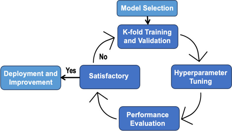
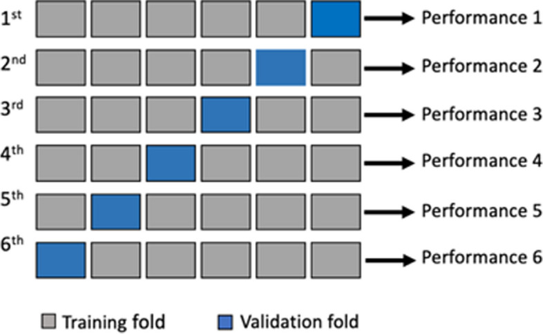
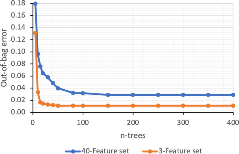
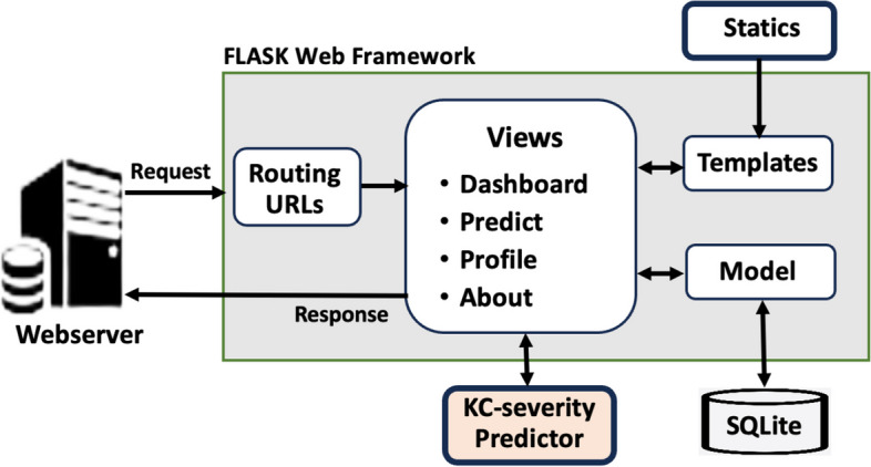
Methods: A clinical dataset is initially obtained from Pentacam corneal tomography imaging devices, which undergoes pre-processing and addresses imbalanced sampling through the application of an oversampling technique for minority classes. Subsequently, a combination of statistical methods, visual analysis, and expert input is employed to identify Pentacam indices most correlated with severity class labels. These selected features are then utilized to develop and validate three distinct machine learning models. The model exhibiting the most effective classification performance is integrated into a real-world web-based application and deployed on a web application server. This deployment facilitates evaluation of the proposed system, incorporating new data and considering relevant human factors related to the user experience.
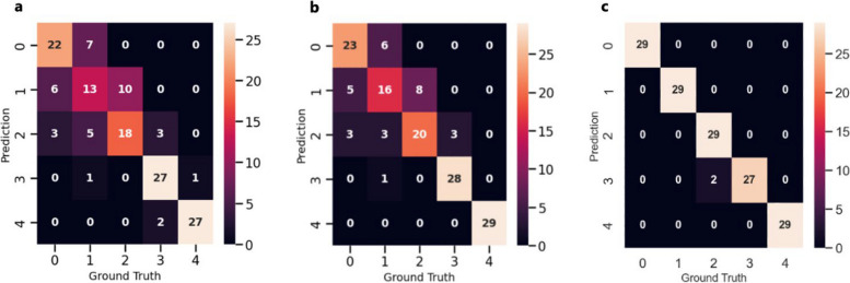
Results: The performance of the developed system is experimentally evaluated, and the results revealed an overall accuracy of 98.62%, precision of 98.70%, recall of 98.62%, F1-score of 98.66%, and F2-score of 98.64%. The application's deployment also demonstrated precise and smooth end-to-end functionality.
Conclusion: The developed decision support system establishes a robust basis for subsequent assessment by ophthalmologists before potential deployment as a screening tool for keratoconus severity detection in a clinical setting.
Keywords: Corneal tomography; Feature selection; Keratoconus; Machine learning; Severity staging; Smart web.
© 2024. The Author(s).
Conflict of interest statement
The authors declare that they have no competing interests.
Figures
References
LinkOut - more resources
Full Text Sources
Miscellaneous
