. 2024 Jul 8;19(1):54.
doi: 10.1186/s13062-024-00498-7.

YTHDC1 aggravates high glucose-induced retinal vascular endothelial cell injury via m6A modification of CDK6

Affiliations

YTHDC1 aggravates high glucose-induced retinal vascular endothelial cell injury via m6A modification of CDK6

Qi Zhou et al. Biol Direct. .

Abstract

Objective: Retinal vascular endothelial cell (RVECs) injury is a major cause of morbidity and mortality among the patients with diabetes. RVECs dysfunction is the predominant pathological manifestation of vascular complication in diabetic retinopathy. N6-methyladenosine (m6A) serves as the most prevalent modification in eukaryotic mRNAs. However, the role of m6A RNA modification in RVECs dysfunction is still unclear.

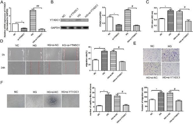
Methods: RT-qPCR analysis and western blot were conducted to detect the change of m6A RNA modification in diabetic retinopathy. CCK-8 assay, transwell experiment, wound healing assay, tube formation experiment, m6A-IP-qPCR were performed to determine the role of YTHDC1 in RVECs. Retinal trypsin digestion test and H&E staining were used to evaluate histopathological changes.

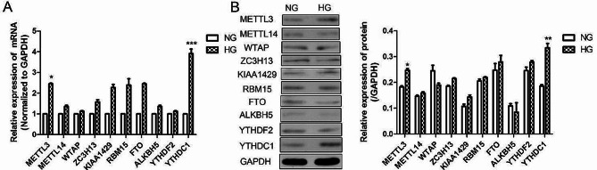
Results: The levels of m6A RNA methylation were significantly up-regulated in HG-induced RVECs, which were caused by increased expression of YTHDC1. YTHDC1 regulated the viability, proliferation, migration and tube formation ability in vitro. YTHDC1 overexpression impaired RVECs function by repressing CDK6 expression, which was mediated by YTHDC1-dependent mRNA decay. Moreover, it showed sh-YTHDC1 inhibited CDK6 nuclear export. Sh-YTHDC1 promotes the mRNA degradation of CDK6 in the nucleus but does not affect the cytoplasmic CDK6 mRNA. In vivo experiments showed that overexpression of CDK6 reversed the protective effect of sh-YTHDC1 on STZ-induced retinal tissue damage.

Conclusion: YTHDC1-mediated m6A methylation regulates diabetes-induced RVECs dysfunction. YTHDC1-CDK6 signaling axis could be therapeutically targeted for treating DR.

Keywords: CDK6; Retinal vascular endothelial cell; YTHDC1; m6A.


Conflict of interest statement

The authors declare no competing interests.

Figures

Fig. 1
Fig. 1
High glucose induced YTHDC1 expression in RVECs. (A) RT-qPCR analysis was used to detect the mRNA expression in RVECs. (B) western blotting assay was applied to detect the protein expressions in RVECs. The data were presented as mean ± SD, n = 3. NG, normal glucose; HG, high glucose. *P < 0.05, **P < 0.01, ***P < 0.001, vs. NG group
Fig. 2
Fig. 2
Silencing of YTHDC1 inhibited the proliferation, migration, invasion and tube formation ability of RVECs. RT-qPCR analysis (A) and western blot (B) were used to detect the mRNA and protein expression of YTHDC1 in different groups. (C) CCK-8 kit was used to detect the cell viability. (D) Wound healing assay was applied to detect the migration in different groups. (E) The invasion in different group was assessed using transwell experiment. (F) Tube formation experiment was used to detect the angiogenic ability. Data were expressed as mean ± SD, n = 3. *P < 0.05, vs. NC group, #P < 0.05, ##P < 0.01, vs. HG + si-NC group
Fig. 3
Fig. 3
Overexpression of YTHDC1 promoted the proliferation, migration, invasion and tube formation ability of RVECs RT-qPCR analysis (A) and western blot (B) were used to detect the mRNA and protein expression of YTHDC1 in different groups. (C) CCK-8 kit was used to detect the cell viability. (D) Wound healing assay was applied to detect the migration in different groups. (E) The invasion in different group was assessed using transwell experiment. (F) Tube formation experiment was used to detect the angiogenic ability. Data were expressed as mean ± SD, n = 3. *P < 0.05, **P < 0.01, vs. NC group, #P < 0.05, ##P < 0.01, vs. HG + vector group
Fig. 4
Fig. 4
Si-YTHDC1 down-regulated CDK6 expression (A) m6A methylation chip was used to analyze the differential genes regulated by YTHDC1. (B) KEGG pathway enrichment analysis. (C) RT-qPCR analysis was used to detect the mRNA expressions of CTR9, IGF2R, ARF6 and CDK6. (D) m6A-IP-qPCR assay was used to detect the CDK6 level. RT-qPCR analysis (E) and western blot (F) were performed to detect the mRNA and protein expresions of CDK6 in different groups. The data were expressed as mean ± SD, n = 3. P < 0.05, **P < 0.01, ***P < 0.001, vs. HG + vector group; #P < 0.05, ##P < 0.01, vs. HG + si-NC group
Fig. 5
Fig. 5
YTHDC1 regulated CDK6 m6A modification. (A) CDK6 m6A level in high glucose-induced RVECs was examined by RIP-qPCR (B) RT-qPCR analysis was used to detect the ratio of CDK6 mRNA in cytoplasm to nucleus (C) RNA-FISH was utilized to examine the co-localization between YTHDC1 (Cy3-labelled) and CDK6 (FITC-labelled) in the nucleus of HRVECs cells. Nuclei were stained with DAPI (blue). RT-qPCR analysis (D) and western blot (E) were used to detecet the mRNA and protein expressions of CDK6 in the cytoplasm and nuclear. The mRNA expressions of CDK6 in cytoplasm (F) and nuclear (G) were measured at different time points after actinomycin D treatment. The data were expressed as mean ± SD, n = 3. **P < 0.01, ***P < 0.001, vs. NG group; ##P < 0.01, vs. si-NC group
Fig. 6
Fig. 6
Silencing CDK6 inhibited the proliferation, migration, invasion and tube formation ability of RVECs induced by HG. RT-qPCR (A) analysis and western blot assay (B) were used to detect the mRNA and protein expression of CDK6 in RVECs. (C) CCK-8 kit was used to detect the cell viability. (D) Wound healing assay was used to detect the migration. (E) The invasion of RVECs was detected using transwell experiment. (F) Tube formation experiment was used to detect the angiogenic capacity in RVECs. The data were expressed as mean ± SD, n = 3. **P < 0.01, ***P < 0.001, vs. NG group; ##P < 0.01, vs. si-NC group
Fig. 7
Fig. 7
YTHDC1 aggravated HG-induced RVECs dysfunction and rescued by CDK6. (A) RT-qPCR analysis was used to detect the mRNA expression of CDK6. The mRNA expression (B) and protein expression (C) of CDK6 in different groups were determined using RT-qPCR analysis and western blot. (D) CCK-8 kit was used to detect the cell viability. (E) Wound healing assay was performed to detect the migration in different groups. The invasion in different groups was detected using Transwell experiment(F). (G) Tube formation experiment was used to detect the angiogenesis ability. The data were expressed as mean ± SD, n = 3,*P < 0.05, **P < 0.01, ***P < 0.001, vs. NC group; #P < 0.05, ##P < 0.01, vs. HG + NC group; &P < 0.05, vs. HG + si-YTHDC1 + vector group
Fig. 8
Fig. 8
Overexpression of CDK6 reversed the protective effect of sh-YTHDC1 on diabetic retinopathy in rats. RT-qPCR analysis was used to detect the mRNA expressions of Ythdc1(A) and Cdk6(B) in retinal tissues. (C) western blot was performed to detect the protein expressions of YTHDC1 and CDK6 in retinal tissues. (D) The representative H&E stained images and quantitative analysis plots. (E) Periodic Acid-Schiff staining was used to detect the morphological changes of retinal capillaries. *P < 0.05, **P < 0.01, ***P < 0.001, vs. sham + AAV-NC group; #P < 0.05, ##P < 0.01, vs. STZ + AAV-NC group; &P < 0.05, vs. STZ + AAV-sh-YTHDC1 group, n = 6

Similar articles

Cited by

References

    1. Nan W et al. Identification and validation of Autophagy-related genes in Diabetic Retinopathy. Front Endocrinol (Lausanne), 2022. 13(0). - PMC - PubMed
    1. Man R, et al. Association between Perceived barriers to Diabetes Self-management and Diabetic Retinopathy in Asian patients with type 2 diabetes. JAMA Ophthalmol. 2017;135(12):1387–93. doi: 10.1001/jamaophthalmol.2017.4888. - DOI - PMC - PubMed
    1. K O et al. IDF Diabetes Atlas: global estimates for the prevalence of diabetes for 2015 and 2040. Diabetes Res Clin Pract, 2017. 128(0). - PubMed
    1. Shafabakhsh R, et al. Association between microRNAs expression and signaling pathways of inflammatory markers in diabetic retinopathy. J Cell Physiol. 2019;234(6):7781–7. doi: 10.1002/jcp.27685. - DOI - PubMed
    1. Jalilian E, Elkin K, Shin S. Novel cell-based and Tissue Engineering Approaches for Induction of Angiogenesis as an alternative therapy for Diabetic Retinopathy. Int J Mol Sci, 2020. 21(10). - PMC - PubMed

Publication types

LinkOut - more resources