[Preprint]. 2024 Jun 24:rs.3.rs-4536350.
doi: 10.21203/rs.3.rs-4536350/v1.

B cell-mediated antigen presentation promotes adverse cardiac remodeling in chronic heart failure

Affiliations

B cell-mediated antigen presentation promotes adverse cardiac remodeling in chronic heart failure

Jana P Lovell et al. Res Sq. .

Abstract

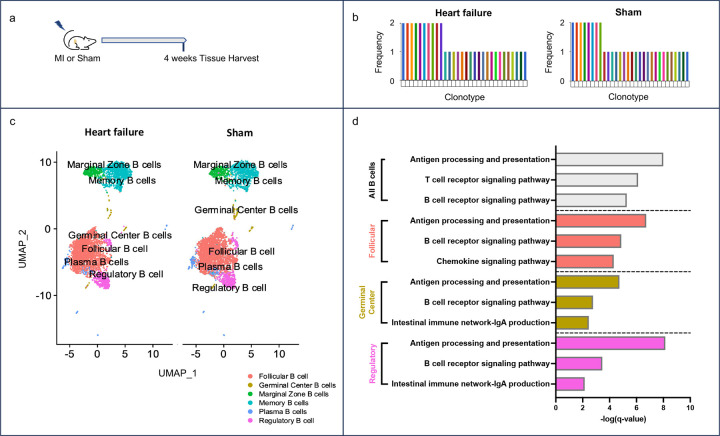
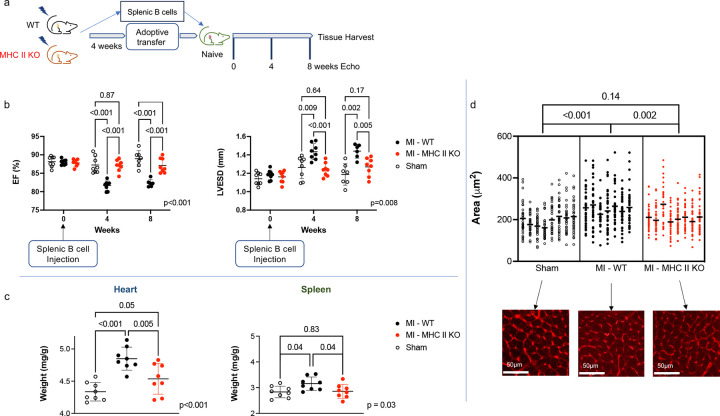
Cardiovascular disease remains the leading cause of death worldwide. A primary driver of cardiovascular mortality is ischemic heart failure, a form of cardiac dysfunction that can develop in patients who survive myocardial infarction. Acute cardiac damage triggers robust changes in the spleen with rapid migration of immune cells from the spleen to the heart. Activating this "cardio-splenic" axis contributes to progressive cardiac dysfunction. The cardio-splenic axis has, therefore, been identified as a promising therapeutic target to prevent or treat heart failure. However, our understanding of the precise mechanisms by which specific immune cells contribute to adverse cardiac remodeling within the cardio-splenic axis remains limited. Here, we show that splenic B cells contribute to the development of heart failure via MHC II-mediated antigen presentation. We found that the adoptive transfer of splenic B cells from mice with ischemic heart failure promoted adverse cardiac remodeling and splenic inflammatory changes in naïve recipient mice. Based on single-cell RNA sequencing analysis of splenic B cells from mice with ischemic heart failure, we hypothesized that B cells contributed to adverse cardiac remodeling through antigen presentation by MHC II molecules. This mechanism was confirmed using transgenic mice with B cell-specific MHC II deletion, and by analyzing circulating B cells from humans who experienced myocardial infarction. Our results broaden our understanding of B lymphocyte biology, reshape current models of immune activation in response to myocardial injury, and point towards MHC II-mediated signaling in B cells as a novel and specific therapeutic target in chronic heart failure.


Figures

Figure 1.
Figure 1.. Splenic B cells promote adverse cardiac remodeling after ischemic myocardial injury.
a) Schema for adoptive transfer studies. Four weeks following permanent coronary artery ligation or sham surgery, unfractionated splenocytes or isolated splenic B cells were transferred to naïve recipient mice. Recipient mice underwent serial transthoracic echocardiography over 8 weeks followed by tissue harvest. b) On echocardiography, adoptive transfer of splenocytes or isolated splenic B cells from heart failure (HF) mice resulted in a reduction in left ventricular ejection fraction (LVEF) and an increase in left ventricular end-systolic diameter (LVESD) in naïve recipient mice over eight weeks. c) Gravimetric data revealed significantly larger hearts and spleens (normalized to body weight) in naive mice eight weeks post-adoptive transfer of isolated splenic B cells from HF mice relative to recipients of B cells from sham-operated mice. d) Representative images of WGA staining of hearts from recipients of isolated splenic B cells from HF or sham-operated mice. Cardiomyocyte area was increased in recipients of splenocytes or isolated splenic B cells from HF mice relative to sham recipients. n = 5–6 male mice per group. Mean values ± SD are represented.
Figure 2.
Figure 2.. Adoptively transferred splenic B cells migrate to the heart and spleen of recipients, and post-MI, induce inflammatory changes in the spleen of naïve mice.
a) Schema for adoptive transfer of isolated splenic CD45.1 B cells 4 weeks following LAD ligation or sham surgery to naïve CD45.2 mice. b) Flow cytometry of peripheral blood from naïve recipient mice revealed the presence of donor CD45.1 B cells 1 week and 8 weeks following adoptive transfer. Donor CD45.1 B cells were also present in the spleen and heart of CD45.2 mice 8 weeks after adoptive transfer. c) Representative gating strategy for flow cytometry of spleens from recipient mice 8 weeks post-adoptive transfer of splenic B cells. Adoptive transfer of splenic B cells from HF mice resulted in increases in lymphocytes, B cells, and CD8+ T cells relative to recipients of sham splenic B cells. The number of cells per mg spleen is reported. n = 6 male mice per group. Median values are represented by horizontal lines.
Figure 3.
Figure 3.. Acute myocardial injury activates antigen processing and presentation pathways in splenic B cells.
a) Schema for scRNA sequencing of B cells from mice 4 weeks following permanent coronary ligation or sham surgery. b) V(D)J analysis of splenic B cells. Myocardial infarction (MI) did not induce splenic B cell clonal expansion. c) UMAP plots of splenic B cells 4 weeks post-MI (“heart failure”) or sham surgery. Splenic B cells were classified into B cell sub-types. d) KEGG pathway analysis of differentially expressed genes in splenic B cells (p<0.05) revealed significant dysregulation of antigen processing and presentation in multiple B cell subtypes post-MI. Top 3 dysregulated immune system pathways are reported for specified B cell subtypes. n = 4–5 mice per group.
Figure 4.
Figure 4.. Isolated splenic B cells deficient in MHC class II do not transfer adverse cardiac remodeling to recipient mice.
a) Schema for adoptive transfer studies of isolated splenic B cells from Cd19tm1(cre)Cgn/−H2-Ab1b-tm1Koni/ b-tm1Koni mice and wildtype mice following permanent coronary artery ligation or sham surgery. b) Naïve recipients of splenic B cells with MHC II deletion from post-MI mice did not experience adverse cardiac remodeling on serial echocardiography over an 8-week period post-adoptive transfer. c) Gravimetric data of recipient mice normalized to body weight. Recipient mice of B cells deficient in MHC II from post-MI mice did not experience splenocyte or cardiac remodeling by gravimetric analysis. d) Representative images of WGA staining of hearts from recipient mice. Transfer of isolated MHC II-deficient splenic B cells did not induce cardiomyocyte hypertrophy. n = 8 male mice per group. Mean values ± SD are represented.
Figure 5.
Figure 5.. Isolated splenic B cells deficient in MHC class II do not induce inflammatory changes in the spleen of naïve mice post-adoptive transfer.
a) Schema for adoptive transfer studies of isolated splenic B cells from Cd19tm1(cre)Cgn/−H2-Ab1b-tm1Koni/ b-tm1Koni mice and wildtype mice following permanent coronary artery ligation or sham surgery. b) Representative gating strategy for flow cytometry of spleens from recipient mice 8 weeks post-adoptive transfer of splenic B cells. Adoptive transfer of splenic B cells with MHC II deletion from post-MI mice did not induce changes in immune cell populations in the spleens of recipient mice. n = 8 male mice per group. Median values are represented by horizontal lines.
Figure 6.
Figure 6.. Antigen processing and presentation is dysregulated in circulating B cells in human patients post-STEMI.
a) scRNA sequencing of peripheral blood B cells from 38 human patients 8 weeks post-STEMI and 38 healthy controls. UMAP plots are shown with B cells identified. b) KEGG pathway analysis of differentially expressed genes (log2fold-change ≥1, p<0.05) revealed significant dysregulation of antigen processing and presentation in peripheral B cells of patients 24 hours and 8 weeks post-STEMI. All immune system pathways with q-value <0.05 are listed. NOD= nucleotide oligomerization domain.
Figure 7.
Figure 7.. Antigen processing and presentation is dysregulated in circulating B cells in humans with ischemic cardiomyopathy.
a) scRNA sequencing of peripheral blood B cells from 3 human patients with ischemic cardiomyopathy (ICM) and 1 healthy patient. UMAP plots are shown with B cells identified. b) KEGG pathway analysis of differentially expressed genes (log2fold-change ≥0.58 p<0.05) revealed significant dysregulation of antigen processing and presentation in peripheral B cells from ICM patients. All immune system pathways with q-value <0.05 are listed.

References

    1. Hofmann U., Frantz S. Immunity strikes: Heart failure as a systemic disease. Eur Heart J 2014:341–3. Doi: 10.1093/eurheartj/eht405. - DOI - PubMed
    1. Adamo L., Rocha-Resende C., Prabhu SD., Mann DL. Reappraising the role of inflammation in heart failure. Nat Rev Cardiol 2020:269–85. Doi: 10.1038/s41569-019-0315-x. - DOI - PubMed
    1. Libby P., Nahrendorf M., Swirski FK. Leukocytes link local and systemic inflammation in ischemic cardiovascular disease an expanded cardiovascular continuum. J Am Coll Cardiol 2016:1091–103. Doi: 10.1016/j.jacc.2015.12.048. - DOI - PMC - PubMed
    1. Nahrendorf M., Swirski FK., Aikawa E., et al. The healing myocardium sequentially mobilizes two monocyte subsets with divergent and complementary functions. Journal of Experimental Medicine 2007;204(12):3037–47. Doi: 10.1084/jem.20070885. - DOI - PMC - PubMed
    1. Murphy SP., Kakkar R., McCarthy CP., Januzzi JL. Inflammation in Heart Failure: JACC State-of-the-Art Review. J Am Coll Cardiol 2020:1324–40. Doi: 10.1016/j.jacc.2020.01.014. - DOI - PubMed

Publication types

LinkOut - more resources