[Preprint]. 2024 Jun 26:rs.3.rs-4510670.
doi: 10.21203/rs.3.rs-4510670/v1.

Characterizing the tumor suppressor activity of FLCN in Birt-Hogg-Dubé syndrome through transcriptiomic and proteomic analysis

Affiliations

Characterizing the tumor suppressor activity of FLCN in Birt-Hogg-Dubé syndrome through transcriptiomic and proteomic analysis

Andrew Tee et al. Res Sq. .

Update in

Abstract

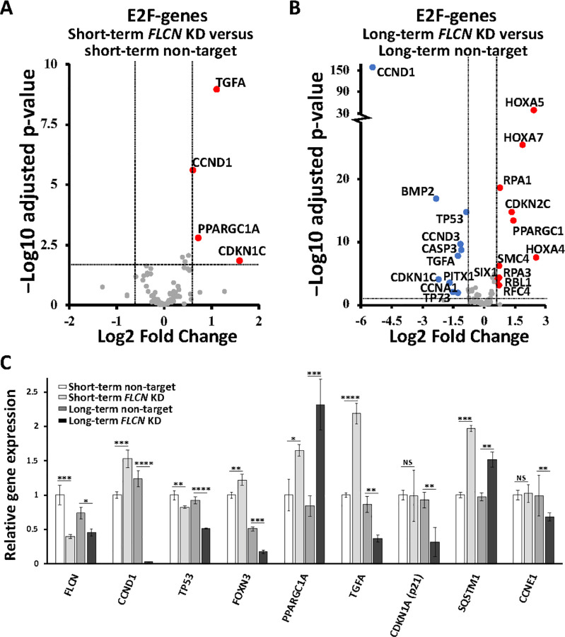
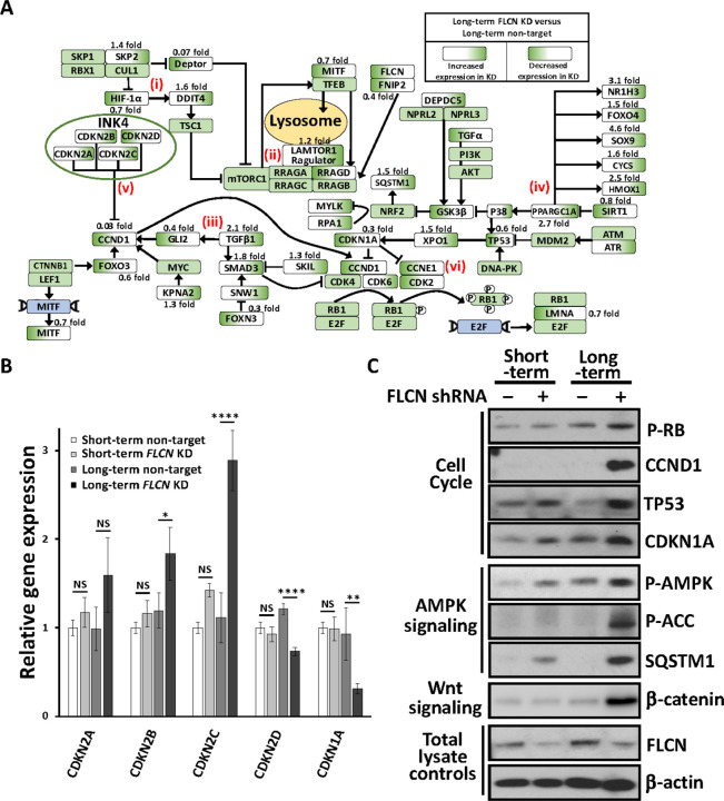
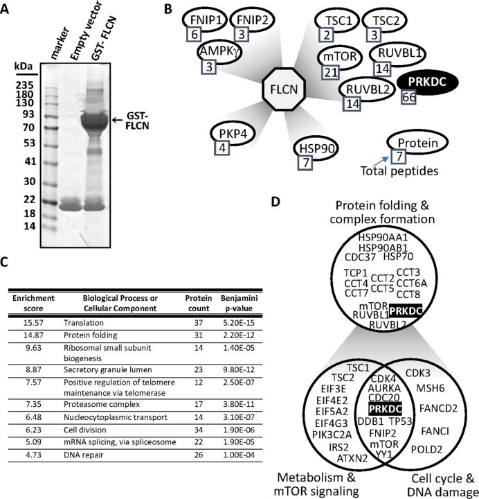
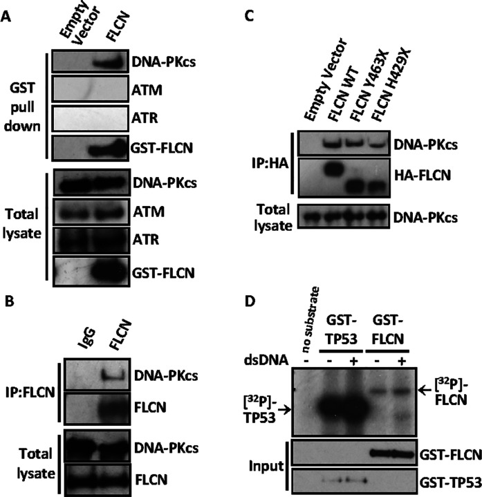
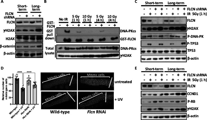
Birt-Hogg-Dubé (BHD) syndrome patients are uniquely susceptible to all renal tumour subtypes. The underlying mechanism of carcinogenesis is unclear. To study cancer development in BHD, we used human proximal kidney (HK2) cells and found that long-term folliculin (FLCN) knockdown was required to increase their tumorigenic potential, forming larger spheroids in non-adherent conditions. Transcriptomic and proteomic analysis uncovered links between FLCN, cell cycle control and the DNA damage response (DDR) machinery. HK2 cells lacking FLCN had an altered transcriptome profile with cell cycle control gene enrichment. G1/S cell cycle checkpoint signaling was compromised with heightened protein levels of cyclin D1 (CCND1) and hyperphosphorylation of retinoblastoma 1 (RB1). A FLCN interactome screen uncovered FLCN binding to DNA-dependent protein kinase (DNA-PK). This novel interaction was reversed in an irradiation-responsive manner. Knockdown of FLCN in HK2 cells caused a marked elevation of γH2AX and RB1 phosphorylation. Both CCND1 and RB1 phosphorylation remained raised during DNA damage, showing an association with defective cell cycle control with FLCN knockdown. Furthermore, Flcn-knockdown C. elegans were defective in cell cycle arrest by DNA damage. This work implicates that long-term FLCN loss and associated cell cycle defects in BHD patients could contribute to their increased risk of cancer.


Conflict of interest statement

The authors declare no competing financial interests. While this study is part-funded by Health and Care Research Wales, the views expressed are those of the author/s and not necessarily those of Health and Care Research Wales or Welsh Government.

Figures

Figure 1
Figure 1. Long-term knockdown of FLCN increases tumorigenesis.
(A) HK2 cells with and without short-term or long-term FLCN shRNA knockdown (compared to non-target shRNA) were grown in soft agar for 21 days. Colony diameter was measured using ImageJ. (v1.53t), and a representative image of tumors is shown and distribution of tumors graphed. n=360 per condition over 6 biological samples. Each data point is a single tumor. Stats: distributed data was not normal (D’Agostino & Pearson test) so was analysed using nonparametric Kruskai-Wallis ANOVA with Dunn’s multiple comparisons. (B) These HK2 cells were also plated in non-adherent conditions and imaged over 14 days, and % change in diameter graphed over time (n=42). RNA sequencing of HK2 cells with either short- or long-term non-target or FLCN shRNA knockdown was carried out (n=3). (C) Differential gene expression was compared in short-term FLCN versus non-target shRNA knockdown and long-term FLCN versus non-target shRNA knockdown HK2 cells. Volcano plots are shown with the following thresholds: ≤ & ≥ 2.5 fold change, padj < 0.05 with false rate discovery (FDR) correction applied. Relative gene expression for selected genes are shown.
Figure 2
Figure 2. Differentially expressed genes linked to E2F and known FLCN regulatory genes after FLCN knockdown.
Differential gene expression of E2F-genes was compared in (A) short-term FLCNversus non-target shRNA knockdown and (B) long-term FLCN versus non-target shRNA knockdown HK2 cells, after RNA sequencing. Volcano plots are shown with the following thresholds: ≤ & ≥ 2.5 fold change, padj < 0.05 with false rate discovery (FDR) correction applied. (C) FLCN linked genes from this RNA sequencing experiment are graphed, and include FLCN, CCND1, TP53, PPARGC1A, TGFA, CDKN1A, SQSTM1 and CCNE1.
Figure 3
Figure 3. Differentially expressed genes and their associated signaling pathways.
(A) Gene expression comparing long-term FLCN shRNA versus non-target shRNA knockdown in HK2 cells, after RNA sequencing. Each gene is depicted in a signaling flow diagram. Known signaling functions of FLCN include: (i) Enhanced HIF-1a activity, as shown by increased HIF-1a target gene expression, SLC2A2 and REDD1. Signaling feedback mechanism to reduce HIF-1a, through reduced HIF-1a expression and upregulation of SKP2, a HIF-1a inhibitor. (ii) Upregulation of mTORC1 through enhanced expression of LAMTOR1 and a marked reduction in DEPTORexpression, an mTORC1 inhibitor. (iii) Upregulation of TGFb/SMAD3 signaling upon knockdown of FLCN. SMAD3 is an inhibitor of CCND1-CDK4/CDK6 complexes. (iv) PGC1a and downstream regulated genes, NR1H3, FOXO4, SOX9, CYCS, HMOX1 are markedly upregulated upon FLCN knockdown. (v) The INK4 family members that inhibit CCND1, CDKN2A-C, are increased while CDKN2D is reduced. (vi) There is reduced expression of CDKN1A that inhibits both CCND1 and CCNE1 activity. This demonstrates a transcriptional feedback mechanism to enhance CDK4/6 and CDK2 activity. Further differences were observed with enhanced CDK6expression and lower expression of CCND1 and CCNE1. (B) Relative gene expression of cyclin dependent kinase inhibitors across the different HK2 cell lines was calculated, (n=3). (C) Protein lysates from spheroids generated from HK2 cells with and without short- or long-term FLCN shRNA knockdown (non-target shRNA used as a control) were probed for phosphorylated RB1, CCND1, TP53, CDKN1A, phosphorylated AMPK and ACC, SQSTM1, b-catenin, FLCN, and b-actin as a loading control (n=3).
Figure 4
Figure 4. FLCN has an extensive PPI network linked to cell cycle and DNA damage.
(A) GST-FLCN was overexpressed in HEK293 cells, purified and interacting protein separated by SDS-PAGE and stained with colloidal blue. The gel was sectioned for mass spectrometry analysis. (B) FLCN protein interaction network is represented showing known FLCN interactions and includes PRKDC. Total peptides of protein sequences identified after mass spectrometry is indicated. (C) FLCN binding proteins identified by mass spectrometry was analyzed by DAVID, and the top ten enriched scored biological processes or cellular component are presented. (D) FLCN binding proteins are grouped together in protein folding and complex formation, metabolism and mTOR signaling, and cell cycle and DNA damage.
Figure 5
Figure 5. FLCN interacts with DNA-PK.
(A) GST-tagged FLCN was overexpressed in HEK293 cells and used as a bait protein to validate protein interactions between FLCN and endogenously expressed DNA damage components (DNA-PKcs, ATM, ATR) by GST-pull down. (B) Endogenous FLCN was immunoprecipitated using an antibody raised against N-terminal FLCN and the interaction of endogenous DNA-PKcs was detected by western blot. (C) HA-tagged FLCN constructs (wild type FLCN (WT), and two patient derived C-terminal truncated mutants (Y463X and H429X) were over-expressed in HEK293 cells, immunoprecipitated using anti-HA antibodies and bound endogenous DNA-PKcs was detected by western blot. (D) DNA-PK kinase assays were performed, using GST-FLCN or GST-TP53 that was overexpressed and purified from HEK293 cells. Incorporation of radiolabelled phosphate [32P] was determined with active DNA-PK that was further induced with supplementation of short double-strand DNA (dsDNA).
Figure 6
Figure 6. FLCN knockdown promotes cell cycle progression.
(A) Serine 139 phosphorylation of histone variant H2AX (γH2AX) was assessed under basal conditions in HK2 cells with and without short-term or long-term FLCN shRNA knockdown (non-target shRNA was used as a control). (B) FLCN/DNA-PKcs interaction was investigated following IR induced DNA damage. GST-tagged FLCN was overexpressed in HEK293 cells that was then subjected to IR (5 or 10 Gy) and left for either 1 or 4 h prior to cellular lysis, as indicated. GST-FLCN was purified and endogenous DNA-PKcs detected by western blot. (C) In the HK2 cells with and without short- or long-term FLCN shRNA knockdown, markers of DNA damage were assessed following 5 Gy IR for 1 h, where indicated. γH2AX, P-DNA-PKcs (Ser2056) and the downstream DNA-PK substrate, P-TP53 (Ser15) are shown, alongside. FLCN and b-actin as controls. (D) Mitotic germ cells were quantified in C. elegans with and without Flcn siRNA knockdown and subjected to UV damage, where indicated. Mitotic germ cells were scored are graphed. Representative images of the mitotic cells are presented, (n=18). (E) Same lysates from panel C were analysed for G1/S phase cell cycle markers, CCND1, and P-RB1, with FLCN, γH2AX and b-actin as controls.

References

    1. Birt AR, Hogg GR, Dube WJ. Hereditary multiple fibrofolliculomas with trichodiscomas and acrochordons. Arch Dermatol. 1977; 1 13:1674–1677. - PubMed
    1. Schmidt LS, Linehan WM. Molecular genetics and clinical features of Birt-Hogg-Dubé syndrome. Nat Rev Urol. 2015;1 2:558–569. - PMC - PubMed
    1. Johannesma PC, van de Beek I, van der Wel TJWT, Reinhard R, Rozendaal L, Starink TM, et al. Renal imaging in 199 Dutch patients with Birt-Hogg-Dube syndrome: Screening compliance and outcome. PLoS One. 2019; 14:e0212952. - PMC - PubMed
    1. Li K, Wada S, Gosis BS, Thorsheim C, Loose P, Arany Z. Folliculin promotes substrate-selective mTORC1 activity by activating RagC to recruit TFE3. PLoS Biol. 2022; 20:e3001594. - PMC - PubMed
    1. Napolitano G, Di Malta C, Esposito A, de Araujo MEG, Pece S, Bertalot G, et al. A substrate-specific mTORC1 pathway underlies Birt-Hogg-Dubé syndrome. Nature. 2020; 585:597–602. - PMC - PubMed

Publication types

LinkOut - more resources