

Sinus Savvy: Exploring the Current Techniques of Maxillary Sinus Augmentation
- PMID: 38978885
- PMCID: PMC11230615
- DOI: 10.7759/cureus.61933
Sinus Savvy: Exploring the Current Techniques of Maxillary Sinus Augmentation
Abstract
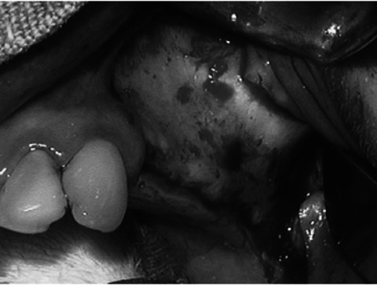
Sinus ridge augmentation is a surgical procedure aimed at increasing the volume of bone in the posterior maxilla to permit successful dental implant placement. The current review article presents an overview of various techniques used for sinus ridge augmentation, including the lateral window technique, crestal approach, transalveolar technique, and piezoelectric osteotomy. The article examines the advantages and limitations of each technique, such as invasiveness, surgical difficulty, and the requirement for additional procedures. Additionally, the article discusses the factors that influence the success of the procedure, including patient age, residual bone height, and the kind of bone graft substance used. The review also emphasizes the importance of proper case selection, surgical planning, and postoperative care to ensure optimal outcomes. Overall, the article provides valuable insights into the current techniques used for sinus ridge augmentation, highlighting the need for further research to improve patient outcomes and the success of placing dental implants over the long run.
Keywords: bone morphogenetic protein (bmp); dental implants; maxillary sinus lift; sinus floor augmentation; sinus floor elevation.
Copyright © 2024, Chittoria et al.
Conflict of interest statement
Conflicts of interest: In compliance with the ICMJE uniform disclosure form, all authors declare the following: Payment/services info: All authors have declared that no financial support was received from any organization for the submitted work. Financial relationships: All authors have declared that they have no financial relationships at present or within the previous three years with any organizations that might have an interest in the submitted work. Other relationships: All authors have declared that there are no other relationships or activities that could appear to have influenced the submitted work.
Figures


Similar articles
-
Indications and Regenerative Techniques for Lateral Window Sinus Floor Elevation With Ridge Augmentation.Clin Implant Dent Relat Res. 2025 Jun;27(3):e70007. doi: 10.1111/cid.70007. Clin Implant Dent Relat Res. 2025. PMID: 40344320 Free PMC article. Review.
-
[Maxillary sinus floor augmentation using the transalveolar technique with simultaneous placement of dental implants: a 5-year clinical retrospective study].Zhonghua Kou Qiang Yi Xue Za Zhi. 2014 Mar;49(3):161-5. Zhonghua Kou Qiang Yi Xue Za Zhi. 2014. PMID: 24820784 Chinese.
-
[Clinical evaluation of two transalveolar methods for sinus augmentation with placing 1 204 implants immediately].Zhonghua Kou Qiang Yi Xue Za Zhi. 2018 Dec 9;53(12):821-825. doi: 10.3760/cma.j.issn.1002-0098.2018.12.006. Zhonghua Kou Qiang Yi Xue Za Zhi. 2018. PMID: 30522205 Chinese.
-
Lateral trap-door window approach with maxillary sinus membrane lifting for dental implant placement in atrophied edentulous alveolar ridge.J Chin Med Assoc. 2015 Feb;78(2):85-8. doi: 10.1016/j.jcma.2014.05.016. Epub 2014 Oct 3. J Chin Med Assoc. 2015. PMID: 25287252 Review.
-
Clinical evaluation of modified transalveolar sinus floor elevation and osteotome sinus floor elevation in posterior maxillae: study protocol for a randomized controlled trial.Trials. 2018 Sep 14;19(1):489. doi: 10.1186/s13063-018-2879-x. Trials. 2018. PMID: 30217227 Free PMC article.
References
-
- Grafting of the maxillary sinus floor with autogenous marrow and bone. Boyne PJ, James RA. https://pubmed.ncbi.nlm.nih.gov/6993637/ J Oral Surg. 1980;38:613–616. - PubMed
-
- Sinus lift procedures: an overview of current techniques. Stern A, Green J. Dent Clin North Am. 2012;56:219–233. - PubMed
-
- Maxillary sinus anatomy: a cadaveric study with clinical implications. Gosau M, Rink D, Driemel O, Draenert FG. Anat Rec (Hoboken) 2009;292:352–354. - PubMed
-
- Endoscopic view of the infraorbital nerve. Yanagisawa E, Yanagisawa K. https://pubmed.ncbi.nlm.nih.gov/10224695/ Ear Nose Throat J. 1999;78:226–228. - PubMed
-
- Analysis of the anatomy of the maxillary sinus septum using 3-dimensional computed tomography. Park YB, Jeon HS, Shim JS, Lee KW, Moon HS. J Oral Maxillofac Surg. 2011;69:1070–1078. - PubMed
Publication types
LinkOut - more resources
Full Text Sources