[Preprint]. 2024 Jun 25:2024.06.21.600001.
doi: 10.1101/2024.06.21.600001.

A broadly-neutralizing antibody against Ebolavirus glycoprotein that potentiates the breadth and neutralization potency of other antibodies

Affiliations

A broadly-neutralizing antibody against Ebolavirus glycoprotein that potentiates the breadth and neutralization potency of other antibodies

Francesca R Donnellan et al. bioRxiv. .

Abstract

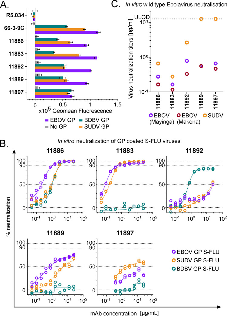
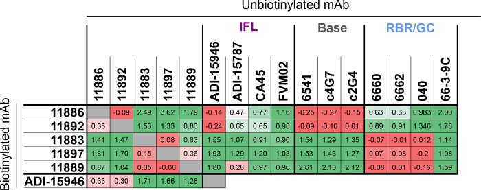
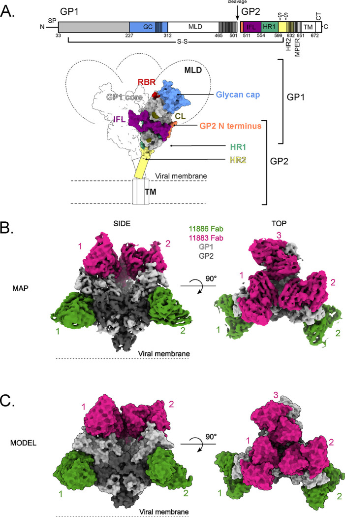
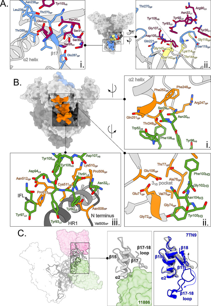
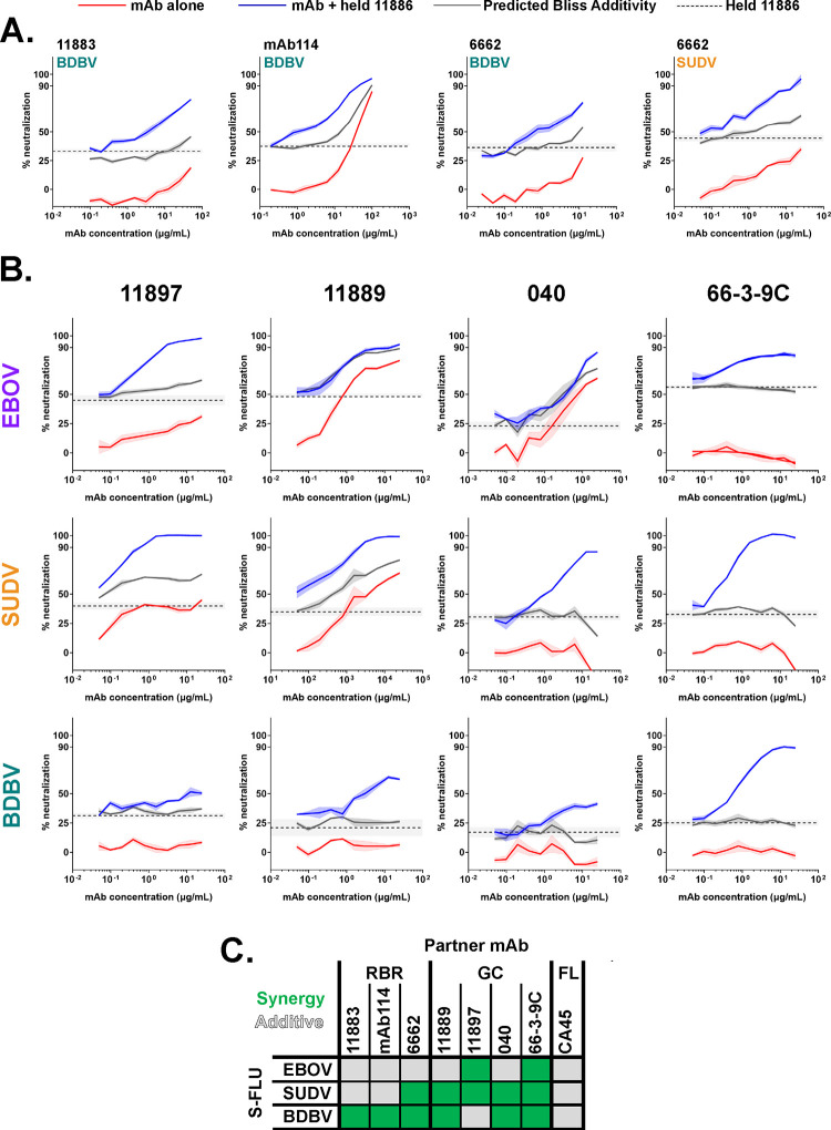
Ebolavirus disease (EVD) is caused by multiple species of Ebolavirus. Monoclonal antibodies (mAbs) against the virus glycoprotein (GP) are the only class of therapeutic approved for treatment of EVD caused by Zaire ebolavirus (EBOV). Therefore, mAbs targeting multiple Ebolavirus species may represent the next generation of EVD therapeutics. Broadly reactive anti-GP mAbs were produced; among these, mAbs 11886 and 11883 were broadly neutralizing in vitro. A 3.0 Å cryo-electron microscopy structure of EBOV GP bound to both mAbs shows that 11886 binds a novel epitope bridging the glycan cap (GC), 310 pocket and GP2 N-terminus, whereas 11883 binds the receptor binding region (RBR) and GC. In vitro, 11886 synergized with a range of mAbs with epitope specificities spanning the RBR/GC, including 11883. Notably, 11886 increased the breadth of neutralization by partner mAbs against different Ebolavirus species. These data provide a strategic route to design improved mAb-based next-generation EVD therapeutics.


Conflict of interest statement

Declaration of Interests DJL, VO’D are employees of UCB Pharma. DJL holds stock and/or stock options in UCB Pharma. FRD, SJD, DJL, VO’D, ART, PR and LS are inventors on patent applications relating to anti-Ebolavirus GP antibodies.

Figures

Figure 1.
Figure 1.. Binding to GP and virus neutralization by rabbit mAb panel.
A) Binding of broadly reactive rabbit mAb panel to full-length transmembrane Ebolavirus GPs. MDCK SIAT-1 cells expressing EBOV GP (purple), SUDV GP (orange), BDBV GP (teal) or parental cells not expressing GP (No GP, grey) were incubated with 20 μg/mL of mAb, and mAb binding detected using an Alexa Fluor 647 anti-IgG conjugate secondary antibody. R5.034 (non-anti-GP mAb) and 66-3-9C (broadly reactive anti-GP mAb) were included as negative and positive controls, respectively. Background (mean of 8 wells per assay plate incubated with relevant secondary only) was subtracted from data. Mean and SEM of duplicates shown for each mAb. B) In vitro neutralization of Ebolavirus GP pseudotyped S-FLU viruses. S-FLU viruses coated with EBOV GP (purple) (NP_066246.1), SUDV GP (orange) (YP_138523.1) and BDBV GP (teal) (YP_003815435.1) were incubated with mAb, and then MDCK SIAT-1 cells were infected with the mAb-virus mixture. Percent neutralization of S-FLU virus by titrated mAb was calculated using maximal (no antibody added) and minimal fluorescence (no virus added) signals within each assay. Duplicates within assay for each mAb test concentration and calculated non-linear regression curves are shown. C) In vitro neutralization of wild-type Ebolaviruses. EBOV Mayinga (purple) (GenBank accession: AF086833), EBOV Makona (maroon) (GenBank accession: KJ660347) or SUDV (orange) (GenBank accession: FJ968794) were incubated with test mAb, and Vero E6 cells then infected with the mAb-virus mixture. Virus neutralization titers (VNT) indicate the lowest concentration of mAb at which inhibition of the cytopathic effect on infected Vero E6 cells was observed. Lower limit of detection = 0.006 μg/mL. Upper limit of detection (ULOD) = 12.5 μg/mL.
Figure 2.
Figure 2.. Competition of mAbs for binding to EBOV GP (Mayinga, NP_066246.1) expressed on cells.
Table summarizes competition experiments between pairs of mAbs for binding to GP: 1 = signal when the biotinylated antibody is incubated with non-GP mAb or PBS (max); 0 = signal when competed against self (min). Values were calculated using the formula: (max-X)/(max-min), where X = mean signal of 6 replicate wells within each assay plate for a given competition pair. Table colored on scale from red (0; strong competition) to green (≥1; no competition). Grey; self-competition. IFL; internal fusion loop. RBR; receptor binding region. GC; glycan cap. Background (mean of 12 wells per assay plate) was subtracted from all data before calculation of competition.
Figure 3.
Figure 3.. Glycan cap dependency of 11886 binding and neutralization.
(A) Antibody-mediated inhibition of thermolysin (THL) cleavage of cell surface-expressed EBOV GP was assessed in an immunofluorescence assay. For each test mAb, binding to GP was tested under three conditions: GP-expressing cells were pre-incubated with test mAbs, then cells were treated with THL (light grey); GP-expressing cells were pre-incubated with test mAbs, then cells were treated with buffer only without enzyme (mid-grey); and GP-expressing cells were pre-incubated with THL to produce GPCL, then incubated with test mAbs (dark grey). mAb binding was then detected using Alexa Fluor 647 conjugate and cells stained with wheat germ agglutinin Alexa Fluor 488 conjugate. Mean and SEM for triplicate wells within the assay are shown. (B) Immunoprecipitation of thermolysin cleaved EBOV GP by 11886 and 11883 shows loss of binding to cleaved GP compared to 11892. GP-expressing cells were treated to biotinylate surface proteins, then incubated with increasing concentrations of THL. Cells were washed then lysed. GP was immunoprecipitated from cell lysates using Protein A Sepharose and anti-GP mAb of interest. Samples were run on reducing SDS-PAGE, and bands were revealed using streptavidin Alexa Fluor 647 conjugate. Band at ~150 kDa is full-length GP; band at ~25 kDa is GPCL (GP1 core and GP2); other bands are products of sequential cleavage of MLD and GC. For immunoprecipitation with 11889, 11897 and 6541 see Figure S3. (C) 11886 and 11883 lose ability to neutralize thermolysin-cleaved EBOV GP S-FLU pseudovirus. The ability of mAbs to neutralize EBOV GP S-FLU virus and THL-treated EBOV GP S-FLU was tested at a single concentration of mAb. Fluorescence intensity (FI) indicates degree of infectivity of the viruses. After background correction, the change in fluorescence intensity was calculated by: FITHL-treated virus – FIUntreated virus. mAbs that neutralize the THL-treated virus better than the untreated virus (green bars) will give negative values. mAbs that neutralize the untreated virus better than the THL-treated virus (red bars) will give positive values. Mean and SEM shown for four replicate wells within each assay.
Figure 4.
Figure 4.. Structural overview of EBOV GP in complex with 11886 and 11883 Fabs.
(A) Schematic representation of EBOV GP. Mucin-like domain (MLD), transmembrane domain (TM) and Cytoplasmic tail (CT) are deleted. Receptor binding region (RBR) and location of cathepsin cleavage loop (CL) are indicated in red and khaki respectively. The glycan cap (GC), GP2 N-terminus, internal fusion loop (IFL), heptad repeat 1 (HR1) and heptad repeat 2 (HR2) are colored blue, coral, purple, green and yellow respectively. GP1 (light grey) and GP2 monomers are linked by a disulphide bond. Hashed areas of schematic indicate areas of disorder in the structure. Signal peptide (SP) and membrane-proximal external region (MPER) are also indicated. (B) The 3.0 Å cryo-EM map of the EBOV GPΔmuc ectodomain in complex with three 11883 Fabs (pink) and two 11886 Fabs (green) with no symmetry applied. GP1 and GP2 are light and dark grey respectively. (C) Surface representation of the top and side views of the model built into the map shown in (B). Fabs simultaneously bound to the GP trimer are numbered.
Figure 5.
Figure 5.. 11883 binds across the receptor binding site and glycan cap, whereas 11886 binds across the glycan cap, 310 pocket and GP2 N-terminus of a GP1/2 protomer.
(A) Surface representation of EBOV GPΔmuc trimer with 11883 epitope footprint highlighted. 11883 contact residues on GP1 are colored blue and NPC1 receptor binding site residues are in red, with residues in common between the two footprints colored yellow. Where shown as sticks in insets, R groups of residues are coloured by heteroatom, with 11883 residues shown in dark pink. (i) Top view of GP showing extent of 11883 epitope footprint across the glycan cap (GC) and (ii) side view showing 11883 contacts in receptor binding site of GP1. (B) Surface representation of EBOV GPΔmuc trimer with the tripartite 11886 epitope footprint highlighted in orange (i) 11886 HC residues (green) interact with the base of the α2 helix and neighbouring residues of the GC. (ii) 11886 HC residues (green) interact with residues in the 310 pocket of GP1. (iii) 11886 LC residues contact the GP2 N-terminus adjacent to the end of the IFL and above HR1. 11886 HC residues 130–132 make additional contacts with GP Lys510. (C) 11886 displaces the β17–18 loop from the 310 pocket. A comparison of the position of 11886 Fab (green) and the start of the largely unresolved and missing β17–18 loop in this model (light grey) with that of the well-resolved β17–18 loop in the model of GP in complex with the Inmazeb mAb cocktail (dark blue) (PDB: 7TN9).
Figure 6.
Figure 6.. 11886 potentiates a range of GC and RBR binding broadly reactive partner antibodies across Ebolavirus species in a pseudovirus neutralization assay.
(A) 11886 potentiates RBR mAbs 11883, mAb114 and 6662. (B) 11886 potentiates non-competing GC mAbs 11897, 11889, 040 and 66-3-9C. For all data, a representative experiment of at least two independent repeats is shown. Dotted line represents the mean inhibition given by a held fixed concentration of 11886 with 95 % confidence limits shown in shaded grey. Solid lines represent mean of triplicate wells within an experiment, with shaded areas indicating the standard error. Red lines indicate the inhibition given by a titration series of the partner mAb alone. Blue lines indicate the inhibition given by a titration series of the partner mAb plus the held concentration of mAb 11886. Grey line represents the calculated Bliss Additivity value which assumes a purely independent and additive interaction between the partner mAb and 11886. Held concentration of 11886 ranged from 0.2–0.6 μg/mL between experiments to achieve target 20–45 % inhibition in a given assay. (C) Summary of interactions between 11886 and partner mAbs tested against EBOV, SUDV and BDBV GP S-FLU pseudoviruses. Green squares indicate a synergistic interaction between 11886 and mAb in neutralizing the virus, grey squares indicate independent and additive interactions.
Figure 7.
Figure 7.. Cocktails of antibodies containing 11886 and 11883 have improved breadth of neutralization of pseudoviruses.
Antibody cocktail mixes were tested for neutralization of EBOV (purple), SUDV (orange) and BDBV (teal) GP coated S-FLU pseudoviruses. Median and 95 % CI of the IC50 values (concentration of antibody required to achieve 50 % virus neutralization) for antibody mixes and 11886 alone were calculated from N = 3 experiments. For comparison, median and 95 % CI of IC50 values of individual antibodies 11883 and mAb114 calculated from N=2 to N=5 experiments run separately are also shown. Open symbols represent results where 50 % neutralization of virus was not achieved at the highest concentration of the test antibody assayed in that experiment. Horizontal dotted lines denote 12.5, 25 and 50 μg/mL of total antibody respectively. Shaded region demarcates IC50 concentration range 0.1–1 μg/mL.

References

    1. Breman J.G., Heymann D.L., Lloyd G., Mccormick J.B., Miatudila M., Murphy F.A., Muyembé-Tamfun J.-J., Piot P., Ruppol J.-F., Sureau P., et al. (2016). Discovery and Description of Ebola Zaire Virus in 1976 and Relevance to the West African Epidemic During 2013–2016. Journal of Infectious Diseases 214, S93–S101. 10.1093/infdis/jiw207. - DOI - PMC - PubMed
    1. Formenty P., Hatz C., Le Guenno B., Stoll A., Rogenmoser P., and Widmer A. (1999). Human Infection Due to Ebola Virus, Subtype Côte d’Ivoire Clinical and Biologic Presentation. The Journal of Infectious Diseases. - PubMed
    1. Towner J.S., Sealy T.K., Khristova M.L., Albariño C.G., Conlan S., Reeder S.A., Quan P.L., Lipkin W.I., Downing R., Tappero J.W., et al. (2008). Newly discovered ebola virus associated with hemorrhagic fever outbreak in Uganda. PLoS Pathog 4, e1000212. 10.1371/journal.ppat.1000212. - DOI - PMC - PubMed
    1. Barrette R.W., Metwally S.A., Rowland J.M., Xu L., Zaki S.R., Nichol S.T., Rollin P.E., Towner J.S., Shieh W.J., Batten B., et al. (2009). Discovery of swine as a host for the Reston ebolavirus. Science 325, 204–206. 10.1126/science.1172705. - DOI - PubMed
    1. Goldstein T., Anthony S.J., Gbakima A., Bird B.H., Bangura J., Tremeau-Bravard A., Belaganahalli M.N., Wells H.L., Dhanota J.K., Liang E., et al. (2018). The discovery of Bombali virus adds further support for bats as hosts of ebolaviruses. Nat Microbiol 3, 1084–1089. 10.1038/s41564-018-0227-2. - DOI - PMC - PubMed

Publication types

LinkOut - more resources