[Preprint]. 2024 Jun 28:2024.06.24.600447.
doi: 10.1101/2024.06.24.600447.

Skp1 is a conserved structural component of the meiotic synaptonemal complex

Affiliations

Skp1 is a conserved structural component of the meiotic synaptonemal complex

Lisa E Kursel et al. bioRxiv. .

Abstract

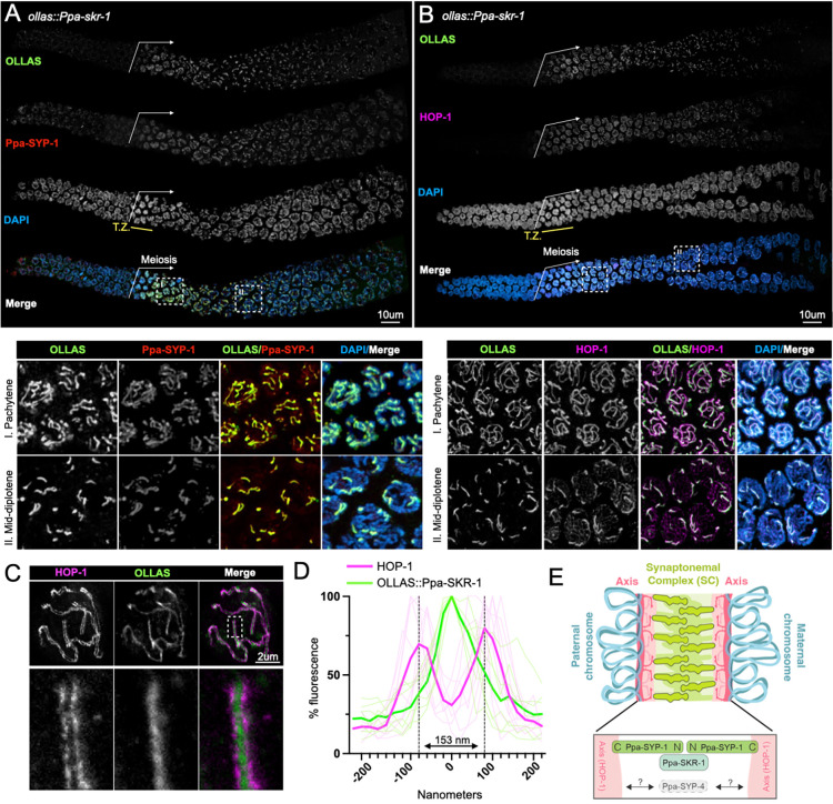
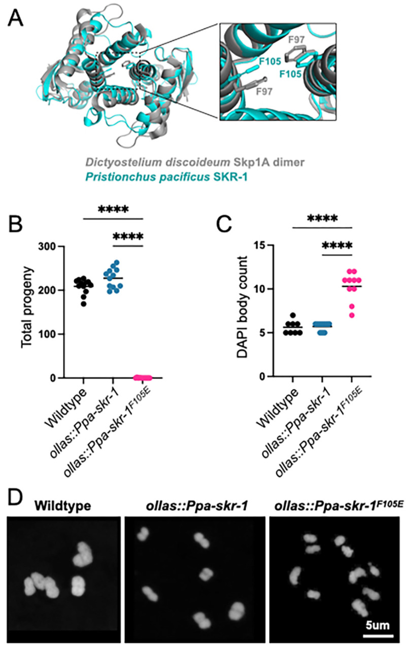
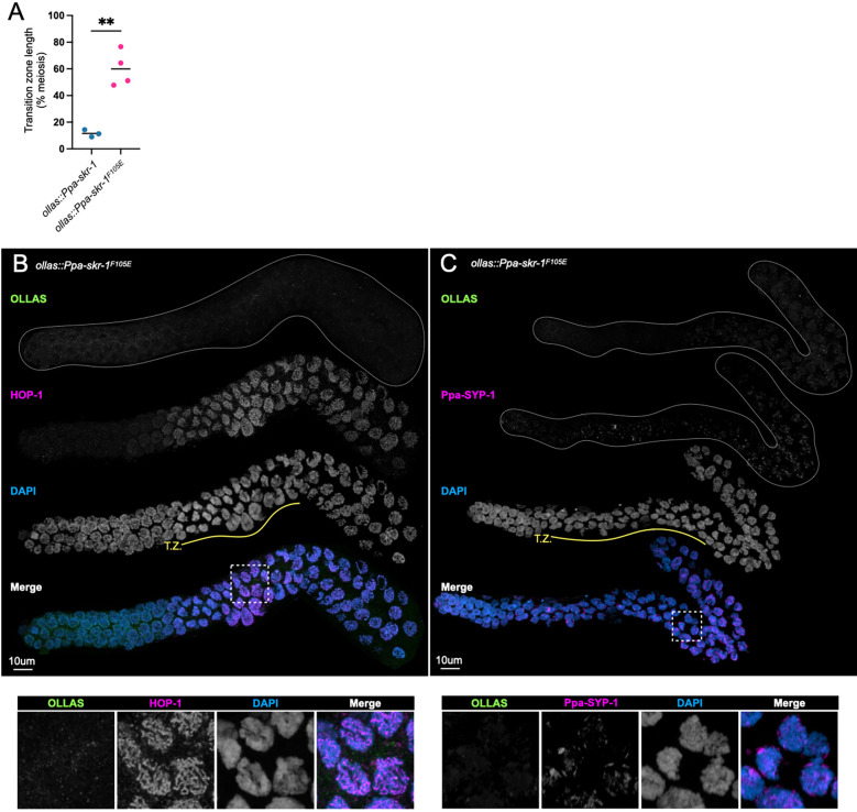
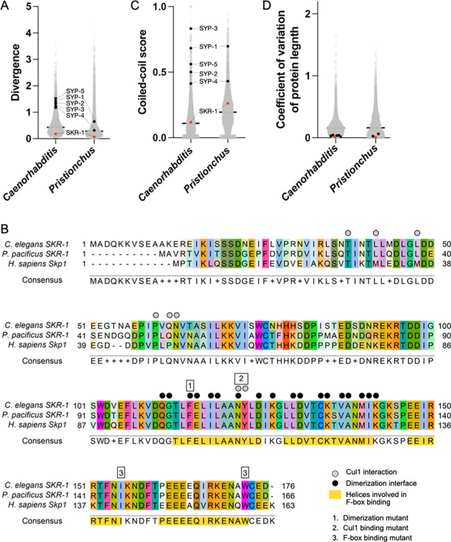
The synaptonemal complex (SC) is a meiotic interface that assembles between parental chromosomes and is essential for the formation of gametes. While the dimensions and ultrastructure of the SC are conserved across eukaryotes, its protein components are highly divergent. Recently, an unexpected component of the SC has been described in the nematode C. elegans: the Skp1-related proteins SKR-1/2, which are components of the Skp1, Cullin, F-box (SCF) ubiquitin ligase. Here, we find that the role of SKR-1 in the SC is conserved in nematodes. The P. pacificus Skp1 ortholog, Ppa-SKR-1, colocalizes with other SC proteins throughout meiotic prophase, where it occupies the middle of the SC. Like in C. elegans, the dimerization interface of Ppa-SKR-1 is required for its SC function. A dimerization mutant, Ppa-skr-1 F105E , fails to assemble SC and is almost completely sterile. Interestingly, the evolutionary trajectory of SKR-1 contrasts with other SC proteins. Unlike most SC proteins, SKR-1 is highly conserved in nematodes. Our results suggest that the structural role of SKR-1 in the SC has been conserved since the common ancestor of C. elegans and P. pacificus, and that rapidly evolving SC proteins have maintained the ability to interact with SKR-1 for at least 100 million years.

Keywords: P. pacificus; SCF; Skp1; evolution; meiosis; nematodes; synaptonemal complex.


Figures

Figure 1:
Figure 1:. Ppa-SKR-1 localizes to the middle of the SC.
(A) Top panel, confocal image of whole gonads from ollas::Ppa-skr-1 stained with anti-OLLAS, anti-SYP-1 and DAPI. Bottom panel, zoom in on pachytene (I) or mid-diplotene (II) nuclei. (B) Confocal image as in (A) except with HOP-1 staining. In (A) and (B), the beginning of the meiotic gonad is indicated with a white arrow and the transition zone is labeled below the DAPI channel in yellow (T.Z.). (C) Super-resolution STED image of a single pachytene nucleus from ollas::Ppa-skr-1 worms stained with anti-OLLAS and anti-HOP-1. Zoom-in panels show OLLAS::Ppa-SKR-1 between parallel HOP-1 tracks. (D) Plot of line scans of pixel intensity for anti-HOP-1 and anti-OLLAS across parallel axes in ollas::Ppa-skr-1 worms. The average distance between parallel axes is 153nm. (E) Cartoon of the P. pacificus synaptonemal complex with the orientation and position of Ppa-SYP-1 and Ppa-SKR-1 relative to HOP-1 indicated in the bottom panel. The relative position of Ppa-SYP-4 is not known (grey arrows and question marks). Adapted from (Kursel, Aguayo Martinez, and Rog 2023).
Figure 2:
Figure 2:. Conserved dimerization interface in SKR-1 is required for P. pacificus meiosis.
(A) Alignment of P. pacificus SKR-1 AlphaFold model (cyan) to Dictyostelium Skp1A dimer NMR structure (PDB structure 6V88, gray). Conserved phenylalanines required for dimerization are labeled in zoom. Dot plot depicting total progeny (B) and DAPI body count (C) for wild-type P. pacificus, ollas::Ppa-skr-1 and ollas::Ppa-skr-1F105E. Asterisks reflect P-values from Tukey’s multiple comparison test where **** indicates P < 0.0001. (D) Representative images of DAPI-stained Meiosis I bivalents (DAPI bodies) from the indicated genotypes.
Figure 3:
Figure 3:. Ppa-SKR-1F105E fails to assemble the SC.
(A) Dot plot showing transition zone length as percent of meiosis. Asterisks reflect the P-value from an unpaired T-test where ** indicates P < 0.01. (B) and (C), Confocal images of whole gonads from P. pacificus ollas::skr-1F105E stained with anti-OLLAS, anti-HOP-1 (B) or anti-SYP-1 (C), and DAPI. Lower panels in (B) and (C) show zoom-in on regions indicated by white, dashed boxes and the transition zone is labeled below the DAPI channel in yellow (T.Z.).
Figure 4:
Figure 4:. SKR-1 has an evolutionary signature distinct from other SC proteins.
(A) Dot plot showing protein divergence for the Caenorhabditis and Pristionchus proteomes. SYP proteins and SKR-1 are indicated (black and pink, respectively). (B) Alignment of Skp1 orthologs from C. elegans and P. pacificus, and H. sapiens with Cul1 interaction, dimerization and F-box binding sites labeled (Zheng et al. 2002; Kim et al. 2020). Additionally, three mutants generated by Blundon and Caesar et al. are indicated by numbered boxes (Blundon et al. 2024). (C) and (D), dot plots showing coiled-coil conservaBon and coefficient of variaBon of protein length for the Caenorhabdi+s and Pris+onchus proteomes. SYP proteins and SKR-1 are indicated as in (A).

Similar articles

References

    1. Adler Margo I., and Bonduriansky Russell. 2014. “Sexual Conflict, Life Span, and Aging.” Cold Spring Harbor Perspectives in Biology 6 (8). 10.1101/cshperspect.a017566. - DOI - PMC - PubMed
    1. Ahuja Jasvinder S., Sandhu Rima, Mainpal Rana, Lawson Crystal, Henley Hanna, Hunt Patricia A., Yanowitz Judith L., and Valentin Börner G.. 2017. “Control of Meiotic Pairing and Recombination by Chromosomally Tethered 26S Proteasome.” Science 355 (6323): 408–11. - PMC - PubMed
    1. Blundon Joshua M., Cesar Brenda I., Bae Jung Woo, Čavka Ivana, Haversat Jocelyn, Ries Jonas, Köhler Simone, and Kim Yumi. 2024. “Skp1 Proteins Are Structural Components of the Synaptonemal Complex in C. Elegans.” Science Advances 10 (7): eadl4876. - PMC - PubMed
    1. Castellanos María Del Pilar, Wickramasinghe Chathuri Devmika, and Betrán Esther. 2024. “The Roles of Gene Duplications in the Dynamics of Evolutionary Conflicts.” Proceedings. Biological Sciences / The Royal Society 291 (2024): 20240555. - PMC - PubMed
    1. Clark Nathan L., Alani Eric, and Aquadro Charles F.. 2012. “Evolutionary Rate Covariation Reveals Shared Functionality and Coexpression of Genes.” Genome Research 22 (4): 714–20. - PMC - PubMed

Publication types

LinkOut - more resources