. 2024 Jul 16;13(14):e032904.
doi: 10.1161/JAHA.123.032904. Epub 2024 Jul 9.

Enhanced Cardiomyocyte NLRP3 Inflammasome-Mediated Pyroptosis Promotes d-Galactose-Induced Cardiac Aging

Affiliations

Enhanced Cardiomyocyte NLRP3 Inflammasome-Mediated Pyroptosis Promotes d-Galactose-Induced Cardiac Aging

Wen-Bin Liu et al. J Am Heart Assoc. .

Abstract

Background: Cardiac aging represents an independent risk factor for aging-associated cardiovascular diseases. Although evidence suggests an association between NOD-, LRR-, and pyrin domain-containing protein 3 (NLRP3) inflammasome formation and numerous cardiovascular diseases, its role in cardiac aging remains largely unclear.

Methods and results: The longevity of mice with wild-type and NLRP3 knockout (NLRP3-/-) genotypes was assessed, with or without d-galactose treatment. Cardiac function was evaluated using echocardiography, and cardiac histopathology was examined through hematoxylin and eosin and Masson's trichrome staining. Senescence-associated β-galactosidase (SA-β-gal) staining was employed to detect cardiac aging. Western blotting was used to assess aging-related proteins (p53, p21) and pyroptosis-related proteins. Additionally, dihydroethidium staining, lactate dehydrogenase release, and interleukin-1β ELISA assays were performed, along with measurements of total superoxide dismutase and malondialdehyde levels. In vitro, H9c2 cells were exposed to d-galactose for 24 hours in the absence or presence of N-acetyl-l-cysteine (reactive oxygen species inhibitor), BAY-117082 (nuclear factor κ-light-chain enhancer of activated B cells inhibitor), MCC950 (NLRP3 inhibitor), and VX-765 (Caspase-1 inhibitor). Immunofluorescence staining was employed to detect p53, gasdermin D, and apoptosis-associated speck-like protein proteins. Intracellular reactive oxygen species levels were assessed using fluorescence microscopy and flow cytometry. Senescence-associated β-galactosidase staining and Western blotting were also employed in vitro for the same purpose. The results showed that NLRP3 upregulation was implicated in aging and cardiovascular diseases. Inhibition of NLRP3 extended life span, mitigated the aging phenotype, improved cardiac function and blood pressure, ameliorated lipid metabolism abnormalities, inhibited pyroptosis in cardiomyocytes, and ultimately alleviated cardiac aging. In vitro, the inhibition of reactive oxygen species, nuclear factor κ-light-chain enhancer of activated B cells, NLRP3, or caspase-1 attenuated NLRP3 inflammasome-mediated pyroptosis.

Conclusions: The reactive oxygen species/nuclear factor κ-light-chain enhancer of activated B cells/NLRP3 signaling pathway loop contributes to d-galactose-treated cardiomyocyte senescence and cardiac aging.

Keywords: NLRP3 inflammasome; ROS; cardiac aging; pyroptosis.


Figures

Figure 1
Figure 1. NLRP3 upregulation is involved in age and CVDs.
(A ) Comparison of PBMCs NLRP3 expression between young and old subjects (GSE63117). ( B) Correlation between PBMCs NLRP3 expression with age (GSE63117). ( C) NLRP3 expression comparison between atherosclerotic lesions and control arteries (GSE100927). (D) NLRP3 expression in human LV free wall heart tissue from individuals with end‐stage HF and healthy heart (GSE84796). (E) NLRP3 expression in atrial myocardium from subjects with AF compared with healthy control (GSE2240). *P<0.05, **P<0.01, and ****P<0.0001. AF indicates atrial fibrillation; AS, atherosclerotic; CVDs, cardiovascular diseases; HF, heart failure; LV, left ventricular; NLRP3, NOD‐, LRR‐, and pyrin domain–containing protein 3; and PBMCs, peripheral blood mononuclear cells.
Figure 2
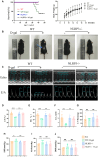
Figure 2. Inhibition of NLRP3 extended lifespan and improved cardiac function and blood pressure in d‐galactose–induced mice.
(A) Kaplan–Meier survival curves (n=8) and body weight of different groups. (B) Representative photographs of 16‐wk‐old mice. Blue arrows indicate back depilation. (C) Representative echocardiographic (Echo) and the ratio of the E/A ratio images (n=6). (D) Statistical results for LVEF, (E) LVFS, (F) E/A ratio, and (G) LV mass (n=6). (H) Statistical results for SBP, (I) MAP, and (J) DBP (n=6). Male, n=3; female, n=3. *P<0.05, **P<0.01, ***P<0.001, and ****P<0.0001. d‐gal indicates d‐galactose; DBP, diastolic blood pressure; E/A, E peak to the A peak; LV, left ventricular; LVEF, left ventricular ejection fraction; LVFS, left ventricular fractional shortening; MAP, mean arterial pressure; NLRP3, NOD‐, LRR‐, and pyrin domain–containing protein 3; SBP, systolic blood pressure; and WT, wild‐type.
Figure 3
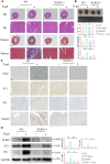
Figure 3. Inhibition of NLRP3 alleviated cardiac aging in d‐galactose–induced mice.
(A) Representative HE and Masson's trichrome staining images (2× and 400×). (B) Representative images of mouse heart in different groups and the heart index (heart weight normalized to body weight); quantitative analysis of cardiomyocyte cross‐sectional area and fibrotic areas with measurements (n=6). (C) Representative β‐galactosidase and immunohistochemistry staining of p53, p21, and NLRP3 images within the mouse heart sections under 400×. (D) Representative immunoblots and the corresponding quantification of the aging‐associated proteins (p53, p21) and NLRP3 in myocardial tissues (n=6). male, n=3; female, n=3; *P<0.05, **P<0.01, ***P<0.001, and ****P<0.0001. d‐gal indicates d‐galactose; HE, hematoxylin and eosin; β‐gal, β‐galactosidase; NLRP3, NOD‐, LRR‐, and pyrin domain–containing protein 3; and WT, wild‐type.
Figure 4
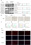
Figure 4. The feedback loop between ROS and the NLRP3 inflammasome contributed to the d‐galactose–induced cardiac aging by promoting pyroptosis.
(A) Representative immunoblots and the corresponding quantification of the indicated proteins in myocardial tissues (n=6). (B) Representative immunohistochemistry staining of ASC images within the mouse heart sections under 400×. (C) Serum level of interleukin‐1β (n=6). (D) Serum level of LDH (n=6). (E) Serum levels of T‐SOD (n=6). (F) Serum levels of MDA (n=6). (G) ROS was detected using a dihydroethidium probe. Representative fluorescence images (red) showed ROS production in different groups (400×). male, n=3; female, n=3; *P<0.05, **P<0.01, and ****P<0.0001. ASC indicates apoptosis‐associated speck‐like protein; d‐gal, d‐galactose; DHE, dihydroethidium; GSDMD, gasdermin D; GSDMD‐NT, gasdermin D N‐terminal cleavage product; IL, interleukin; LDH, lactate dehydrogenase; MDA, malondialdehyde; NLRP3, NOD‐, LRR‐, and pyrin domain–containing protein 3; ROS, reactive oxygen species; T‐SOD, total superoxide dismutase; and WT, wild‐type.
Figure 5
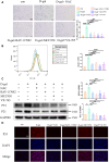
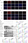
Figure 5. Inhibition of ROS, NF‐κB, NLRP3, or caspase‐1 alleviates d‐galactose–treated H9c2 cardiomyocytes senescence.
(A) The H9c2 cardiomyocytes senescence treated by d‐galactose was identified by β‐galactosidase staining (100×) (n=3). (B) Flow cytometry analysis was used to detect the β‐galactosidase MFI in different groups (n=3). (C) Representative immunoblots and the corresponding quantification of the aging‐associated proteins (p53, p21) in cardiomyocytes (n=3). (D) Representative immunofluorescence staining (red) of p53 images under 100×. *P<0.05, **P<0.01, ***P<0.001, and ****P<0.0001. DAPI indicates 4′,6‐diamidino‐2‐phenylindole; d‐gal, d‐galactose; MFI, mean fluorescence intensity; NF‐κB, nuclear factor κ‐light‐chain enhancer of activated B cells; NLRP3, NOD‐, LRR‐, and pyrin domain–containing protein 3; and ROS, reactive oxygen species.
Figure 6
Figure 6. Inhibition of ROS, NF‐κB, NLRP3, or caspase‐1 attenuates NLRP3 inflammasome‐mediated pyroptosis in cardiac aging.
(A) Representative immunofluorescence staining (red) of GSDMD images under 100×. (B) Representative immunofluorescence staining (red) of ASC images under 100×. (C) Representative immunoblots and the corresponding quantification of the indicated proteins in H9c2 cardiomyocytes (n=3). (D) IL‐1β levels in supernatant (n=3). (E) LDH levels in supernatant (n=3). *P<0.05, **P<0.01, ***P<0.001 and ****P<0.0001. GSDMD indicates gasdermin D; IL‐1β, interleukin‐1β; LDH, lactate dehydrogenase; NF‐κB, nuclear factor κ‐light‐chain enhancer of activated B cells; NLRP3, NOD‐, LRR‐, and pyrin domain–containing protein 3; and ROS, reactive oxygen species.
Figure 7
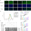
Figure 7. The ROS/NF‐κB/NLRP3 signaling pathway loop contributes to the d‐galactose–treated cardiomyocyte senescence.
(A) Representative fluorescence images (green) show ROS production in different groups (100×). (B) Intracellular ROS was quantified by flow cytometry (n=3). (C) Representative immunoblots and the corresponding quantification of P‐NF‐κB and in H9c2 cardiomyocytes (n=3). *P<0.05, **P<0.01, ***P<0.001, and ****P<0.0001. d‐gal indicates d‐galactose; NF‐κB, nuclear factor κ‐light‐chain enhancer of activated B cells; NLRP3, NOD‐, LRR‐, and pyrin domain–containing protein 3; P‐NF‐κB, ; ROS, reactive oxygen species; and (Total NF‐κB) T‐NF‐κB,.
Figure 8
Figure 8. Possible molecular mechanism of d‐galactose–induced cardiac aging and pyroptosis.
d‐galactose leads to increased ROS production in cardiomyocytes. ROS mediates NLRP3 inflammasome activation by contributing to the priming signal of NLRP3 inflammasome via ROS‐dependent transcriptional factor NF‐κB, leading to the activation of caspase‐1. Cleaved‐caspase‐1 promotes maturation and release of the inflammatory cytokines interleukin‐1β and interleukin‐18. In addition, cleaved‐caspase‐1 can cleave GSDMD to yield the GSDMD‐NT, which induces pyroptosis by forming plasma membrane pores. ASC indicates apoptosis‐associated speck‐like protein; d‐gal, d‐galactose; GSDMD, gasdermin D; GSDMD‐NT, gasdermin D N‐terminal cleavage product; IL, interleukin; NF‐κB, nuclear factor κ‐light‐chain enhancer of activated B cells; NLRP3, NOD‐, LRR‐, and pyrin domain–containing protein 3; and ROS, reactive oxygen species.

References

    1. Donato AJ, Machin DR, Lesniewski LA. Mechanisms of dysfunction in the aging vasculature and role in age‐related disease. Circ Res. 2018;123:825–848. doi: 10.1161/circresaha.118.312563 - DOI - PMC - PubMed
    1. Ren J, Zhang Y. Targeting autophagy in aging and aging‐related cardiovascular diseases. Trends Pharmacol Sci. 2018;39:1064–1076. doi: 10.1016/j.tips.2018.10.005 - DOI - PMC - PubMed
    1. Bergsbaken T, Fink SL, Cookson BT. Pyroptosis: host cell death and inflammation. Nat Rev Microbiol. 2009;7:99–109. doi: 10.1038/nrmicro2070 - DOI - PMC - PubMed
    1. Hsu SK, Li CY, Lin IL, Syue WJ, Chen YF, Cheng KC, Teng YN, Lin YH, Yen CH, Chiu CC. Inflammation‐related pyroptosis, a novel programmed cell death pathway, and its crosstalk with immune therapy in cancer treatment. Theranostics. 2021;11:8813–8835. doi: 10.7150/thno.62521 - DOI - PMC - PubMed
    1. Yu P, Zhang X, Liu N, Tang L, Peng C, Chen X. Pyroptosis: mechanisms and diseases. Signal Transduct Target Ther. 2021;6:128. doi: 10.1038/s41392-021-00507-5 - DOI - PMC - PubMed

MeSH terms

LinkOut - more resources