

NUSAP1 Promotes Immunity and Apoptosis by the SHCBP1/JAK2/STAT3 Phosphorylation Pathway to Induce Dendritic Cell Generation in Hepatocellular Carcinoma
- PMID: 38980111
- PMCID: PMC11753460
- DOI: 10.1097/CJI.0000000000000531
NUSAP1 Promotes Immunity and Apoptosis by the SHCBP1/JAK2/STAT3 Phosphorylation Pathway to Induce Dendritic Cell Generation in Hepatocellular Carcinoma
Abstract
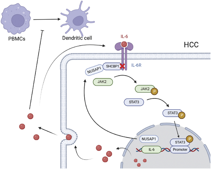
Hepatocellular carcinoma (HCC) is the most common type of liver cancer and is associated with high morbidity and mortality rates. The aims of this study were to investigate the immune-promoting action of nucleolar and spindle-associated protein 1 (NUSAP1) and identify an immunotherapy target for HCC. The Cancer Genome Atlas (TCGA) was used to analyze interaction molecules and immune correlation. The interaction between NUSAP1 and SHC binding and spindle associated 1 (SHCBP1) was examined. The role of the SHCBP1/Janus kinase 2/signal transducer and activator of transcription 3 (SHCBP1/JAK2/STAT3) pathway in this process was explored. After co-culture with HCC cell lines, the differentiation of peripheral blood mononuclear cells (PBMCs) into dendritic cells (DC) was evaluated by measuring the expression of surface factors CD1a and CD86. Pathological tissues from 50 patients with HCC were collected to validate the results of cell experiments. The expression levels of CD1a and CD86 in tissues were also determined. The results show that NUSAP1 interacted with SHCBP1 and was positively correlated with DC. In HCC cell lines, an interaction was observed between NUSAP1 and SHCBP1. It was verified that NUSAP1 inhibited the JAK2/STAT3 phosphorylation pathway by blocking SHCBP1. After co-culture, the levels of CD1a and CD86 in PBMC were elevated. In the clinical specimens, CD1a and CD86 expression levels were significantly higher in the high-NUSAP1 group versus the low-NUSAP1 group. In Summary, NUSAP1 enhanced immunity by inhibiting the SHCBP1/JAK2/STAT3 phosphorylation pathway and promoted DC generation and HCC apoptosis. NUSAP1 may be a target of immunotherapy for HCC.
Copyright © 2024 The Author(s). Published by Wolters Kluwer Health, Inc.
Conflict of interest statement
None reported. All authors have declared there are no financial conflicts of interest with regard to this work.
Figures


Similar articles
-
Yu-Ping-Feng-San improve the immunosuppression of microenvironment in hepatocellular carcinoma by promoting the maturation of DCs through the JAK2-STAT3 pathway.Sci Rep. 2024 Dec 28;14(1):31522. doi: 10.1038/s41598-024-83197-7. Sci Rep. 2024. PMID: 39733089 Free PMC article.
-
LncRNA 00152 promotes the development of hepatocellular carcinoma by activating JAK2/STAT3 pathway.Eur Rev Med Pharmacol Sci. 2019 Feb;23(3):1038-1046. doi: 10.26355/eurrev_201902_16991. Eur Rev Med Pharmacol Sci. 2019. PMID: 30779070
-
Circ_0072088 promotes progression of hepatocellular carcinoma by activating JAK2/STAT3 signaling pathway via miR-375.IUBMB Life. 2021 Sep;73(9):1153-1165. doi: 10.1002/iub.2520. Epub 2021 Jun 25. IUBMB Life. 2021. PMID: 34148288
-
Curcumin Inhibits the Growth of Hepatocellular Carcinoma via the MARCH1-mediated Modulation of JAK2/STAT3 Signaling.Recent Pat Anticancer Drug Discov. 2025;20(2):145-157. doi: 10.2174/0115748928261490231124055059. Recent Pat Anticancer Drug Discov. 2025. PMID: 38243928
-
STAT3 orchestrates immune dynamics in hepatocellular carcinoma: A pivotal nexus in tumor progression.Crit Rev Oncol Hematol. 2025 Mar;207:104620. doi: 10.1016/j.critrevonc.2025.104620. Epub 2025 Jan 14. Crit Rev Oncol Hematol. 2025. PMID: 39818308 Review.
Cited by
-
MiR-490-3p promotes cell apoptosis and cell-cycle arrest in osteosarcoma via the modulation of CDCA8/ATF3 by targeting NUSAP1.Transl Pediatr. 2024 Dec 31;13(12):2242-2253. doi: 10.21037/tp-2024-529. Epub 2024 Dec 27. Transl Pediatr. 2024. PMID: 39823004 Free PMC article.
-
Cancer metastasis: molecular mechanisms and therapeutic interventions.Mol Biomed. 2025 Apr 7;6(1):20. doi: 10.1186/s43556-025-00261-y. Mol Biomed. 2025. PMID: 40192949 Free PMC article. Review.
-
The role and therapeutic value of NUSAP1 in human cancers.J Transl Med. 2025 Jul 2;23(1):725. doi: 10.1186/s12967-025-06746-2. J Transl Med. 2025. PMID: 40605080 Free PMC article. Review.
-
Integrated machine learning to predict the prognosis of lung adenocarcinoma patients based on SARS-COV-2 and lung adenocarcinoma crosstalk genes.Cancer Sci. 2025 Jan;116(1):95-111. doi: 10.1111/cas.16384. Epub 2024 Nov 3. Cancer Sci. 2025. PMID: 39489517 Free PMC article.
-
Research progress on NUSAP1 and its role in digestive system neoplasms.Front Oncol. 2025 Jun 4;15:1582361. doi: 10.3389/fonc.2025.1582361. eCollection 2025. Front Oncol. 2025. PMID: 40535133 Free PMC article. Review.
References
-
- Siegel RL, Miller KD, Jemal A. Cancer statistics, 2019. CA Cancer J Clin. 2019;69:7–34. - PubMed
-
- Marquardt JU, Andersen JB, Thorgeirsson SS. Functional and genetic deconstruction of the cellular origin in liver cancer. Nat Rev Cancer. 2015;15:653–667. - PubMed
-
- Llovet JM, Kelley RK, Villanueva A, et al. . Hepatocellular carcinoma. Nat Rev Dis Primers. 2021;7:6. - PubMed
-
- Forner A, Reig M, Bruix J. Hepatocellular carcinoma. Lancet. 2018;391:1301–1314. - PubMed
MeSH terms
Substances
LinkOut - more resources
Full Text Sources
Medical
Research Materials
Miscellaneous
