Review
. 2024 Sep 3;36(9):2931-2975.
doi: 10.1093/plcell/koae193.

The lowdown on breakdown: Open questions in plant proteolysis

Affiliations
Review

The lowdown on breakdown: Open questions in plant proteolysis

Nancy A Eckardt et al. Plant Cell. .

Abstract

Proteolysis, including post-translational proteolytic processing as well as protein degradation and amino acid recycling, is an essential component of the growth and development of living organisms. In this article, experts in plant proteolysis pose and discuss compelling open questions in their areas of research. Topics covered include the role of proteolysis in the cell cycle, DNA damage response, mitochondrial function, the generation of N-terminal signals (degrons) that mark many proteins for degradation (N-terminal acetylation, the Arg/N-degron pathway, and the chloroplast N-degron pathway), developmental and metabolic signaling (photomorphogenesis, abscisic acid and strigolactone signaling, sugar metabolism, and postharvest regulation), plant responses to environmental signals (endoplasmic-reticulum-associated degradation, chloroplast-associated degradation, drought tolerance, and the growth-defense trade-off), and the functional diversification of peptidases. We hope these thought-provoking discussions help to stimulate further research.


Conflict of interest statement

Conflict of interest statement. None declared.

Figures

Figure 1.
Figure 1.
Regulation and possible roles of FBL17 in Arabidopsis. A) During the G1/S-phase, E2Fa-DPa directly activates the transcription of FBL17. The degradation of KRPs by FBL17-mediated ubiquitylation and degradation would release the activity of CDKA/CYCD. Whether FBL17 recognizes its substrate via a phospho-degron is unknown. B) During replication stress, WEE1 phosphorylates FBL17 and the APC10 subunit of the APC/C, which promotes FBL17 ubiquitylation and degradation by the proteasome.
Figure 2.
Figure 2.
Proteasome- and vacuole-dependent protein degradation in the plant DDR. After DNA damage, a plant cell launches a DNA damage response (DDR), which includes the transcriptional upregulation of certain genes and the targeted degradation of proteins. The two major protein degradation systems, the UPS and the vacuole-dependent system (autophagy) both appear to be involved in the DDR. The diagram shows a prototypical proteasome target (green X). Under non-stress conditions (gray arrows), X is marked by K48 polyubiquitin chains and subsequently degraded by the proteasome. After DNA damage, X becomes stabilized and participates in the DDR. Examples of X are the transcriptional repressor of cell proliferation MYB3R3 and the RMI1 removing factor KNO1. Another and previously not DDR-associated degradation pathway is shown for the orange-marked protein Y, a protein that interferes with efficient DDR. Under non-damaging conditions, Y is present. Under damaging conditions, Y is polyubiquitylated via K63 chains marking it for autophagy-dependent degradation via the cytoplasm. The only example for Y so far is the RTR-complex scaffolding component RMI1, which becomes polyubiquitylated via K63, targeting it for removal to the cytoplasm and degradation via autophagy. The observation that mutants in the macroautophagy pathway (such as ATG mutants) are sensitive to various DNA-damage-inducing drugs indicates that macroautophagy also degrades other proteins after DNA damage and likely plays a major role in the DDR of plants.
Figure 3.
Figure 3.
Proteolytic networks across different plant mitochondrial compartments. Protein quality control (PQC in red dotted lines) that includes the disassembly, unfolding, and degradation of proteins and complexes is carried out by various proteases as indicated. PQC is carried out at various pathways including protein import, organello translation, and assembly. Preproteins are imported from the cytosol through the translocase of the outer membrane (TOM) complex. Preproteins targeted to the outer membrane (OM) and the IMS undergo PQC by various IMS-facing proteases, such as overlapping with m-AAA protease OMA1 and rhomboid-like (RBL) protease. Preproteins targeted to the inner membrane (IM) and the matrix are translocated by the translocase of the inner membrane (TIM) complex. Preproteins then undergo the maturation process to remove the N-terminal targeting signal and destabilizing residues by mitochondrial processing peptidase (MPP) embedded within Complex III (CIII) in plants, octapeptidyl peptidase OCT1, and intermediate cleavage peptidase ICP55. Proteins assembled within a complex, such as the oxidative phosphorylation (OXPHOS) supercomplex SC I + III2 + IV (or the respirasome) may undergo PQC by the IM-embedded proteases like the matrix-facing FTSH3/FTSH10 and the IMS-facing FTSH4/FTSH11. Nascent polypeptides translated by the mitoribosome may be regulated by FTSH3/10, while PQC of matrix-facing and matrix-located proteins is regulated by the matrix-located CLPP2 and LON1 proteases. Free peptides generated from both targeting peptide cleavage and protein degradation (blue dashed lines) can be further degraded into single amino acids in the peptide processing pathway. This multi-step pathway uses multiple peptidases, including presequence peptidases (PREP), organellar oligopeptidase (OOP), and various aminopeptidases (AP), including alanyl aminopeptidase (AAP), leucyl aminopeptidase (LAP), aspartyl aminopeptidase (DAP), and prolyl aminopeptidase (PAP).
Figure 4.
Figure 4.
The relationship between NTA and protein stability. A) The conditionality of Ac/N-degrons and their link to protein quality control. Acetylated (Ac) N-termini are often shielded through internal protein folding (i) or protein–protein interactions (ii) but can be exposed through protein misfolding or if there is an excess of a particular protein complex subunit. This leads to exposure of the acetylated N-terminus, which can act as a specific degron for proteasomal degradation via the Ac/N-degron pathway (Shemorry et al. 2013). B) Hypothetical indirect effects of NTA on protein stability. NTA can increase protein-interaction affinities, to create more stable complexes. A lack of NTA can lead to reduced thermostability, complex breakdown, and the consequent degradation of non-bound and potentially misfolded subunits via then UPS (e.g. as has been shown for cytosolic ribosomes in yeast; Guzman et al. 2023). C) NATA-mediated NTA (potentiated by HYPK in plants and mammals) was shown to promote broad proteome stabilization in diverse eukaryotic taxa. In plants, drought-induced downregulation of NATA activity leads to reduced NTA of NATA substrates and an increase in their degradation via exposed “non-Ac/N-degrons” (Linster et al. 2015, 2022). This suggests that NATs may integrate stress signals to control proteome turnover.
Figure 5.
Figure 5.
Enzymes and substrates of plant Arg/N-degron pathways. A) Schematic of N-recognins in plants and mammals showing substrate recognition and E3 ligase domains (drawn approximately to scale; hRING, hemi-RING). B) Destabilizing residues and specificity of Arabidopsis N-recognins, deduced from model substrates (Garzón et al. 2007; Graciet et al. 2010). In these examples, Ub fusion protein is cleaved in planta by deubiquitylating enzymes (DUBs) to produce glucuronidase (GUS) or luciferase (LUC) with Nt destabilizing residues (shown in single letter amino acid code). Proteins with primary destabilizing residues (R, F, L) are targeted for proteasomal degradation by known and unknown N-recognins. Tertiary destabilizing residues N and Q are converted to D and E by Nt(Asn) amidase (NTAN) and Nt(Gln) amidase (NTAQ), respectively. These secondary destabilizing residues are then Nt-arginylated by arginyl-tRNA-transferases (ATEs), enabling PRT6-mediated degradation. C) Physiological substrates of the Arg/N-degron pathway. Met-Cys-initiating proteins VRN2, ZPR2, and ERFVIIs undergo co-translational Met excision by methionine amino peptidases (MetAPs) to reveal Nt-Cys, a tertiary destabilizing residue. Following Nt oxidation by plant cysteine oxidases (PCOs) and arginylation by ATEs, they become substrates for PRT6. BIG also mediates the degradation of ERFVIIs and VRN2.
Figure 6.
Figure 6.
Schematic view of the N-degron pathway for degradation by the chloroplast Clp chaperone-protease system. Proteins can be converted into substrates for the Clp system by various events including protein complex disassembly and aggregation, different stresses such as heat and radical oxygen species (ROS), or through metabolic feedback (e.g. known to occur in the chlorophyll synthesis pathway). These changes to proteins can result in the generation of a degradation signal known as a degron, either by simply exposing (“unmasking”) the N-terminus of the protein or by a post-translational modification (PTM). Examples of such PTMs are phosphorylation, acetylation, oxidation, or the addition of an amino acid to the N-terminus. This N-degron is then recognized by the ClpS1 recognin (and possibly also ClpF), which delivers the bound substrate to the ClpC or ClpD chaperones for ATP-dependent unfolding and concomitant threading into the Clp protease complex. The unfolded substrates are degraded within the Clp proteolytic chamber resulting in the release of degradation products in the form of small peptides. However, the in vivo nature of these chloroplast N-degrons is yet to be determined. Elucidation of these N-degrons and the molecular players involved in their generation and recognition is a major challenge to be addressed.
Figure 7.
Figure 7.
Regulation of light-signaling components through the UPS. Photoreceptors, various light-promoting transcription factors (inside the yellow circle on right), and light repressors (inside the blue circle on left) are regulated by UPS through indicated CRL E3 ligase complexes.
Figure 8.
Figure 8.
Proteolysis of core ABA signaling components occurs in different subcellular compartments. The inset shows that ABA is perceived through dimeric or monomeric receptors (blue), which triggers the formation of ternary complexes with clade A PP2Cs (red), and relief of inhibition of SnRK2.2/2.3/2.6 (pink) kinase activity. Nuclear, cytosolic, and PM targeting pathways of core components are indicated. RSL1 illustrates the targeting of ABA receptors at the PM, which promotes endosome-mediated vacuolar degradation via the ESCRT machinery, whereas PUB12 and AIRP3 might target PP2Cs in the proximity of the PM and follow either cytosolic or vacuolar degradation pathways. Nuclear degradation of ABA receptors, PP2Cs, and OST1 involves the multimeric CRL3, CRL4, and RING-type COP1 E3 ligases, among others (see text for details). Nuclear and cytosolic 26S proteasomes and the vacuole participate in the degradation of core signaling components, which might influence signaling, desensitization, or resetting of the ABA pathway. The lytic vacuole also receives cargo for degradation via autophagy but data linking ABA with autophagy are limited. The figure was created using BioRender (https://biorender.com).
Figure 9.
Figure 9.
Regulation of SL signaling through protein degradation: (i) SL (light pink) is perceived by the receptor D14 (blue). (ii) The activated SL receptor then binds to D3/MAX2 F-box (gray) as part of the SCFD3/MAX2 complex. The presence of citrate or citrate-like molecules (red) triggers a conformational change in the C-terminal helix, CTH (red) of D3/MAX2 (gray), causing it to dislodge. (iii) The dislodged CTH of D3/MAX2-D14 complex subsequently loads the transcriptional repressor, D53/SMXLs (purple), through its D2 domain leading to polyubiquitylation (Ub, yellow). The binding of D53/SMXLs reactivates the hydrolysis of SL by D14 either during or after the polyubiquitylation of D53/SMXLs. (iv) The hydrolysis of SLs triggers a conformational change of D14 and restores the CTH of D3/MAX2 to its engaged conformation and subsequently triggers the release of polyubiquitylated D53/SMXLs from the D14-D3/MAX2 to proteasomal degradation. (v) D14 undergoes ubiquitylation and proteasomal degradation completing the feedback regulation of the SL signaling cascade. Interestingly, before D53/SMXLs are released to the 26S proteasome, their transient interaction with D3 alters D14 inhibition and gradually restores SL hydrolysis (Shabek et al. 2018; Tal et al. 2022). This restored activity can effectively “reset” the SL signal by depleting the hormone and degrading the D14 receptor until the next cue. This E3 ligase domain plasticity provides an additional level of signaling control and represents a unique mode of targeting substrates for proteasomal degradation in the realm of phytohormone and UPS signaling. The figure was created using BioRender (https://biorender.com).
Figure 10.
Figure 10.
Interplay between carbon-containing metabolites and autophagy. The figure was created using BioRender (https://biorender.com).
Figure 11.
Figure 11.
Fruit plastid evolution along ripening progression and their possible delivery to vacuoles. A to E) Transmission electron micrographs of ultrathin sections of tomato pericarp cells from three ripening stages as indicated. A) Chloroplast (arrow). B) Chromoplast (arrow). C) Vacuoles contain structures likely to be plastids (arrowheads) by judging the electron density of the cytoplasmic plastids in their vicinity (arrows in the inset). D) A chromoplast with signs of internal degradation (arrow). E) Vacuolar inclusions of what appear to be plastids remain (judging the internal plastoglobules). C, Cytoplasm. V, Vacuole lumen. Image credits: S. Mursalimov, A. Upcher, S. Michaeli.
Figure 12.
Figure 12.
The role of ERAD in plant growth, crop yield, and stress response. A) The role of ERAD in stress response and phytohormone signaling in Arabidopsis. The leucine-rich repeat receptor-like kinase (LRR-RLK) CEPR2 phosphorylates ABA importer NRT1.2/NPF4.6, inhibiting its ability to import ABA. The phosphorylated NRT1.2/NPF4.6 is then transported to the ER for ubiquitylation and degradation, mediated by UBC32 and its homologs UBC33 and UBC34. ABA inhibits the CEPR2-mediated phosphorylation of NRT1.2/NPF4.6. ABA receptor PYL recognizes ABA and initiates the transduction of ABA signaling. UBC27 and AIRP3 act as an E2–E3 pair to activate ABA signaling and enhance drought tolerance by promoting the ubiquitylation and degradation of ABI1. Moreover, UBC32 collaborates with AtEMR1 to facilitate the degradation of misfolded BRI1, thereby influencing BR signaling under ER stress conditions. During drought stress, Rma1 and UBC32 work together to enhance drought tolerance by promoting the degradation of phosphorylated aquaporin PIP2; 1. B) The role of ERAD in crop yield and disease resistance. The ERAD-related E2-E3 pair, OsUBC45/SiUBC32-DGS1/SGD1 in rice and millet, enhances yield by regulating BR signaling via distinct mechanisms. They enhance BR signaling by reducing the protein level of misfolded BR receptor BRI1 (in rice) or increasing the protein level of folded BRI1 (in millet). Additionally, the E2–E3 pair also promotes the Ub-dependent degradation of OsGSK3, a negative regulator of BR signaling. Under fungi attack, OsPIP2; 1 facilitates the translocation of H2O2 from the cytoplasm to the apoplast, negatively regulating pattern-triggered immunity (PTI). OsUBC45 and DGS1 promote the degradation of OsPIP2; 1, enhancing rice resistance to disease.
Figure 13.
Figure 13.
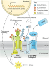
Possible mechanisms of regulation of chloroplast-associated protein degradation. CHLORAD is a UPS pathway that selectively degrades chloroplast-resident proteins, including the TOC apparatus that is responsible for protein import. The SP1 Ub E3 ligase recruits E2 Ub-conjugating enzyme [via its RING finger (RNF) domain] to direct the ubiquitylation of TOC proteins, which are then degraded through the combined action of the SP2 retrotranslocation channel, the CDC48 ATPase motor, and the cytosolic 26S proteasome (26SP); in this, the activity of SP1 is modulated by the action of SP1-like components (SPLs). Thus, CHLORAD exerts important control over protein import and the organelle's proteome and functions. Such control is responsive to developmental and environmental cues through unclear mechanisms. Under different conditions, phytohormone, Ca2+, or reactive oxygen species (ROS) signaling might regulate the activity of CHLORAD, and this is possibly mediated through post-translational modification of the CHLORAD machinery or its TOC apparatus targets, and/or through retrograde signaling and stress-responsive proteins. Post-translational modifications that are potentially involved in such regulation are indicated.
Figure 14.
Figure 14.
Possible mechanisms by which autophagy regulates drought tolerance. Autophagy is activated by drought stress via the TOR complex, SnRK1, and transcriptional pathways. Activation of autophagy may lead to increased degradation of protein aggregates and aquaporins, and decreased growth and stomatal aperture, in turn aiding tolerance of drought conditions.
Figure 15.
Figure 15.
The proteasome influences the growth-defense trade-off. A) The proteasome degrades substrates from various cellular compartments and organelles to maintain cell survival and optimal growth. B) Proteins that accumulate due to stress conditions or accumulating preproteins from organelles or microbes as well as chemical inhibitors can interfere with proteasome function leading to proteotoxic stress. This will cause growth penalties, impact survival, and in the case of microbes cause disease. C) On the one hand, a natural allele of proteasome maturation factor UMP1R2115 results in more proteasome abundance and activity improving resistance to multiple pathogens without growth penalties. On the other hand, proteasome activation can be achieved by the transcription factor pair NAC53 and NAC78. Whether this transcriptional activation of the proteasome results in resistance to pathogens and how it impacts plant growth remains to be discovered. The figure was created using BioRender (https://biorender.com).

References

    1. Acosta JAT, Fowke LC, Wang H. Analyses of phylogeny, evolution, conserved sequences and genome-wide expression of the ICK/KRP family of plant CDK inhibitors. Ann Bot. 2011:107(7):1141–1157. 10.1093/aob/mcr034 - DOI - PMC - PubMed
    1. Adachi S, Minamisawa K, Okushima Y, Inagaki S, Yoshiyama K, Kondou Y, Kaminuma E, Kawashima M, Toyoda T, Matsui M, et al. . Programmed induction of endoreduplication by DNA double-strand breaks in Arabidopsis. Proc Natl Acad Sci U S A. 2011:108(24):10004–10009. 10.1073/pnas.1103584108 - DOI - PMC - PubMed
    1. Adams EHG, Spoel SH. The ubiquitin—proteasome system as a transcriptional regulator of plant immunity. J Exp Bot. 2018:69(19):4529–4537. 10.1093/jxb/ery216 - DOI - PubMed
    1. Agbemafle W, Wong MM, Bassham DC. Transcriptional and post-translational regulation of plant autophagy. J Exp Bot. 2023:74(19):6006–6022. 10.1093/jxb/erad211 - DOI - PMC - PubMed
    1. Aguilar Lucero D, Cantoia A, Sanchez-Lopez C, Binolfi A, Mogk A, Ceccarelli EA, Rosano GL. Structural features of the plant N-recognin ClpS1 and sequence determinants in its targets that govern substrate selection. FEBS Lett. 2021:595(11):1525–1541. 10.1002/1873-3468.14081 - DOI - PubMed