. 2024 Jul 1;13(7):7.
doi: 10.1167/tvst.13.7.7.

Lipid Nanoparticle-Mediated Delivery of mRNA Into the Mouse and Human Retina and Other Ocular Tissues

Affiliations

Lipid Nanoparticle-Mediated Delivery of mRNA Into the Mouse and Human Retina and Other Ocular Tissues

Cheri Z Chambers et al. Transl Vis Sci Technol. .

Abstract

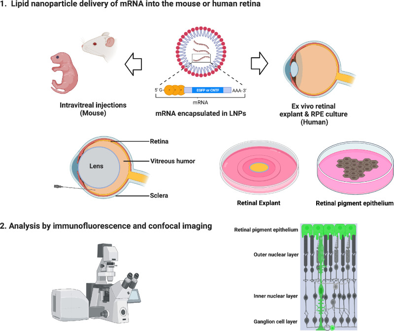
Purpose: Lipid nanoparticles (LNPs) show promise in their ability to introduce mRNA to drive protein expression in specific cell types of the mammalian eye. Here, we examined the ability of mRNA encapsulated in LNPs with two distinct formulations to drive gene expression in mouse and human retina and other ocular tissues.

Methods: We introduced mRNA-carrying LNPs into two biological systems. Intravitreal injections were tested to deliver LNPs into the mouse eye. Human retinal pigment epithelium (RPE) and retinal explants were used to assess mRNA expression in human tissue. We analyzed specificity of expression using histology, immunofluorescence, and imaging.

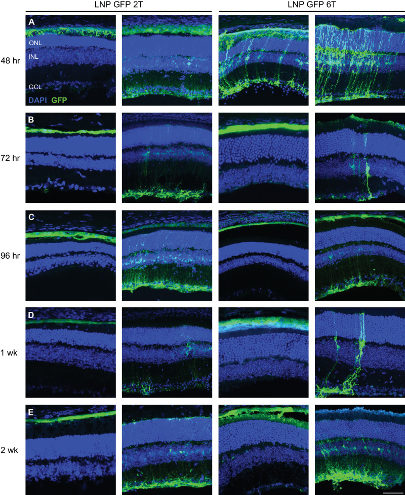
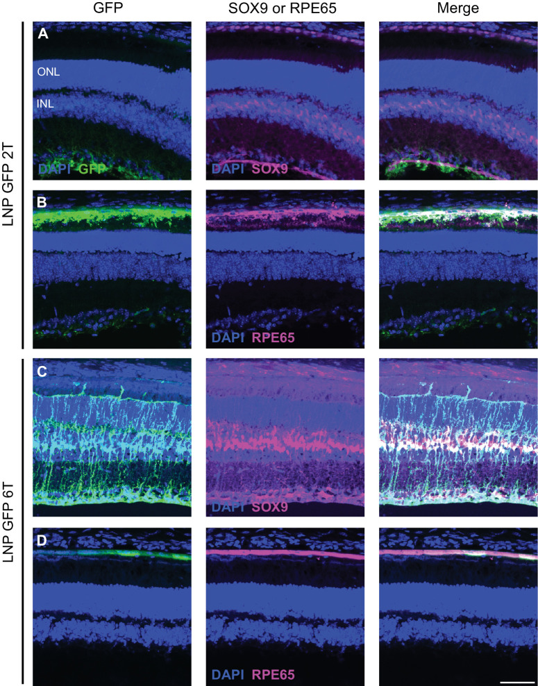
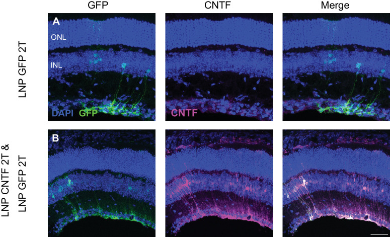
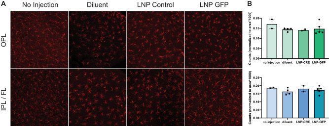
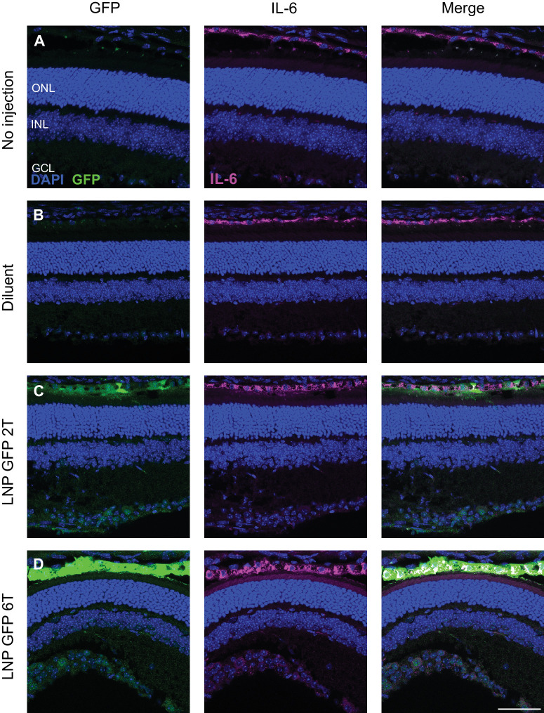
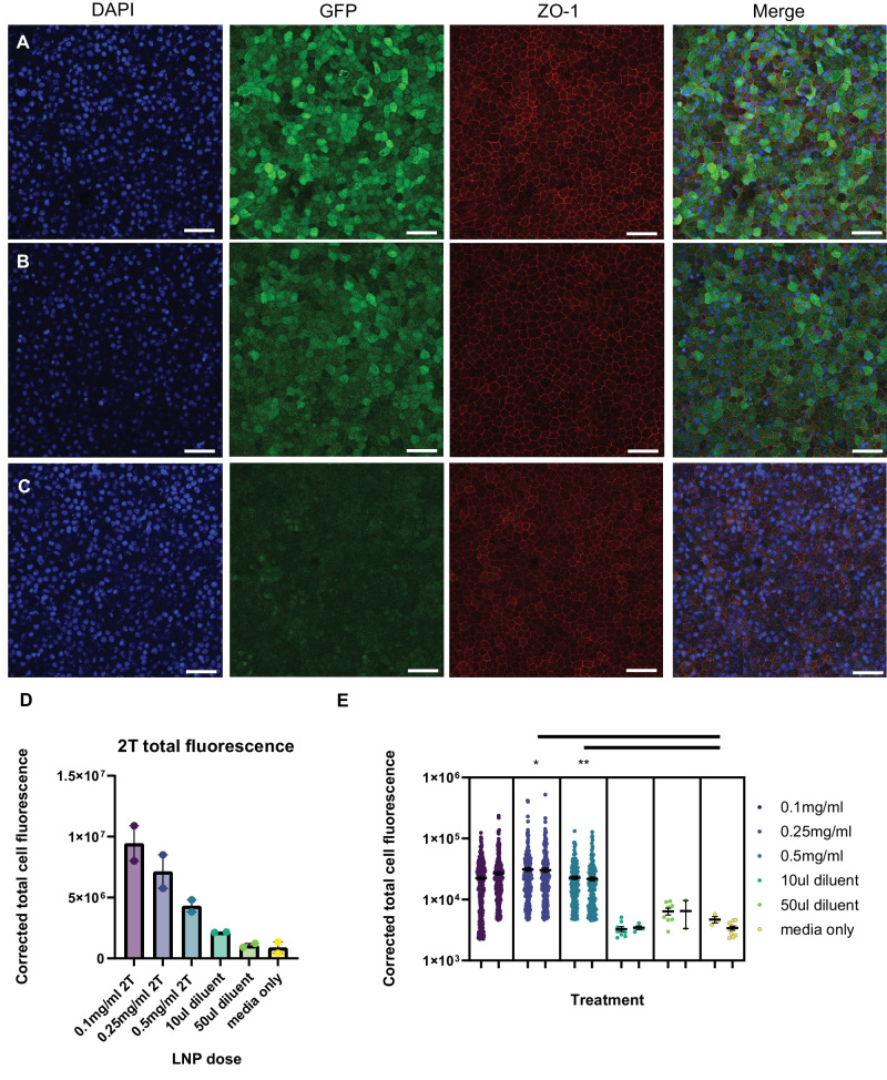
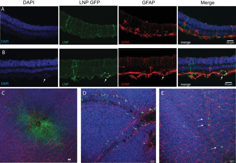
Results: In mice, mRNAs encoding GFP and ciliary neurotrophic factor (CNTF) were specifically expressed by Müller glia and RPE. Acute inflammatory changes measured by microglia distribution (Iba-1) or interleukin-6 (IL-6) expression were not observed 6 hours post-injection. Human RPE also expressed high levels of GFP. Human retinal explants expressed GFP in cells with apical and basal processes consistent with Müller glia and in perivascular cells consistent with macrophages.

Conclusions: We demonstrated the ability to reliably transfect subpopulations of retinal cells in mouse eye tissues in vivo and in human ocular tissues. Of significance, intravitreal injections were sufficient to transfect the RPE in mice. To our knowledge, we demonstrate delivery of mRNA using LNPs in human ocular tissues for the first time.

Translational relevance: Ocular gene-replacement therapies using non-viral vector methods are a promising alternative to adeno-associated virus (AAV) vectors. Our studies show that mRNA LNP delivery can be used to transfect retinal cells in both mouse and human tissues without inducing significant inflammation. This methodology could be used to transfect retinal cell lines, tissue explants, mice, or potentially as gene-replacement therapy in a clinical setting in the future.


Conflict of interest statement

Disclosure: C.Z. Chambers, None; G.L. Soo, None; A.L. Engel, None; I.A. Glass, None; A. Frassetto, Moderna, Inc. (E, F, I); P.G.V. Martini, Moderna, Inc. (E, F, I); T.J. Cherry, None

Figures

Figure 1.
Figure 1.
Schematic of experimental workflow to deliver mRNA encapsulated in LNPs. Adult and neonatal CD-1 mice were used for in vivo experiments. Intravitreal injections of mRNA LNPs were performed to deliver the reagents to the vitreous chamber. Ex vivo experiments were performed using human retinal explants or human RPE cultures. After histological processing, analysis was completed using immunofluorescence and confocal imaging to determine transfection cell type expression and duration.
Figure 2.
Figure 2.
Intravitreal injections of LNP-packaged EGFP mRNA transfects retinal cells in the mouse. Two amino lipid formulations, 2T and 6T, were compared for their ability to transfect retinal cells at various time points. Confocal images of retinal cross-sections with DAPI and anti-GFP immunostaining are shown. EGFP is expressed in similar cell populations at (A) 48 hours, (B) 72 hours, (C) 96 hours, (D) 1 week, and (E) 2 weeks for both 2T and 6T LNPs. Images shown are representative of at least three retinas (scale bar = 50 µm).
Figure 3.
Figure 3.
LNP delivery of EGFP mRNA transfects Müller glia and RPE cells in the mouse retina. Retinal cross-sections were co-stained with DAPI, anti-GFP, and either anti-SOX9 or anti-RPE65 immunofluorescence 48 hours after intravitreal injections of EGFP mRNA encapsulated in 2Tor 6T LNPs. (A) For the 2T formulation, cells labelled with anti-GFP colocalize with nuclei labelled with anti-SOX9 in the inner nuclear layer, indicating EGFP expression in the Müller glia. (B) Cells that express EGFP also express RPE65, indicating EGFP expression in the RPE. Similarly, for 6T, there is co-expression of EGFP and (C) SOX9 and (D) RPE65. Images shown are representative of at least three retinas (scale bar = 50 µm).
Figure 4.
Figure 4.
CNTF mRNA in 2T LNP transfects mouse Müller glia cells. Intravitreal injections of EGFP or both EGFP and CNTF mRNA were done. (A) Control EGFP injections show EGFP expression in the Müller glia and basal CNTF expression in the GCL based on immunofluorescence staining. (B) When EGFP and CNTF mRNA were co-delivered, EGFP mRNA expression is in the Müller glia. CNTF expression is increased and in Müller glia cells as well. There is only partial overlap of EGFP and CNTF expression, indicating that there are populations of Müller glia cells that express either EGFP only, CNTF only, or both. Images shown are representative of at least three retinas (scale bar = 50 µm).
Figure 5.
Figure 5.
Distribution of microglia in LNP EGFP injected and control retinas. (A) Microglial cells were visualized using anti-Iba1 immunostaining of flatmounted, uninjected mouse retinas and retinas injected with LNP GFP 6T, LNP CRE-recombinase (Control), or 1xPBS (Diluent) with Fast Green dye. Images represent maximum intensity projections of volumes acquired in the outer plexiform layer (OPL) or the inner plexiform layer and fiber layer (IPL/FL). (B) Quantification of Iba1+ microglial cells in the OPL or IPL/FL normalized to area (n > 2 retinas per condition). Each point on the bar graph represents a single retina where the number of microglia in two independent mid-peripheral regions were quantified and normalized by area. Error bars represent standard deviation.
Figure 6.
Figure 6.
Intravitreal injections of EGFP mRNA LNPs does not affect the localization of cytokine IL-6. Expression of interleukin-6 in the RPE was similar comparing 4 conditions: (A) no injection, (B) PBS controls, (C) EGFP 2T LNP, or (D) EGFP 6T LNP at 6 hours post-intravitreal injection in mouse retinas. Images shown are representative of at least three retinas (scale bar = 50 µm).
Figure 7.
Figure 7.
LNP delivery of EGFP mRNA transfects human fRPE in vitro. Human fRPE cells were treated with EGFP mRNA encapsulated in 2T LNPs for 2 hours prior to an overnight washout, followed by fixation and immunofluorescence for GFP and ZO-1. Cells were treated with (A) 0.1 mg/mL EGFP mRNA 2T-LNP, (B) 0.5 mg/mL mRNA 2T-LNP, or (C) diluent only (same volume as (A) Images shown are representative of two biological replicates of each treatment (scale bar = 50 µm). (D) Total GFP signal was quantified for fRPE treated with 0.1, 0.25, or 0.5 mg/mL LNP GFP and volume matched diluent or media controls (n = 2 biological replicates per condition). (E) Distribution of segmented GFP signal per cell for each of two biological replicates per treatment (* P < 0.05, ** P < 0.01; nested t-test).
Figure 8.
Figure 8.
EGFP mRNA encapsulated in LNPs transfects adult human retina explants. Adult human post-mortem eye globes were treated with EGFP mRNA LNPs for 4 hours. (A) Confocal images of human retina cross-section treated with EGFP mRNA encapsulated in 6T LNPs and then stained with DAPI, anti-GFP, and anti-GFAP. GFP is expressed in cells with apical and basal processes. (B) Cross-section of human retina treated with EGFP mRNA encapsulated in 2T LNPs. GFP is expressed in perivascular cells surrounding a blood vessel, indicated by the arrow, and in cells with apical and basal processes. (C) Flat mount of parafoveal retina treated with EGFP mRNA in 6T LNPs. GFP is expressed in cells in the fovea. (D) Flat mount of retina with RPE and choroid treated with EGFP mRNA in 2T. Perivascular cells express GFP. (E) Flat mount of retina treated with 6T. Arrows indicate GFP positive cells along a small blood vessel. Images shown are representative of retinas and RPE from two globes from a single donor (scale bars = 50 µm).

Update of

References

    1. Retinal Information Network (RetNet). Available at: https://web.sph.uth.edu/RetNet/. 2022.
    1. Hanany M, Rivolta C, Sharon D, Daiger SP. Worldwide carrier frequency and genetic prevalence of autosomal recessive inherited retinal diseases, doi:10.1073/pnas.1913179117/-/DCSupplemental. - DOI - PMC - PubMed
    1. Gong J, Cheung S, Fasso-Opie A, et al. .. The impact of inherited retinal diseases in the US and Canada from a cost-of-illness perspective. Clin Ophthalmol. 2021; 15: 2855–2866. - PMC - PubMed
    1. Fenner BJ, Tan TE, Barathi AV, et al. .. Gene-based therapeutics for inherited retinal diseases. Front Genet. 2022; 12: 1–17. Preprint at 10.3389/fgene.2021.794805. - DOI - PMC - PubMed
    1. Zhou R, Caspi RR. Ocular immune privilege. F1000 Biol Rep. 2010; 2: 1–3. - PMC - PubMed

Publication types

MeSH terms

LinkOut - more resources