. 2024 Nov 1;14(11):2109-2121.
doi: 10.1158/2159-8290.CD-23-1393.

Rational Protein Engineering to Enhance MHC-Independent T-cell Receptors

Affiliations

Rational Protein Engineering to Enhance MHC-Independent T-cell Receptors

Ju-Fang Chang et al. Cancer Discov. .

Abstract

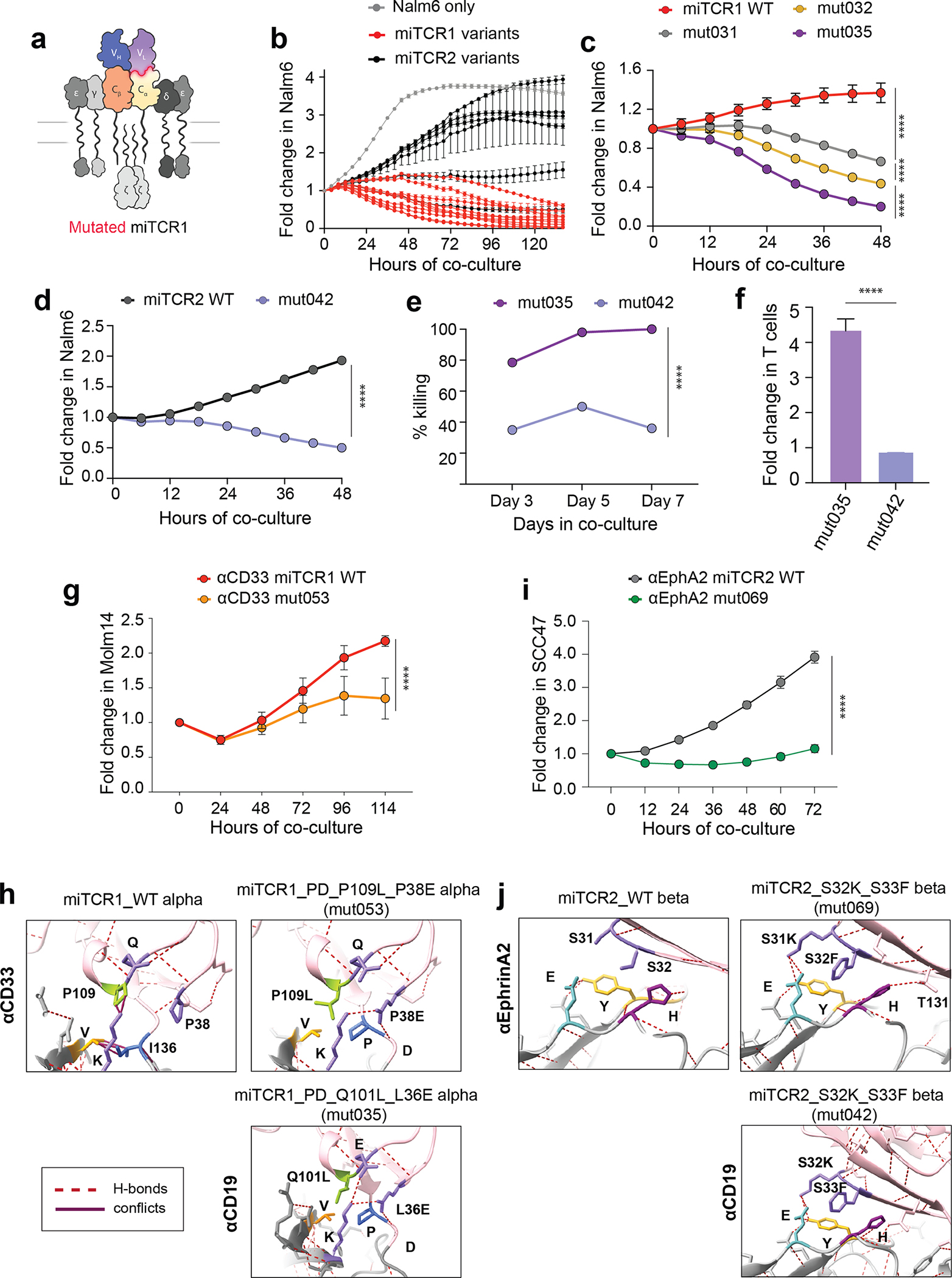
Chimeric antigen receptor (CAR)-based therapies have pioneered synthetic cellular immunity but remain limited in their long-term efficacy. Emerging data suggest that dysregulated CAR-driven T-cell activation causes T-cell dysfunction and therapeutic failure. To re-engage the precision of the endogenous T-cell response, we designed MHC-independent T-cell receptors (miTCR) by linking antibody variable domains to T-cell receptor constant chains. Using predictive modeling, we observed that this standard "cut and paste" approach to synthetic protein design resulted in myriad biochemical conflicts at the hybrid variable-constant domain interface. Through iterative modeling and sequence modifications, we developed structure-enhanced miTCRs which significantly improved receptor-driven T-cell function across multiple tumor models. We found that 41BB costimulation specifically prolonged miTCR T-cell persistence and enabled improved leukemic control in vivo compared with classic CAR T cells. Collectively, we have identified core features of hybrid receptor structure responsible for regulating function. Significance: Improving the durability of engineered T-cell immunotherapies is critical to enhancing efficacy. We used a structure-informed design to evolve improved miTCR function across several models. This work underscores the central role of synthetic receptor structure in T-cell function and provides a framework for improved receptor engineering.


Conflict of interest statement

Competing interests: J.-F.C. and N.S. have submitted patent applications related to this work. N.S. is an inventor on patents related to adoptive cell therapies, held by Washington University and the University of Pennsylvania (some licensed to Novartis). Unrelated to this work, N.S. has served as a consultant for several companies involved in cell therapies and is a board member for Phoreus Biotech. J.F.D. receives research funding from Amphivena Therapeutics, NeoImmuneTech, Macrogenics, Incyte, Bioline Rx, Wugen; has equity ownership in Magenta Therapeutics, Wugen; consults for Incyte, RiverVest Venture Partners, hC Bioscience, Inc.; and is a board member for RiverVest Venture Partners, Magenta Therapeutics.

Figures

Figure 1 |
Figure 1 |. MHC-independent TCR V-C interface contains inherent biochemical conflicts.
a, Comparison of TCR (cryo-EM) and antibody (crystal) variable and constant chain structures. b, Design of MHC-independent TCRs by swapping TCR variable regions with antibody variable regions to create two receptor formats. c-d, Cytotoxicity of miTCR1, miTCR2 and 19/28ζ CAR T cells against CD19+ Nalm6 cells c, after 6 days of co-culture at various E:T ratios and d, over time at an E:T ratio of 1:4. e, Percent of T cells that bound CD19-APC at various CD19 concentrations. f, Predictive modeling of miTCR1 paired α and β chains. g, Detailed analysis of cryo-EM resolved variable-constant (V-C) interface of the native TCR (PDB: 6JXR). h, Predictive modeling of the miTCR1α V-C interface. i, Rotation of the V-C interface to isolate predicted biochemical and structural conflicts. j, Predictive modeling of the miTCR1α V-C interface with PD insertion and k, Q101L and L36E mutations. Modeling performed using Phyre2 or AlphaFold2. c-e are representative data of five independent donors. **P < 0.01, ***P < 0.001, ****P < 0.0001 by two-way ANOVA.
Figure 2 |
Figure 2 |. Targeted V-C interface alterations improve miTCR function.
a, Design of interface altered miTCR1. b, Functional screen of cytotoxicity against Nalm6 by human T cells engineered to express miTCR1 and miTCR2 variants. c-d, Cytotoxicity of lead c, miTCR1 and e, miTCR2 variants against Nalm6. e, Cytotoxicity of miTCR1 mut035 and mut042 against high burden Nalm6WT (E:T 1:8) over time. f, T cell expansion of mut035 and mut042 at the conclusion of 7 day co-cultures. g, Cytotoxicity of lead αCD33 miTCR variant (mut053) compared to its parental αCD33 miTCR1 WT against the AML cell line Molm14. h, Modeling of the interface alterations made to generate mut053 compared to the alterations in αCD19 miTCR1 variant mut035. i, Cytotoxicity of lead αEphrinA2 miTCR variant (mut069) compared to its parental αEphrinA2 miTCR2 WT against the squamous cell carcinoma cell line SCC47. j, Modeling of the interface alterations made to generate mut069 compared to the alterations in αCD19 miTCR2 variant mut042. Screening studies in b performed using two independent donors. c-f are representative data from three independent donors, g-i are representative of data from two independent donors. Modeling in h-i performed using Phyre2. *P < 0.05, **P < 0.01, ***P < 0.001, ****P < 0.0001 by two-way ANOVA (c-e,g-i) or by Student’s t-test (f).
Figure 3 |
Figure 3 |. Distinct T cell biology driven by miTCR variant mut035.
a, Predictive modeling of the V-C interface of mut035 with depicted model confidence. b, Predictive modeling and overlay of mut031 and mut035 α chains. Modeling done using AlphaFold2. c-d, CD19 binding capacity of miTCR1 WT, mut031 and mut035 measured by percent of T cells that c, bound CD19-APC at various CD19 concentrations and d, MFI of bound CD19. e, Memory phenotypes of miTCR1 WT and mut035 T cells one day after clearance of Nalm6 (TN, CD45RA+CD62L+; TCM, CD45RACD62L+; TEM, CD45RACD62L; Teff, CD45RA+CD62L). f-h, MFI of f, pCD3ζ, g, pErk and h, pAkt in Jurkat cells engineered to express miTCR1 WT, mut035 or 19/BBζ CAR following various durations of co-culture with Nalm6. i-j, Representative expression of i, pErk and j, pAkt in human T cells engineered to express miTCR1 WT, mut035 or 19/BBζ CAR at peak phosphorylation (Erk, 5 minutes; Akt, 45 minutes). c-d representative of three independent donors; e, data from one donor; i-j, representative data from two independent donors. ****P < 0.0001 by two-way ANOVA (c-h).
Figure 4 |
Figure 4 |. Costimulation from 41BB significant enhances miTCR function.
a, Cytotoxicity, as measured by fold change in Nalm6 growth during co-culture, of human T cells expressing miTCR1 or miTCR2 against either Nalm6WT or Nalm6triple. b, T cell expansion, as measured by fold change in engineered T cells during co-culture, of human T cells expressing miTCR1 or miTCR2 against Nalm6WT or Nalm6triple. c, Cytotoxicity and d, expansion of miTCR1 and miTCR2 variant expressing human T cells against Nalm6triple. e-g, Cytotoxicity of miTCR1 WT and mut035 T cells against e, Nalm6triple, f, Raji and g, K562CD19. h, Cytotoxicity (left) and T cell expansion (right) of mut035 T cells against various Nalm6 targets. i, Cytotoxicity (left) and T cell expansion (right) of mut035 T cells upon re-challenge with Nalm6WT targets. j, Leukemic control, measured by Nalm6 cells per T cell, over time during chronic stimulation co-cultures. k, Change in in vivo disease burden over time as measured by bioluminescent signal and l, survival of NSG mice engrafted with either Nalm6WT or Nalm641BBL and treated with low-dose (2.5×10⁵) T cells. For a-e, h-I, representative data from three independent donors; f-g, representative data from two donors; j, representative data from five donors. k-l, n=8 animals per group. ****P < 0.0001; all significance testing determined by two-way ANOVA (a-k) or Log-Rank test (l).

References

    1. Good CR, Aznar MA, Kuramitsu S, Samareh P, Agarwal S, Donahue G, et al. An NK-like CAR T cell transition in CAR T cell dysfunction. Cell. 2021;184(25):6081–100 e26. - PMC - PubMed
    1. Selli ME, Landmann JH, Terekhova M, Lattin J, Heard A, Hsu YS, et al. Costimulatory domains direct distinct fates of CAR-driven T-cell dysfunction. Blood. 2023;141(26):3153–65. - PMC - PubMed
    1. Fraietta JA, Lacey SF, Orlando EJ, Pruteanu-Malinici I, Gohil M, Lundh S, et al. Determinants of response and resistance to CD19 chimeric antigen receptor (CAR) T cell therapy of chronic lymphocytic leukemia. Nat Med. 2018;24(5):563–71. - PMC - PubMed
    1. Deng Q, Han G, Puebla-Osorio N, Ma MCJ, Strati P, Chasen B, et al. Characteristics of anti-CD19 CAR T cell infusion products associated with efficacy and toxicity in patients with large B cell lymphomas. Nat Med. 2020;26(12):1878–87. - PMC - PubMed
    1. Davenport AJ, Cross RS, Watson KA, Liao Y, Shi W, Prince HM, et al. Chimeric antigen receptor T cells form nonclassical and potent immune synapses driving rapid cytotoxicity. Proc Natl Acad Sci U S A. 2018;115(9):E2068–E76. - PMC - PubMed

Substances