. 2024 Jul 16;121(29):e2400883121.
doi: 10.1073/pnas.2400883121. Epub 2024 Jul 9.

Palmitoylation at a conserved cysteine residue facilitates gasdermin D-mediated pyroptosis and cytokine release

Affiliations

Palmitoylation at a conserved cysteine residue facilitates gasdermin D-mediated pyroptosis and cytokine release

Zhonghua Liu et al. Proc Natl Acad Sci U S A. .

Abstract

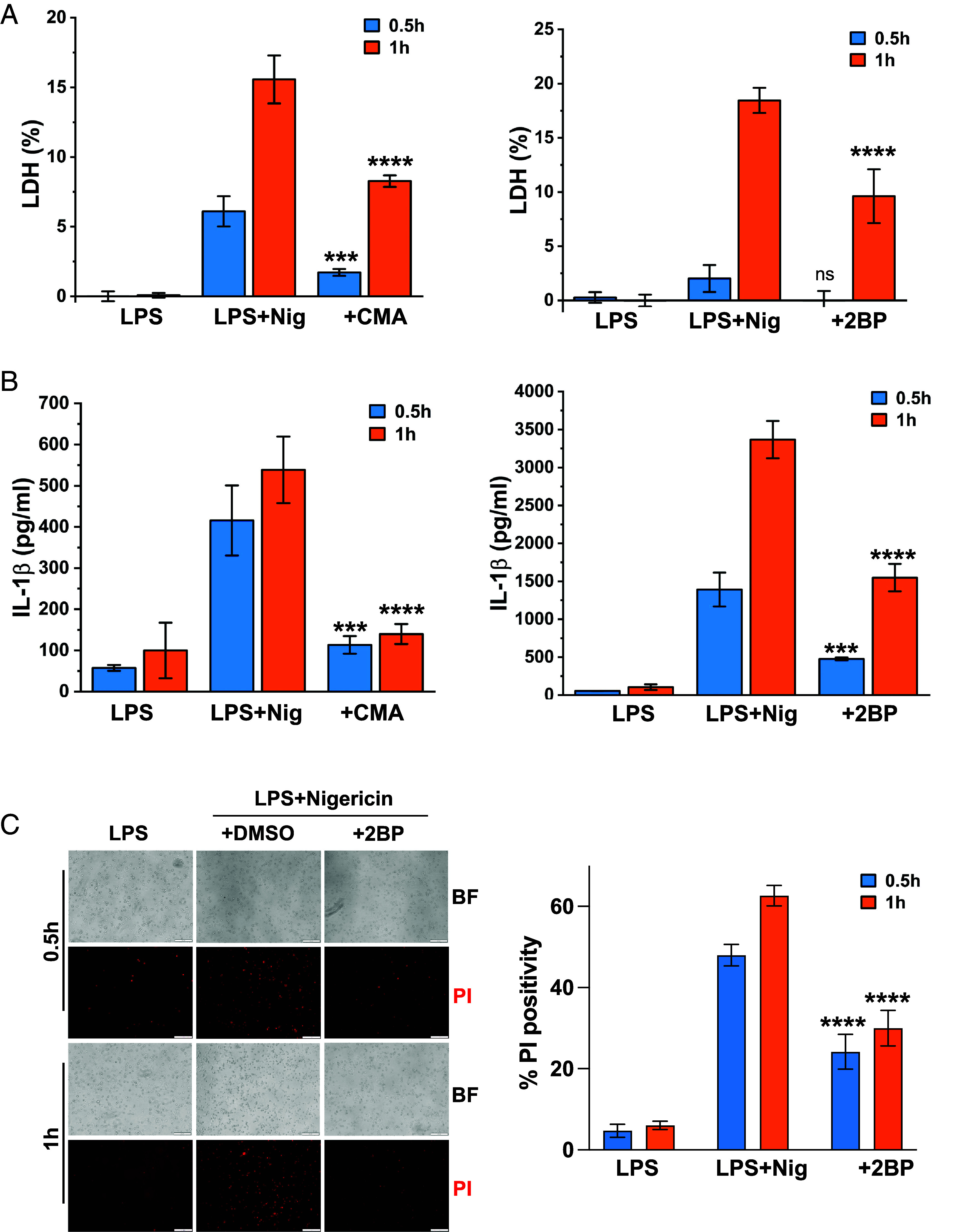
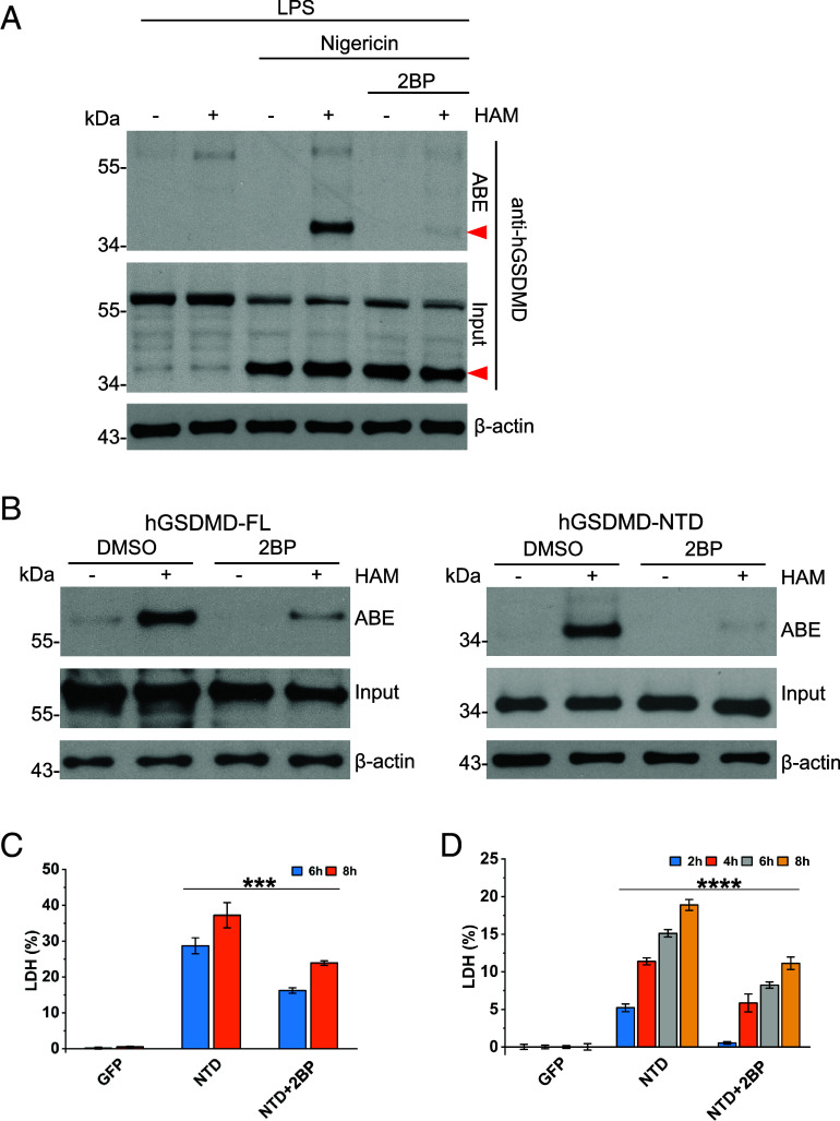
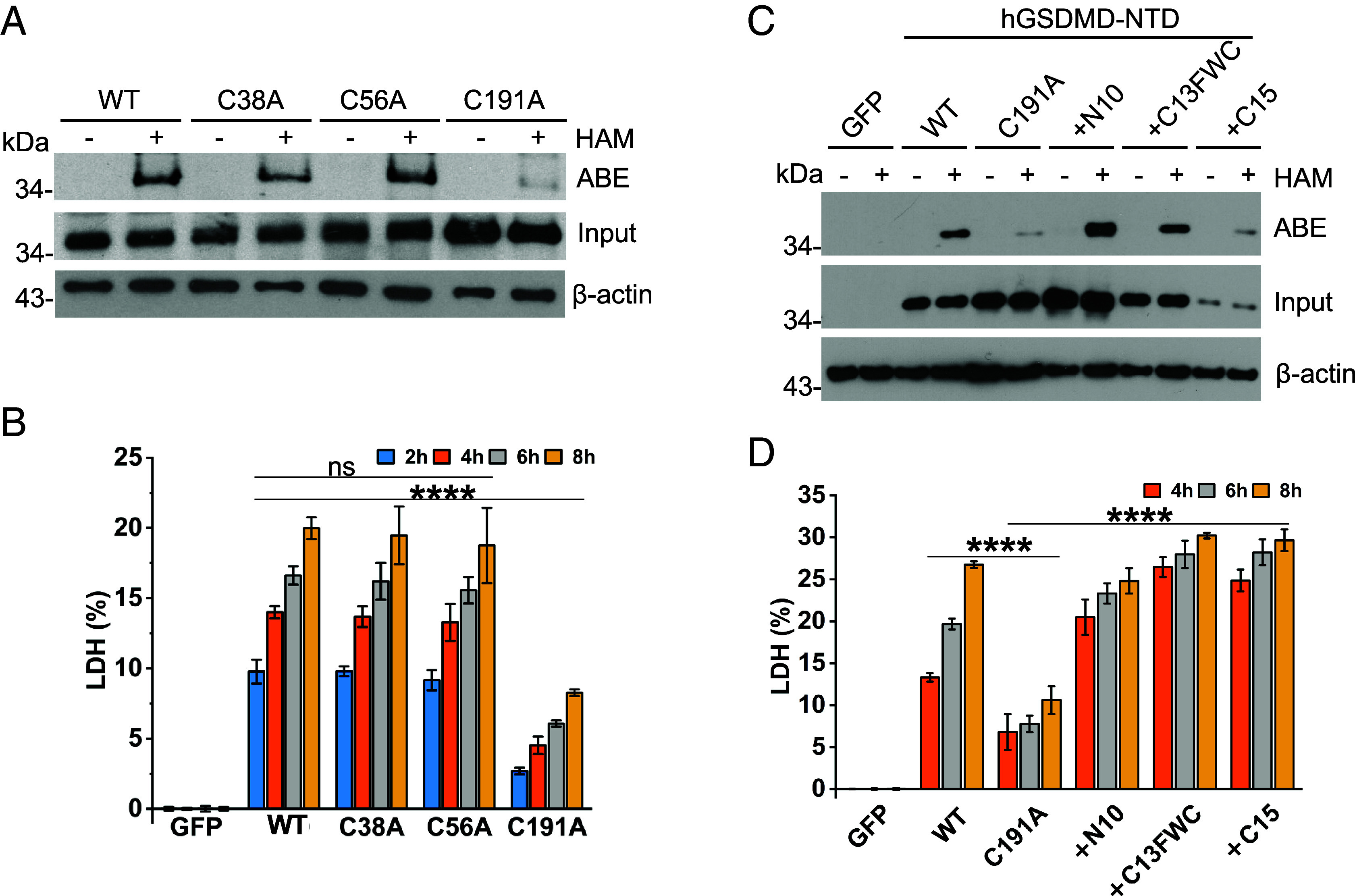
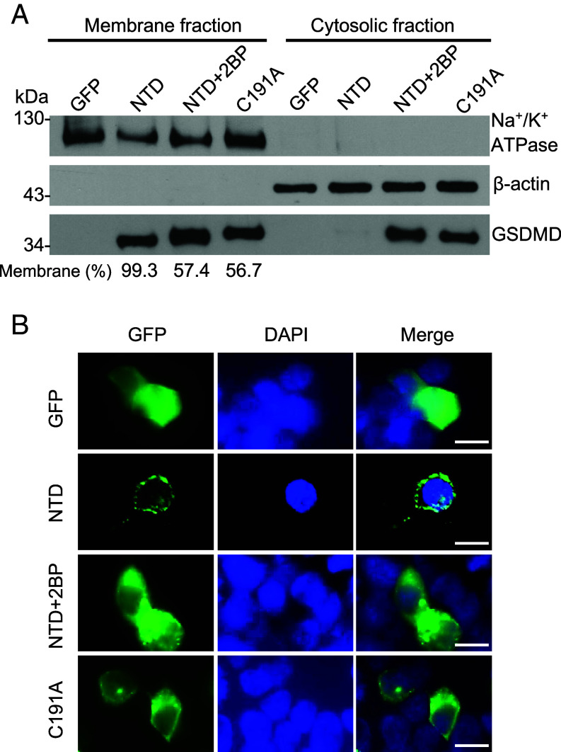
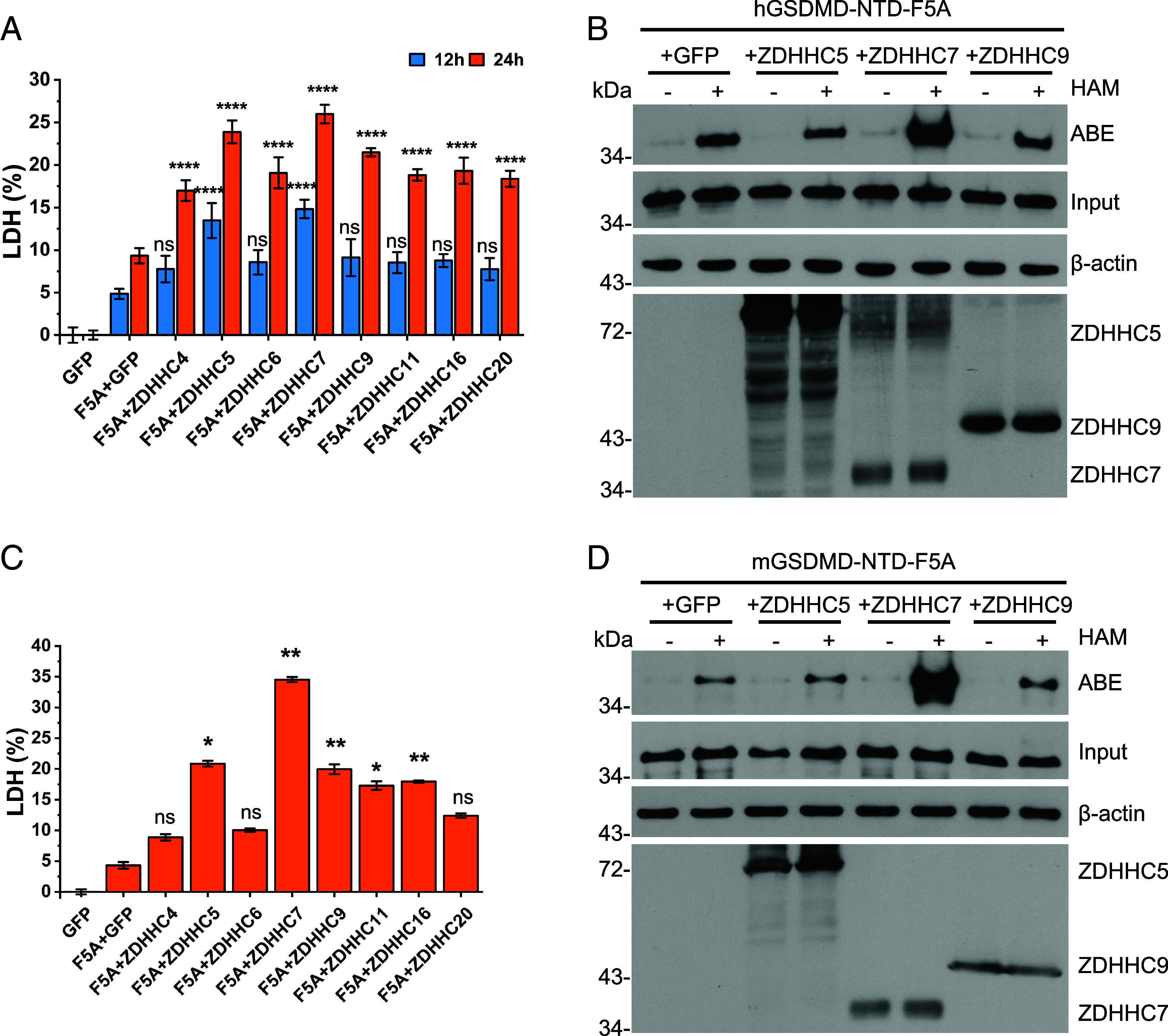
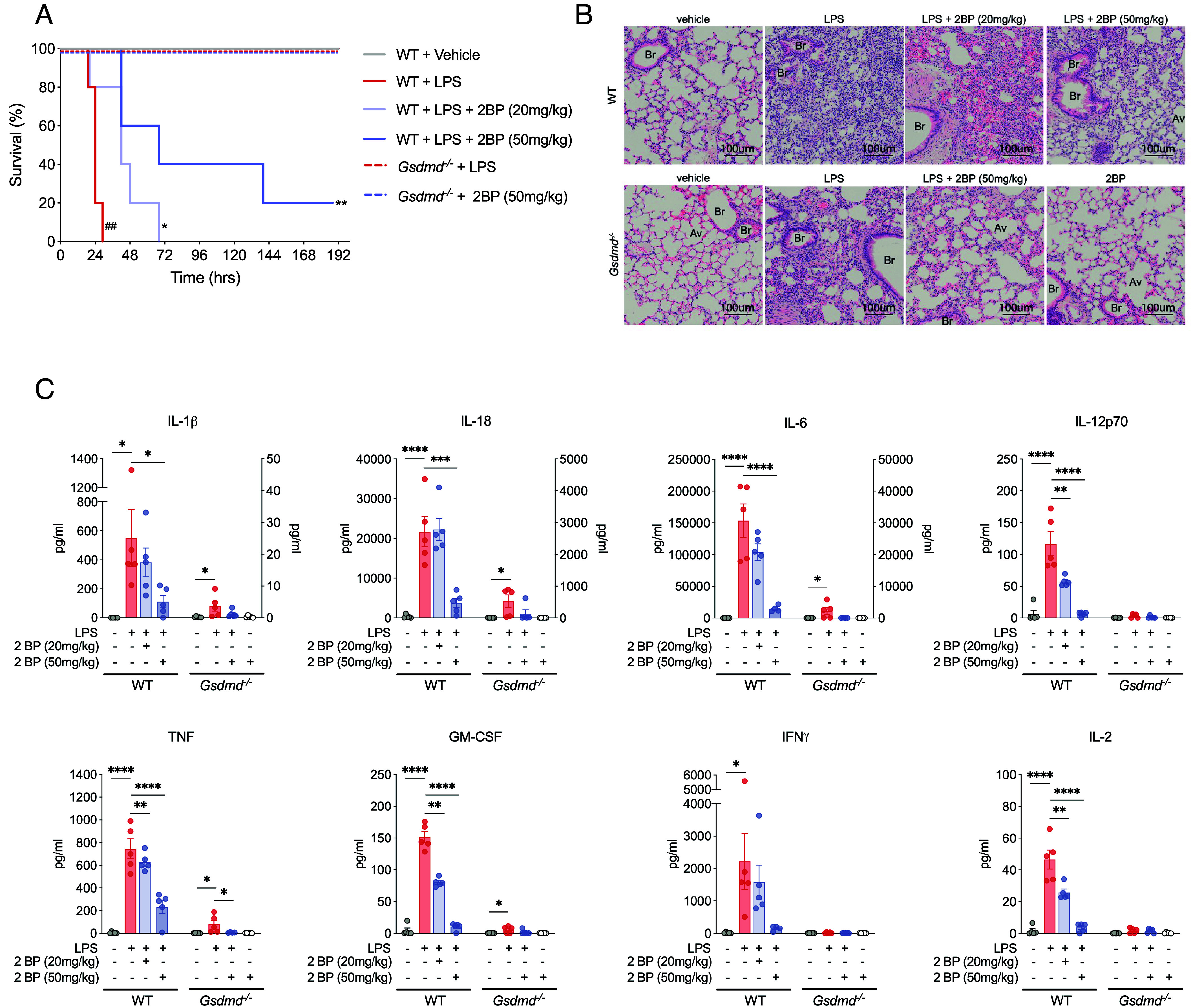
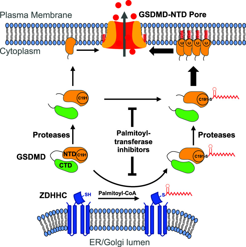
Gasdermin D (GSDMD)-mediated pyroptotic cell death drives inflammatory cytokine release and downstream immune responses upon inflammasome activation, which play important roles in host defense and inflammatory disorders. Upon activation by proteases, the GSDMD N-terminal domain (NTD) undergoes oligomerization and membrane translocation in the presence of lipids to assemble pores. Despite intensive studies, the molecular events underlying the transition of GSDMD from an autoinhibited soluble form to an oligomeric pore form inserted into the membrane remain incompletely understood. Previous work characterized S-palmitoylation for gasdermins from bacteria, fungi, invertebrates, as well as mammalian gasdermin E (GSDME). Here, we report that a conserved residue Cys191 in human GSDMD was S-palmitoylated, which promoted GSDMD-mediated pyroptosis and cytokine release. Mutation of Cys191 or treatment with palmitoyltransferase inhibitors cyano-myracrylamide (CMA) or 2-bromopalmitate (2BP) suppressed GSDMD palmitoylation, its localization to the membrane and dampened pyroptosis or IL-1β secretion. Furthermore, Gsdmd-dependent inflammatory responses were alleviated by inhibition of palmitoylation in vivo. By contrast, coexpression of GSDMD with palmitoyltransferases enhanced pyroptotic cell death, while introduction of exogenous palmitoylation sequences fully restored pyroptotic activities to the C191A mutant, suggesting that palmitoylation-mediated membrane localization may be distinct from other molecular events such as GSDMD conformational change during pore assembly. Collectively, our study suggests that S-palmitoylation may be a shared regulatory mechanism for GSDMD and other gasdermins, which points to potential avenues for therapeutically targeting S-palmitoylation of gasdermins in inflammatory disorders.

Keywords: S-palmitoylation; ZDHHC palmitoyltransferase; gasdermin D; inflammasome; pyroptosis.


Conflict of interest statement

Competing interests statement:The authors declare no competing interest.

Figures

Fig. 1.
Fig. 1.
Inhibition of palmitoylation suppresses GSDMD-mediated pyroptosis and cytokine release. (A) PMA differentiated THP-1 cells were treated with 1 μg/mL LPS for 4 h followed by 20 μM nigericin in the presence or absence of 100 μM CMA or 2BP. LDH release was measured at 0.5 or 1 h after nigericin treatment. (B) THP-1 cells were treated as in (A). IL-1β secretion was measured after nigericin treatment. (C) THP-1 cells treated in (A) were stained with propidium iodide after 0.5 or 1 h of nigericin treatment. “BF” denotes bright-field and “PI” denotes staining with propidium iodide. (Scale bars, 200 μm.) Quantitation of the PI-positive cells is shown on the right. Data are plotted as mean ± SD from at least three independent experiments. Statistical analyses were performed using two-way ANOVA with Bonferroni’s multiple comparisons test versus the “LPS+Nig” samples. *P < 0.05, **P <0.01, ***P < 0.001, ****P < 0.0001. See also SI Appendix, Fig. S1.
Fig. 2.
Fig. 2.
S-palmitoylation of GSDMD in THP-1 and HEK293T cells. (A) PMA differentiated THP-1 cells were treated with LPS and nigericin, in the presence or absence of 100 μM 2BP. After 1 h of nigericin treatment, ABE assays were performed and GSDMD was probed with anti-hGSDMD antibody. The red arrows mark the hGSDMD-NTD. (B) FLAG-tagged full-length (“FL,” Left panel) and N-terminal domain (“NTD,” Right panel) from human GSDMD expressed in HEK293T cells were subjected to ABE assays, in the absence or presence of 100 μM 2BP. (C) LDH release from HEK293T cells transiently expressing hGSDMD-NTD in the presence or absence of 2BP was analyzed at 6 or 8 h posttransfection. (D) LDH release from HEK293T cells expressing hGSDMD-NTD in a tetracycline-inducible system was analyzed 2, 4, 6, or 8 h following tetracycline induction. Data are plotted as mean ± SD from at least three independent experiments. Statistical analyses were performed using two-way ANOVA with Bonferroni’s multiple comparisons versus the NTD samples. *P < 0.05, **P < 0.01, ***P < 0.001, ****P < 0.0001. See also SI Appendix, Fig. S2.
Fig. 3.
Fig. 3.
Cysteine 191 of hGSDMD is palmitoylated. (A) The wildtype hGSDMD-NTD (denoted “WT”) or C38A, C56A, and C191A mutants were expressed in HEK293T cells and ABE assays were performed to probe potential palmitoylation. (B) Time course of LDH release from cells in (A) expressing the wildtype or mutant hGSDMD-NTDs. (C) ABE assays were performed with HEK293T cells expressing hGSDMD-NTD in the wildtype form, C191A mutant form, and the C191A mutant chimera forms containing palmitoylation sequences at the N or C terminus of hGSDMD-NTD. The exogenous palmitoylation sequences are as follows: N10 is from the N terminus of human GAP43 protein with the sequence MLCCMRRTKQ; C13FWC is from the C terminus of yeast CAN1 protein with the sequence DHEPKTFWDKFWNFWC; C15 is from the C terminus of yeast GAP1 protein with the sequence MATKPRWYRIWNFWC. (D) Time course of LDH release from cells in (C) expressing the wildtype or C191A mutant hGSDMD-NTD, or mutant chimeras harboring the exogenous palmitoylation sequences. Data are plotted as mean ± SD from at least three independent experiments. Statistical analyses were performed using two-way ANOVA with Bonferroni’s multiple comparisons versus the WT or “C191A” samples. *P < 0.05, **P < 0.01, ***P < 0.001, ****P < 0.0001, ns: not significant. See also SI Appendix, Fig. S3.
Fig. 4.
Fig. 4.
Palmitoylation of hGSDMD facilitates its localization to the plasma membrane. (A) Analysis of the membrane or cytosolic fractions of HEK293T cells expressing the wildtype or C191A mutant of hGSDMD-NTD. The percentage of GSDMD in the membrane fractions for the NTD, NTD+2BP, and C191A samples is marked. (B) Subcellular localization of GFP-tagged wildtype or C191A mutant of hGSDMD-NTD with or without 2BP treatment. (Scale bars, 5 μm.)
Fig. 5.
Fig. 5.
ZDHHC palmitoyltransferases catalyze the palmitoylation of hGSDMD and mGSDMD. (A) LDH release from HEK293T cells expressing the F5A mutant of hGSDMD-NTD along with various ZDHHC palmitoyltransferases, 12 or 24 h posttransfection. (B) ABE assays were performed for HEK293T cells coexpressing hGSDMD-NTD F5A mutant and ZDHHC5, 7, or 9 enzymes, 24 h posttransfection. (C) LDH release from HEK293T cells expressing the F5A mutant of mGSDMD-NTD along with various ZDHHC palmitoyltransferases, 24 h posttransfection. (D) ABE assays were performed for HEK293T cells coexpressing mGSDMD-NTD F5A mutant and ZDHHC5, 7, or 9 enzymes, 24 h posttransfection. Data are plotted as mean ± SD from at least three independent experiments. Statistical analyses were performed using two-way (A) or one-way (C) ANOVA with Bonferroni’s multiple comparisons test versus the “F5A+GFP” samples. *P < 0.05, **P < 0.01, ***P < 0.001, ****P < 0.0001, ns: not significant. See also SI Appendix, Fig. S4.
Fig. 6.
Fig. 6.
Inhibition of palmitoylation mitigates inflammation and death of LPS-injected mice. (A) C57BL6/J (WT) and Gsdmd−/− mice were pretreated with 2BP or vehicle, and challenged 4 h later with LPS or vehicle. After administration of LPS/vehicle, survival was monitored twice daily for 5 d (120 h) and once daily for remaining 3 d. The Kaplan–Meier survival curve is representative of 4 to 5 experiments (n ≥ 3/experiment); ##P < 0.01 vs. vehicle, *P < 0.05, **P < 0.01 vs. LPS alone by log-rank (Mantel-Cox) test. (B) In separate experiments, mice were killed 12 h after LPS/vehicle, and the representative histologic images of H&E-stained lungs are shown. Severely damaged alveoli with intra-alveolar edema, mixed inflammatory infiltrates consisting of neutrophils, lymphocytes, macrophages, occasional plasma cells, and mildly thickened bronchial walls were observed in WT mice treated with LPS (Upper, Middle Left panel). Pretreatment with low dose 2BP improved LPS-induced lung damage, but alveolar hemorrhage with red blood cells and lymphocytic infiltration occupying alveolar spaces/interstitial tissues were still evident (Upper, Middle Right panel), while high dose 2BP had a dramatic effect, largely restoring alveolar structure with only scattered lymphocytic infiltrates (Upper, Far Right panel). In contrast to the WT mice, Gsdmd−/− mice were largely protected from LPS-induced lung injury, but showed moderate alveolar damage with interstitial inflammation (Lower, Middle Left panel), while alveolar structure was generally normal/preserved with high dose 2BP (Lower, Middle Right panel). Control Gsdmd−/− mice administered 2BP only (Lower, Far Right panel) and vehicle-treated mice (Upper/Lower, Far Left panels) showed normal lung histology with intact alveolar structures. Original mag: X20 (n = 5); Av, alveolar space; Br, bronchiole. (C) Serum cytokine levels were measured 12 h after LPS/vehicle treatment and plotted for WT and Gsdmd−/− or Gsdmd−/− only on either Left or Right (when present) y axes, respectively. Data presented as mean ± SEM (n = 5); *P < 0.05, **P < 0.01, ***P < 0.001, ****P < 0.0001 by post hoc multiple comparisons using Šídák’s test following significant (P < 0.05) one-way ANOVA.
Fig. 7.
Fig. 7.
Palmitoylation facilitates GSDMD-mediated pore formation and release of inflammatory cytokines. A model of how palmitoylation facilitates GSDMD-NTD localization to the membrane, thus promoting pyroptosis and cytokine release.

Similar articles

Cited by

References

    1. Schroder K., Tschopp J., The inflammasomes. Cell 140, 821–832 (2010). - PubMed
    1. Latz E., Xiao T. S., Stutz A., Activation and regulation of the inflammasomes. Nat. Rev. Immunol. 13, 397–411 (2013). - PMC - PubMed
    1. Guo H., Callaway J. B., Ting J. P. Y., Inflammasomes: Mechanism of action, role in disease, and therapeutics. Nat. Med. 21, 677–687 (2015). - PMC - PubMed
    1. Shi J., et al. , Cleavage of GSDMD by inflammatory caspases determines pyroptotic cell death. Nature 526, 660–665 (2015). - PubMed
    1. Kayagaki N., Stowe I. B., Lee B. L., O’Rourke K., Caspase-11 cleaves gasdermin D for non-canonical inflammasome signalling. Nature 526, 666–671 (2015). - PubMed

LinkOut - more resources