. 2024 Jul 9;15(7):491.
doi: 10.1038/s41419-024-06887-0.

IL-27 disturbs lipid metabolism and restrains mitochondrial activity to inhibit γδ T17 cell-mediated skin inflammation

Affiliations

IL-27 disturbs lipid metabolism and restrains mitochondrial activity to inhibit γδ T17 cell-mediated skin inflammation

Mingyue Zhang et al. Cell Death Dis. .

Abstract

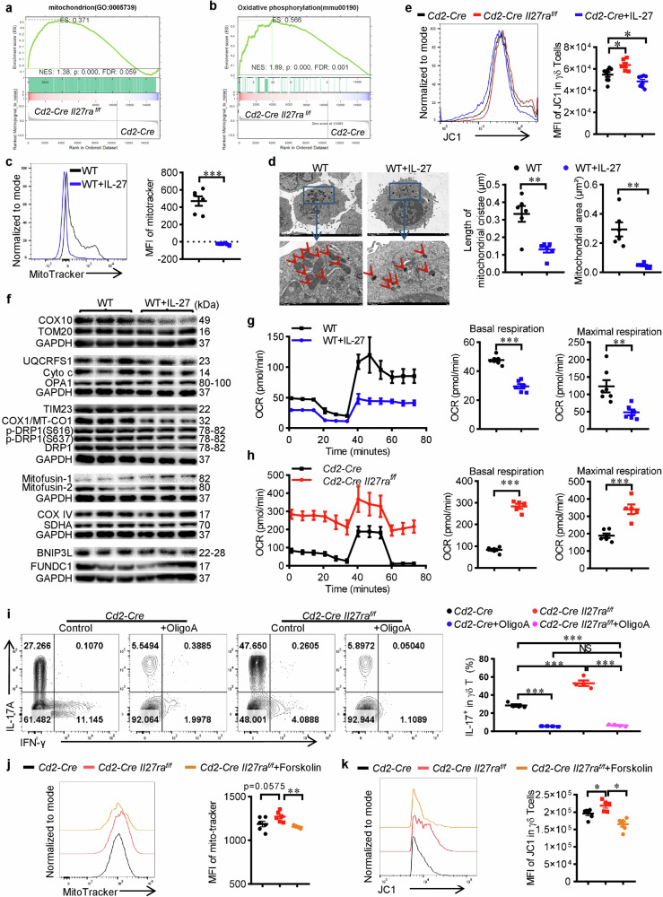
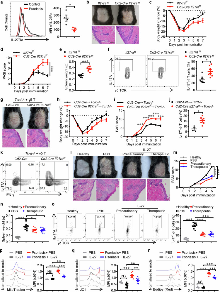
IL-17+ γδ T cells (γδ T17) are kick-starters of inflammation due to their strict immunosurveillance of xenobiotics or cellular damages and rapid response to pro-inflammatory stimulators. IL-27 is a well-recognized pleiotropic immune regulator with potent inhibitory effects on type 17 immune responses. However, its actions on γδ T17 mediated inflammation and the underlying mechanisms are less well understood. Here we find that IL-27 inhibits the production of IL-17 from γδ T cells. Mechanistically, IL-27 promotes lipolysis while inhibits lipogenesis, thus reduces the accumulation of lipids and subsequent membrane phospholipids, which leads to mitochondrial deactivation and ensuing reduction of IL-17. More importantly, Il27ra deficient γδ T cells are more pathogenic in an imiquimod-induced murine psoriasis model, while intracutaneous injection of rmIL-27 ameliorates psoriatic inflammation. In summary, this work uncovered the metabolic basis for the immune regulatory activity of IL-27 in restraining γδ T17 mediated inflammation, which provides novel insights into IL-27/IL-27Ra signaling, γδ T17 biology and the pathogenesis of psoriasis.


Conflict of interest statement

The authors declare no competing interests.

Figures

Fig. 1
Fig. 1. IL-27 signaling inhibits the generation of γδ T17 cells.
a The expression of IL-27Ra in CD4+, CD8+ and γδ T cells from the thymus, spleen or lymph nodes of C57BL/6 J mice was detected by flow cytometry (n = 3). b Flow cytometry analysis of IL-17A production by spleen γδ T cells from Cd2-Cre and Cd2-Cre Il27raflox/flox mice (n = 5). c Flow cytometry analyses of RORγt+ IL-17A+ proportion in spleen γδ T cells from Cd2-Cre and Cd2-Cre Il27raflox/flox mice (n = 5). d, e Splenocytes from WT mice were cultured under γδ T17 priming conditions with or without rmIL-27 (50 ng/ml) for 3 days and then expended till Day 5 (e) or Day 6 (d), the production of IL-17A (d), or RORγt+ IL-17A+ percentages (e) in γδ T cells were detected and shown (n = 6). f Naive γδ T cells (CD44low CD62Lhigh) were sorted from spleens and peripheral lymph nodes of WT mice and polarized toward the γδ T17 condition with or without rmIL-27 (50 ng/ml). Flow cytometry analysis of IL-17 production (n = 3). Data were presented as mean ± SEM, statistical differences were performed using Two-tailed unpaired student’s t-test (b)–(f) or one-way ANOVA (a). **p < 0.01, ***p < 0.001, NS, not significant.
Fig. 2
Fig. 2. IL-27 disturbs lipid metabolism and restrains the generation of phospholipids in γδ T17 cells.
a, b RNA-sequencing analysis was performed using in vitro differentiated γδ T17 cells from the spleen of Cd2-Cre Il27raflox/flox or Cd2-Cre mice. GSEA analysis of genes in lipid metabolic process (a) and lipid homeostasis (b) were shown. c Immunoblotting of in vitro differentiated γδ T17 cells from the spleen of Cd2-Cre Il27raflox/flox or Cd2-Cre mice (n = 3). dg Lipids were extracted from in vitro differentiated γδ T17 cells from the spleen of Cd2-Cre Il27raflox/flox or Cd2-Cre mice and used for lipidomic analysis via LC-MS (n = 6). d Pie chart showing the proportions of each type of lipid component. TG triglyceride, So sphingosine, SM Sphingomyelin, PS Phosphatidylserine, PI phosphatidylinositol, PEt Phosphatidylethanol, PE phosphatidylethanolamines, PC phosphatidylcholine, LPE Lyso phosphatidyl ethanolami, LPC Lyso phosphatidylcholine, dMePE Dimethylphosphatidyleth anolamine. e, f Metabolite set enrichment analysis of significantly altered lipids involved in lipolysis (e) and glycerophospholipids (f). g Heatmap of significantly altered phospholipids belonging to PC, PE and PI. h Flow cytometry analysis of IL-17 in γδ T cells from Cd2-Cre and Cd2-Cre Il27raflox/flox mice that were cultured under γδ T17 priming condition with or without Forskolin (10 μM) (n = 4). Data were presented as mean ± SEM, statistical differences were performed using One-way ANOVA (h). **p < 0.01, ***p < 0.001.
Fig. 3
Fig. 3. IL-27 reduces mitochondrial mass and impedes oxidative phosphorylation in γδ T17 cells.
RNA-sequencing analysis was performed using in vitro differentiated γδ T17 cells from the spleen of Cd2-Cre Il27raflox/flox or Cd2-Cre mice. GSEA analysis of genes in mitochondrion (a) and oxidative phosphorylation (b) were shown. c MitoTracker staining of WT γδ T cells that were differentiated in vitro under γδ T17 condition with or without rmIL-27 (50 ng/ml) (n = 6). d Transmission electron microscopy of WT γδ T cells that were differentiated in vitro under γδ T17 condition with or without rmIL-27 (50 ng/ml). The size and morphology of mitochondria were determined and shown (n = 6). e JC-1 staining of γδ T cells from Cd2-Cre Il27raflox/flox (n = 6) or Cd2-Cre mice (n = 8) that were differentiated in vitro under γδ T17 condition with or without rmIL-27 (50 ng/ml). f Immunoblotting of mitochondrial proteins in WT γδ T cells that were differentiated in vitro under γδ T17 condition with or without rmIL-27 (50 ng/ml) (n = 3). g Oxygen consumption rate (OCR) of WT γδ T cells that were differentiated in vitro under γδ T17 condition with or without rmIL-27 (50 ng/ml) was determined via the Seahorse assay. Cumulative data for the basal and maximal respiration were shown. h Oxygen consumption rate (OCR) of in vitro differentiated γδ T17 cells from Cd2-Cre Il27raflox/flox (n = 5) or Cd2-Cre (n = 6) mice was determined via the Seahorse assay. Cumulative data for the basal and maximal respiration were shown. i Flow cytometry of IL-17 levels in γδ T cells from Cd2-Cre Il27raflox/flox or Cd2-Cre mice that were differentiated in vitro under γδ T17 condition in the presence or absence of Oligomycin A (10 μM) (n = 4). j MitoTracker staining of γδ T cells from Cd2-Cre (n = 6) or Cd2-Cre Il27raflox/flox mice that were differentiated in vitro with (n = 4) or without (n = 6) Forskolin (10 μM). k JC-1 staining of γδ T cells from Cd2-Cre (n = 6) or Cd2-Cre Il27raflox/flox mice that were differentiated in vitro with (n = 6) or without (n = 5) Forskolin (10 μM). Data were presented as mean ± SEM, statistical differences were performed using Two-tailed unpaired student’s t-test (c, d, g, h) or One-way ANOVA (e, ik). *p < 0.05, **p < 0.01, ***p < 0.001, NS not significant.
Fig. 4
Fig. 4. IL-27 restrains lipid anabolism and mitochondrial activity partially via STAT1.
a Freshly isolated γδ T cells from the spleen of WT mice were treated with rmIL-27 (50 ng/ml) for 0, 5, 10, 30, 60, and 120 min and used for the immunoblotting analysis of phosphorylation of STAT1, STAT3 and p38-MAPK. b, c Flow cytometry of IL-17 levels in WT γδ T cells that were differentiated in vitro under γδ T17 priming conditions in the presence or absence of STAT1 inhibitor (Fludarabine, 10 μM) with or without rmIL-27 (50 ng/ml) (n = 6). d Flow cytometry analysis of cellular lipid contents in WT γδ T cells that were differentiated in vitro under γδ T17 priming conditions in the presence or absence of STAT1 inhibitor (Fludarabine, 10 μM) with or without rmIL-27 (50 ng/ml) (n = 6). e MitoTracker or (f) JC-1 staining of WT γδ T cells that were differentiated in vitro under γδ T17 priming conditions in the presence or absence of STAT1 inhibitor (Fludarabine, 10 μM) with or without rmIL-27 (50 ng/ml) (n = 6). Data were presented as mean ± SEM, statistical differences were performed using One-way ANOVA (b-f). *p < 0.05, **p < 0.01, ***p < 0.001, NS not significant.
Fig. 5
Fig. 5. IL-27 ameliorates γδ T17 cells mediated psoriatic skin inflammation.
a Flow cytometry of IL-27Ra levels in γδ T cells isolated from the skin tissues of healthy or IMQ-induced psoriasis mice. bf Cd2-Cre and Cd2-Cre Il27raflox/flox mice (n = 7) were treated with IMQ daily for 7 days. b Representative images of dander and H&E-stained sections were shown. Scale bar, 100μm. c Body weight change. d PASI score. e Spleen weight. f Flow cytometry analysis of IL-17 in skin γδ T cells. gk In vitro cultured γδ T cells from Cd2-Cre (n = 4) or Cd2-Cre Il27raflox/flox (n = 6) mice that were differentiated under γδ T17 priming condition were transferred into TCRδ-KO mice (5 × 105 cells/mouse) two days before induction of psoriasis via IMQ. g Representative images of dander and H&E-stained sections were shown. Scale bar, 100 μm. h Body weight change. i PASI score. j, k Flow cytometry analyses of IL-17 in skin γδ T cells. lo WT mice were intracutaneous administrated with rmIL-27 precautionarily (33.3 ng/kg/dose on Day −1, Day 1 and Day 3) or therapeutically (a single dose of injection on Day 3, 100 ng/kg) for the treatment of IMQ-induced psoriasis (n = 9 for PBS and n = 10 for the other groups). l Representative images of dander and H&E-stained sections were shown. Scale bar, 100 μm. m PASI score. n Spleen weight. o Flow cytometry analysis of IL-17 in skin γδ T cells. pr Naïve or psoriatic mice were intracutaneous administrated with rmIL-27 on Day 3 post IMQ treatment (100 ng/kg), skin lymphocytes were isolated and dermal γδ T cells were analyzed (n = 7). Representative FACS plots and statistical analysis of mean fluorescent intensity for MitoTracker (p), JC-1 (q) and Bidipy (r). Data were presented as mean ± SEM, statistical differences were performed using two-tailed unpaired student’s t-test (a, e, f, j), One-way ANOVA (n–r) or Two-way ANOVA (c, d, h, i, m). *p < 0.05, **p < 0.01, ***p < 0.001, NS not significant.

References

    1. Nielsen MM, Witherden DA, Havran WL. Gammadelta T cells in homeostasis and host defence of epithelial barrier tissues. Nat Rev Immunol. 2017;17:733–45. doi: 10.1038/nri.2017.101. - DOI - PMC - PubMed
    1. Ribot JC, Lopes N, Silva-Santos B. Gammadelta T cells in tissue physiology and surveillance. Nat Rev Immunol. 2021;21:221–32. doi: 10.1038/s41577-020-00452-4. - DOI - PubMed
    1. Majumder S, McGeachy MJ. IL-17 in the pathogenesis of disease: good intentions gone awry. Annu Rev Immunol. 2021;39:537–56. doi: 10.1146/annurev-immunol-101819-092536. - DOI - PMC - PubMed
    1. Kang I, Kim Y, Lee HK. Double-edged sword: gammadelta T cells in mucosal homeostasis and disease. Exp Mol Med. 2023;55:1895–904. doi: 10.1038/s12276-023-00985-3. - DOI - PMC - PubMed
    1. Papotto PH, Ribot JC, Silva-Santos B. IL-17(+) gammadelta T cells as kick-starters of inflammation. Nat Immunol. 2017;18:604–11. doi: 10.1038/ni.3726. - DOI - PubMed

Substances

LinkOut - more resources