. 2024 Jul 10;14(1):15878.
doi: 10.1038/s41598-024-66791-7.

Administration of an antibody against apoptosis inhibitor of macrophage prevents aortic aneurysm progression in mice

Affiliations

Administration of an antibody against apoptosis inhibitor of macrophage prevents aortic aneurysm progression in mice

Taro Fujii et al. Sci Rep. .

Abstract

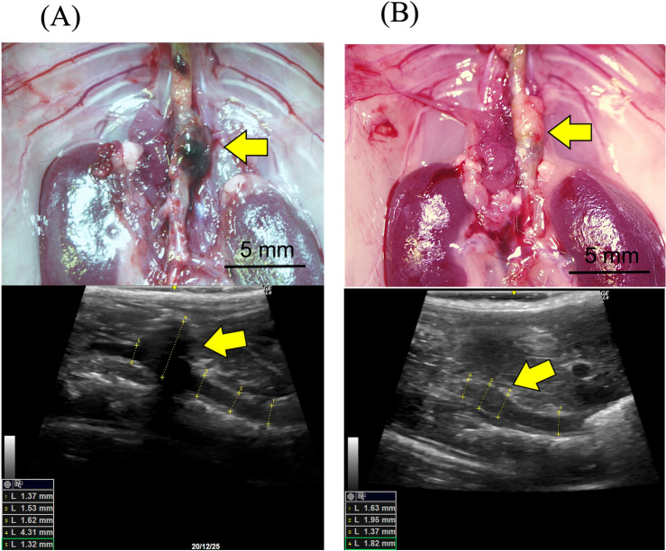
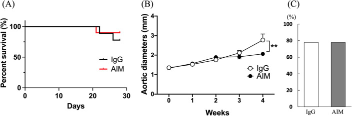
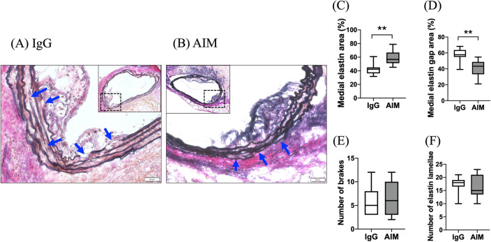
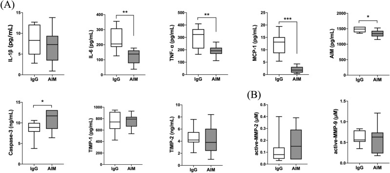
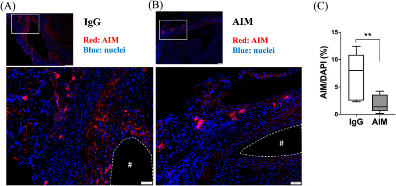
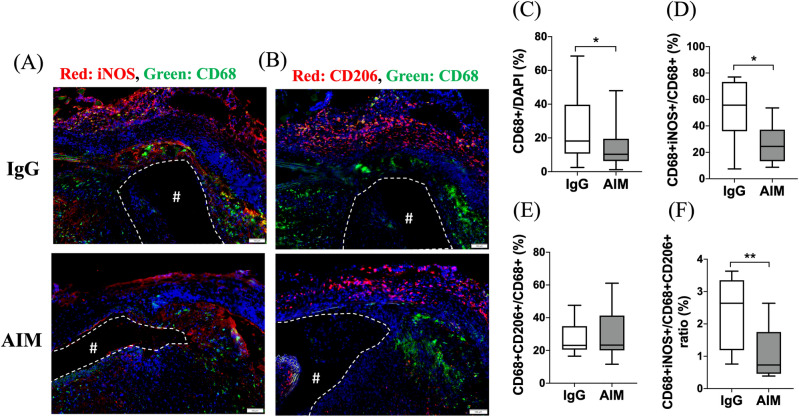
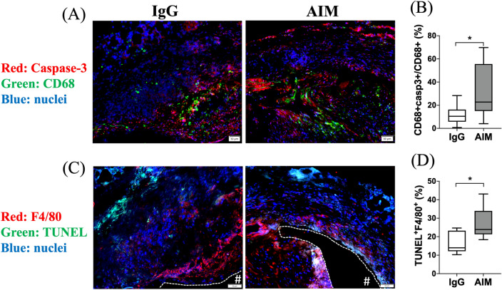
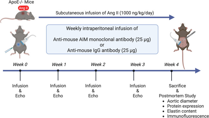
Apoptosis inhibitor of macrophage (AIM) is known to induce apoptosis resistance in macrophages and to exacerbate chronic inflammation, leading to arteriosclerosis. The role of AIM in aortic aneurysm (AA) remains unknown. This study examined the effects of an anti-AIM antibody in preventing AA formation and progression. In apolipoprotein E-deficient mice, AA was induced by subcutaneous angiotensin II infusion. Mice were randomly divided into two groups: (i) AIM group; weekly anti-murine AIM monoclonal antibody injection (n = 10), and (ii) IgG group; anti-murine IgG antibody injection as control (n = 14). The AIM group, compared with the IgG group, exhibited reduced AA enlargement (aortic diameter at 4 weeks: 2.1 vs. 2.7 mm, respectively, p = 0.012); decreased loss of elastic lamellae construction; reduced expression levels of IL-6, TNF-α, and MCP-1; decreased numbers of AIM-positive cells and inflammatory M1 macrophages (AIM: 1.4 vs. 8.0%, respectively, p = 0.004; M1 macrophages: 24.5 vs. 55.7%, respectively, p = 0.017); and higher expression of caspase-3 in the aortic wall (22.8 vs. 10.5%, respectively, p = 0.019). Our results suggest that administration of an anti-AIM antibody mitigated AA progression by alleviating inflammation and promoting M1 macrophage apoptosis.

Keywords: Aortic aneurysm; Apoptosis inhibitor of macrophage; Inflammation; Macrophage.


Conflict of interest statement

The authors declare no competing interests.

Figures

Figure 1
Figure 1
Representative microscopic and echography images of the aorta in each group. (A) 4 weeks in the IgG group, and (B) 4 weeks in the AIM group. Arrows indicate AAs. Scale bars = 5 mm.
Figure 2
Figure 2
(A) Kaplan–Meier survival curves show the survival rates in the two groups. (B) Aortic diameters over time were assessed by two-way ANOVA followed by Tukey’s multiple comparisons test. Data are means ± SEM. **p < 0.01 vs IgG. (C) Fisher’s exact test was used to assess the incidence of AA.
Figure 3
Figure 3
(A,B) EVG staining shows destruction of the elastic lamellae in the aortic wall at 8 weeks. The blue arrows indicate breaks in the elastic lamellae. Scale bars = 50 μm. (C,D) The medial and medial elastin gap areas are significantly different between groups. (E,F) The numbers of breaks and elastin lamellae are not significantly different. Mann–Whitney test, **p < 0.01.
Figure 4
Figure 4
Quantitative analysis of protein expression levels in AA. (A) ELISA analysis of inflammatory cytokines (IL-1β, IL-6, TNF-α), a chemokine (MCP-1), caspase-3, and TIMPs (TIMP-1, TIMP-2). (B) Measurement of active MMP-2 and -9. Data are means ± SEM. *p < 0.05, **p < 0.01, and ***p < 0.001 assessed by the Mann–Whitney U test.
Figure 5
Figure 5
AIM distribution. (A,B) Representative images of immunofluorescence staining for AIM in the IgG and AIM groups. AIM (red) and cell nuclei (blue). Scale bars = 50 μm. (C) The ratio of the AIM-positive area to the DAPI-positive area, expressed as a percentage. Data are means ± SEM. **p < 0.01 assessed by the Mann–Whitney U test. The “#” symbols enclosed by dashed lines indicate the vessel lumens.
Figure 6
Figure 6
Macrophage polarization. (A) iNOS (red) and CD68 (green); (B) CD206 (red), CD68 (green), and cell nuclei (blue). Scale bars = 100 μm. (C) The percentage of CD68 + cells to DAPI + cells. (D,E) The percentage of the CD68 + population consisting of iNOS + /CD206 + cells. (F) The percent ratio of CD206 + cells to iNOS + cells in the CD68 + population. Data are means ± SEM. *p < 0.05 and **p < 0.01 assessed by the Mann–Whitney U test. iNOS + /CD68 + cells indicate M1 macrophages, while CD206 + /CD68 + cells indicate M2 macrophages. The “#” symbols enclosed by dashed lines indicate the vessel lumens.
Figure 7
Figure 7
Macrophage apoptosis. (A) Representative images of immunofluorescence staining for apoptosis in the AIM and IgG groups. Caspase-3 (red), CD68 (green), and cell nuclei (blue). (B) The percentage of the CD68 + population consisting of CD68 + /caspase-3 + cells. (C) Representative images of immunofluorescence staining for apoptosis in the AIM and IgG groups. F4/80 (red), TUNEL (green), and cell nuclei (blue). (D) The percentages of cells that are TUNEL + F4/80 + . Scale bars = 50 μm. Data are means ± SEM. *p < 0.05 assessed by the Mann–Whitney U test. The “#” symbols enclosed by dashed lines indicate the vessel lumens.
Figure 8
Figure 8
The timeline of this study. Continuous subcutaneous Ang II infusion for 28 days induced AA. Anti-mouse AIM monoclonal antibody or anti-mouse IgG antibody was injected weekly intraperitoneally. Mice of each group were sacrificed after evaluation at 0, 1, 2, and 4 weeks. Image created with BioRender.com. AIM apoptosis inhibitor of macrophage, Ang angiotensin, ApoE apolipoprotein E.

Similar articles

References

    1. Moulakakis KG, et al. Open repair of thoracoabdominal aortic aneurysms in experienced centers. J. Vasc. Surg. 2018;68:634–645.e612. doi: 10.1016/j.jvs.2018.03.410. - DOI - PubMed
    1. Daye D, Walker TG. Complications of endovascular aneurysm repair of the thoracic and abdominal aorta: Evaluation and management. Cardiovasc. Diagn. Ther. 2018;8:S138–s156. doi: 10.21037/cdt.2017.09.17. - DOI - PMC - PubMed
    1. Ulug P, Powell JT, Martinez MA, Ballard DJ, Filardo G. Surgery for small asymptomatic abdominal aortic aneurysms. Cochrane Database Syst. Rev. 2020;7:Cd001835. doi: 10.1002/14651858.CD001835.pub5. - DOI - PMC - PubMed
    1. Li R, Liu Y, Jiang J. Research advances in drug therapy for abdominal aortic aneurysms over the past five years: An updated narrative review. Int. J. Cardiol. 2023;372:93–100. doi: 10.1016/j.ijcard.2022.11.058. - DOI - PubMed
    1. Sprague AH, Khalil RA. Inflammatory cytokines in vascular dysfunction and vascular disease. Biochem. Pharmacol. 2009;78:539–552. doi: 10.1016/j.bcp.2009.04.029. - DOI - PMC - PubMed

MeSH terms