. 2024 Jul 9;7(1):837.
doi: 10.1038/s42003-024-06523-9.

Identification of an H-Ras nanocluster disrupting peptide

Affiliations

Identification of an H-Ras nanocluster disrupting peptide

Candy Laura Steffen et al. Commun Biol. .

Abstract

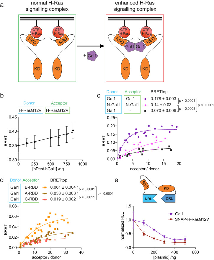
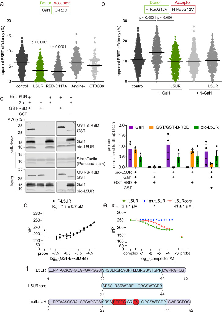
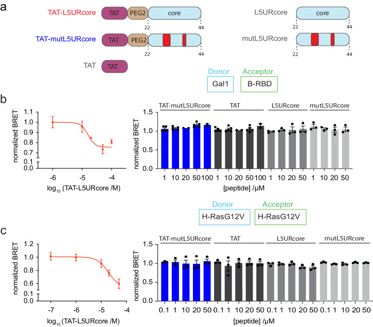
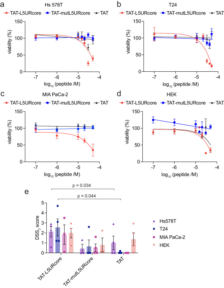
Hyperactive Ras signalling is found in most cancers. Ras proteins are only active in membrane nanoclusters, which are therefore potential drug targets. We previously showed that the nanocluster scaffold galectin-1 (Gal1) enhances H-Ras nanoclustering via direct interaction with the Ras binding domain (RBD) of Raf. Here, we establish that the B-Raf preference of Gal1 emerges from the divergence of the Raf RBDs at their proposed Gal1-binding interface. We then identify the L5UR peptide, which disrupts this interaction by binding with low micromolar affinity to the B- and C-Raf-RBDs. Its 23-mer core fragment is sufficient to interfere with H-Ras nanoclustering, modulate Ras-signalling and moderately reduce cell viability. These latter two phenotypic effects may also emerge from the ability of L5UR to broadly engage with several RBD- and RA-domain containing Ras interactors. The L5UR-peptide core fragment is a starting point for the development of more specific reagents against Ras-nanoclustering and -interactors.


Conflict of interest statement

The authors declare no competing interests.

Figures

Fig. 1
Fig. 1. The B-Raf preference of the H-Ras nanocluster scaffold Gal1 emerges within the RBD.
a Schematic of our model for Gal1 stabilized H-Ras nanocluster. b Dose-dependent effect of human Gal1 expression (48 h) on H-RasG12V nanoclustering-BRET (donor:acceptor plasmid ratio = 1:5); n = 4. c BRET-titration curves of the Gal1/ Gal1-interaction as compared to that of dimer-interface mutated N-Gal1. RLuc8-Gal1 was titrated with GFP2 as a control (black); n = 3. d BRET-titration curves of the Gal1-interaction with the RBDs of A-, B-, and C-Raf; n = 3. e Split-luciferase KinCon B-Raf biosensor response after expression of SNAP-H-RasG12V or Gal1; n = 3.
Fig. 2
Fig. 2. The L5UR-peptide binds to the Raf-RBD and disrupts the Raf-RBD/ Gal1-complex.
a Effect of L5UR expression (24 h) on Gal1/C-RBD FRET (donor:acceptor plasmid ratio = 1:3); n = 3. b Effect of L5UR expression (24 h) on Gal1-augmented H-RasG12V nanoclustering-FRET (donor:acceptor plasmid ratio = 1:3); n = 3. c Immunoblot data from pull-down assay with biotinylated L5UR and purified Gal1, GST-B-RBD or GST-only control with example blots (left) and quantification of repeat data (right); n = 3. d Binding of 10 nM F-L5UR to GST-B-RBD detected in a fluorescence polarization assay; n = 3. e Displacement of F-L5UR (10 nM) from GST-B-RBD (15 µM) by L5UR-derived peptides; n = 3. f Sequences of L5UR-derived peptides as used for in vitro and in cellulo assays. The stretch of the core peptide is highlighted in blue, mutations are in red.
Fig. 3
Fig. 3. The L5UR and L5UR-SNAP peptides disrupt H-RasG12V nanoclustering.
a Schematics of L5UR derived constructs expressed in cellular assays. The stretch of the core peptide is highlighted in blue, loss-of-function mutations are indicated red. b Effect of expression of L5UR constructs (48 h) on Gal1/B-RBD BRET (donor:acceptor plasmid ratio = 1:10); n = 3. c Effect of L5UR construct expression (48 h) on H-RasG12V nanoclustering-BRET with co-transfection of 200 ng Gal1 plasmid (donor:acceptor plasmid ratio = 1:5); n = 3. Statistical comparison was done against the SNAP-only sample. d Electron microscopy-based analysis of H-RasG12V nanoclustering in BHK cells showing the effects of L5UR-construct expression and controls; n = 15. Higher Lmax values indicate higher nanoclustering. e Immunoblot data from pull-down assays with L5UR-SNAP and control constructs from HEK cells co-expressing Gal1 with example blots (left) and quantification of repeat data (right); n = 4 (left).
Fig. 4
Fig. 4. The TAT-tagged L5URcore peptide disrupts H-RasG12V nanoclustering.
a Schematics of TAT-functionalized L5URcore-derived peptides and controls as applied in cellular assays. Loss-of-function mutations of L5UR are indicated in red. Non-TAT peptides are acetylated at the N-terminus. b, c Effect of cell-penetrating derivatives of L5URcore and control peptides on Gal1/B-RBD BRET (b donor:acceptor plasmid ratio = 1:10; n  ≥ 2) or H-RasG12V nanoclustering-BRET (c donor:acceptor plasmid ratio = 1:5, co-transfection of 200 ng Gal1 plasmid; n = 3). After 24 h expression of plasmids, peptides were added to cells at specified concentrations and incubated for 2 h.
Fig. 5
Fig. 5. The TAT-tagged L5URcore peptide impacts on Ras-signaling.
ah Immunoblot analysis of lysates from Hs 578 T (a, e), T24 (b, f), MIA PaCa-2 (c, g), and HEK (d, h) cells after EGF-stimulation and treatment with L5URcore-derived peptides with and without TAT-tag or control compound, trametinib (Tra), for 2 h; n = 3–8.
Fig. 6
Fig. 6. HRAS-mutant cancer cell proliferation is decreased by TAT-L5UR peptides.
ad 2D cell viability of Hs 578 T (a), T24 (b), MIA PaCa-2 (c), and HEK (d) cells in response to 48 h treatment with TAT-L5URcore peptides and TAT-control; n = 3. e Drug sensitivity score (DSS3), an area under the curve metric, calculated for the viability data in (ad). A higher value indicates a stronger anti-proliferative effect. TAT-control was used as a reference for statistical comparisons.

References

    1. Steffen CL, Kaya P, Schaffner-Reckinger E, Abankwa D. Eliminating oncogenic RAS: back to the future at the drawing board. Biochem. Soc. Trans. 2023;51:447–456. doi: 10.1042/BST20221343. - DOI - PMC - PubMed
    1. Punekar SR, Velcheti V, Neel BG, Wong KK. The current state of the art and future trends in RAS-targeted cancer therapies. Nat. Rev. Clin. Oncol. 2022;19:637–655. doi: 10.1038/s41571-022-00671-9. - DOI - PMC - PubMed
    1. Simanshu DK, Nissley DV, McCormick F. RAS proteins and their regulators in human disease. Cell. 2017;170:17–33. doi: 10.1016/j.cell.2017.06.009. - DOI - PMC - PubMed
    1. Spiegel J, Cromm PM, Zimmermann G, Grossmann TN, Waldmann H. Small-molecule modulation of Ras signaling. Nat. Chem. Biol. 2014;10:613–622. doi: 10.1038/nchembio.1560. - DOI - PubMed
    1. Smith MJ. Defining bone fide effectors of RAS GTPases. Bioessays. 2023;45:e2300088. doi: 10.1002/bies.202300088. - DOI - PubMed

Publication types

LinkOut - more resources