

An analysis of residual lung volume changes after segmentectomy based on three-dimensional computed tomography
- PMID: 38983136
- PMCID: PMC11228706
- DOI: 10.21037/jtd-24-83
An analysis of residual lung volume changes after segmentectomy based on three-dimensional computed tomography
Abstract
Background: Based on the results of JCOG0802 and CALGB studies, segmentectomy has considered to be a standard procedure for early-stage non-small cell lung cancer (NSCLC). After lobectomy, the residual cavity is filled with mediastinal and diaphragmatic deviations, and compensatory volume changes are present in the residual lungs. In this study, we examined the efficacy of segmentectomy, a surgical procedure, by focusing on its impact on postoperative lung volume and function.
Methods: We enrolled 77 patients who underwent segmentectomy as their initial surgical procedure, excluding those with additional lung resections and those who lacked postoperative computed tomography imaging. The predicted residual volume (mL) was defined as the total lung volume before surgery minus the volume of the resected area. Using the predicted residual volume (mL) and postoperative total lung volume (mL), we calculated the rate of postoperative lung volume increase [(postoperative total lung volume/predicted residual volume) × 100] (%). We also classified 52 cases with a rate of postoperative lung volume increase of ≥100% into a compensatory group, while those with a rate of <100% were classified into a non-compensatory group.
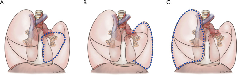
Results: The average postoperative lung volume increase was 104.6% among 77 cases. Age ≥65 years, pack year index ≥27.5, ≥3 resected segments, and use of electrocautery for intersegmental plane division were significantly associated with compensatory group classification. In 20 compensatory cases with preoperative and postoperative pulmonary function tests, postoperative vital capacity and forced expiratory volume in one second values exceeded the preoperative predictions. This study further examined the areas responsible for postoperative compensatory lung volume increase. In the compensatory group, significant expansion was observed in the ipsilateral lobes, excluding the resected segment and contralateral lung, while no significant changes were noted in the volume of the lobe, including the resected segment. Conversely, the non-compensatory group showed a significant volume decrease in the resected lobe, but no significant increase in other areas.
Conclusions: This study emphasizes the importance of preserving lung segments in segmentectomy. The study demonstrates extensive compensatory volume changes in the ipsilateral lung and contralateral lung. There was no significant volume decrease in any residual segment. This underlines the potential of segmentectomy to maintain lung function and expand treatment options post-surgery. In addition, the compensated group included patients with a lower pack-year index and younger patients. These results suggest that postoperative compensatory lung expansion includes not only hyperinflation of the remaining lung, but also an increase in the functional lung parenchyma.
Keywords: Segmentectomy; lung cancer; lung volume change; sublobar resection; three-dimensional computed tomography (3D-CT).
2024 Journal of Thoracic Disease. All rights reserved.
Conflict of interest statement
Conflicts of Interest: All authors have completed the ICMJE uniform disclosure form (available at https://jtd.amegroups.com/article/view/10.21037/jtd-24-83/coif). The authors have no conflicts of interest to declare.
Figures

Similar articles
-
Effect of Resected Lung Volume on Pulmonary Function and Residual Lung Volume in Patients Undergoing Segmentectomy: A Retrospective Study.Ann Surg Oncol. 2024 Oct;31(10):6645-6651. doi: 10.1245/s10434-024-15550-z. Epub 2024 Jun 12. Ann Surg Oncol. 2024. PMID: 38864984
-
Evaluation of the Residual Lung Function After Thoracoscopic Segmentectomy Compared With Lobectomy.Ann Thorac Surg. 2019 Nov;108(5):1543-1550. doi: 10.1016/j.athoracsur.2019.05.052. Epub 2019 Jul 11. Ann Thorac Surg. 2019. PMID: 31302085
-
Thoracoscopic left S1 + 2 segmentectomy as a good resolution for preserving pulmonary function.Interact Cardiovasc Thorac Surg. 2020 Sep 1;31(3):331-338. doi: 10.1093/icvts/ivaa105. Interact Cardiovasc Thorac Surg. 2020. PMID: 32747959
-
Lung Segmentectomy in NSCLC Surgery.Life (Basel). 2023 May 30;13(6):1284. doi: 10.3390/life13061284. Life (Basel). 2023. PMID: 37374067 Free PMC article. Review.
-
Anatomical thoracoscopic segmentectomy for lung cancer.Gen Thorac Cardiovasc Surg. 2014 Oct;62(10):586-93. doi: 10.1007/s11748-014-0409-7. Epub 2014 May 3. Gen Thorac Cardiovasc Surg. 2014. PMID: 24791926 Review.
Cited by
-
Imaging technologies in experimental pulmonary fibrosis research: essential tool for enhanced translational relevance.Eur Respir Rev. 2025 Sep 3;34(177):250012. doi: 10.1183/16000617.0012-2025. Print 2025 Jul. Eur Respir Rev. 2025. PMID: 40903049 Free PMC article. Review.
-
Morphological and Functional Analysis of Residual Lung After Pneumonectomy in Lung Cancer Surgery via 3D-CT Method.Life (Basel). 2025 Aug 10;15(8):1265. doi: 10.3390/life15081265. Life (Basel). 2025. PMID: 40868913 Free PMC article.
References
-
- Hoshino H, Koba H, Sekine K, et al. Compensatory increases in residual lobar volume following lung resection. Nihon Kokyuki Gakkai Zasshi. 1999;37:783-9. - PubMed
LinkOut - more resources
Full Text Sources