. 2024 Jun 30;16(6):3740-3752.
doi: 10.21037/jtd-23-1986. Epub 2024 Jun 11.

Thoracoscopic pulmonary resection combined with real-time image-guided percutaneous ablation for multiple pulmonary nodules: a novel surgical approach and literature review

Affiliations

Thoracoscopic pulmonary resection combined with real-time image-guided percutaneous ablation for multiple pulmonary nodules: a novel surgical approach and literature review

Yi Tian et al. J Thorac Dis. .

Abstract

Background: Due to the widespread use of computed tomography (CT) screening and advances in diagnostic techniques, an increasing number of patients with multiple pulmonary nodules are being detected and pathologically diagnosed as synchronous multiple primary lung cancers (sMPLC). It has become a new challenge to treat multiple pulmonary nodules and obtain a favorable prognosis while minimizing the perioperative risk for patients. The purpose of this study was to summarize the preliminary experience with a hybrid surgery combining pulmonary resection and ablation for the treatment of sMPLC and to discuss the feasibility of this novel procedure with a literature review.

Methods: This is a retrospective non-randomized controlled study. From January 1, 2022 to July 1, 2023, four patients underwent hybrid surgery combining thoracoscopic pulmonary resection and percutaneous pulmonary ablation for multiple pulmonary nodules. Patients were followed up at 3, 6 and 12 months postoperatively and the last follow-up was on November 30, 2023. Clinical characteristics, perioperative outcomes, pulmonary function recovery and oncologic prognosis were recorded. Meanwhile we did a literature review of studies on hybridized pulmonary surgery for the treatment of multiple pulmonary nodules.

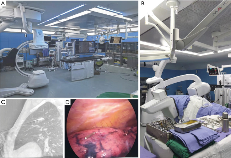
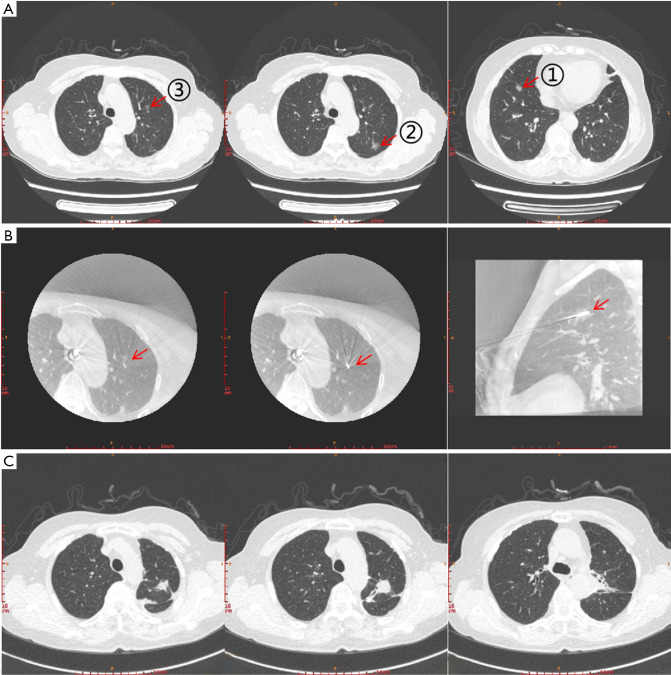
Results: All the four patients were female, aged 52 to 70 years, and had no severe cardiopulmonary dysfunction on preoperative examination. Hybrid surgery of simultaneous pulmonary resection and ablation were performed in these patients to treat 2 to 4 pulmonary nodules, assisted by intraoperative real-time guide of C-arm X-ray machine. The operation time was from 155 to 240 minutes, and intraoperative blood loss was from 50 to 200 mL. Postoperative hospital stay was 2 to 7 days, thoracic drainage duration was 2 to 6 days, and pleural drainage volume was 300-1,770 mL. One patient presented with a bronchopleural fistula due to pulmonary ablation; the fistula was identified and sutured during thoracoscopic surgery and the patient recovered well. No postoperative 90-day complications occurred. After 3 months postoperatively, performance status scores for these patients recovered to 80 to 100. No tumor recurrence or metastasis was detected during the follow-up period.

Conclusions: Hybrid procedures combining minimally invasive pulmonary resection with ablation are particularly suitable for the simultaneous treatment of sMPLC. Patients had less loss of pulmonary function, fewer perioperative complications, and favorable oncologic prognosis. Hybrid surgery is expected to be a better treatment option for patients with sMPLC.

Keywords: Synchronous multiple primary lung cancers (sMPLC); hybrid surgery; novel surgical strategy; percutaneous microwave ablation.


Conflict of interest statement

Conflicts of Interest: All authors have completed the ICMJE uniform disclosure form (available at https://jtd.amegroups.com/article/view/10.21037/jtd-23-1986/coif). All authors report that this study was supported by the National High Level Hospital Clinical Research Funding (No. BJ-2022-185). The authors have no other conflicts of interest to declare.

Figures

Figure 1
Figure 1
Hybrid operation room and procedures. (A) Hybrid operation room in our hospital. (B) C-arm X-ray machine (the Discovery IGS 7 angiography system, GE) which was used for supporting intraoperative localization procedures in our cases. Meanwhile, this machine was also used for localizing small nodules in VATS. (C) One of CT images of chest which were taken by our C-arm X-ray machine in our ablation procedures to help adjusting the location of microwave ablation needle. (D) In our previous research, an advanced way to localize small peripheral pulmonary nodules for surgical resection was reported (8). This figure shows that a titanium clip (Ethicon endo-surgery 1 ligaclip extra ligating clip cartridge 6 M titanium clips LT 300) was clipped at the visceral pleura under VATS observation to localize small peripheral pulmonary nodule with the help of baseline scan and preoperative CT imaging which was taken by our C-arm X-ray machine in the surgery. VATS, video-assistant thoracic surgery; CT, computed tomography.
Figure 2
Figure 2
Images of chest CT scanning in case 1. (A) Pre-surgery scanning showed nodule 1 to 3 (n1 to n3, red arrows, marked as ①②③). Nodule 1 is a pure GGN and located in the anterior segment of left upper lobe, with maximum diameter of 0.7 cm. Nodule 2 is also a mixed GGN with maximum diameter of 1.2 cm, which located in the apicoposterior segment of left upper lobe. Nodule 3 is a mixed GGN located in the anterior basal segment of right lower lobe, with maximum diameter of 1.2 cm. (B) Images of intraoperative localization of n1 under real-time guide by a C-arm X-ray. This nodule was treated by microwave ablation with the help of intraoperative localization under real-time guide by a C-arm X-ray machine. Red arrows show the position of the ablation needle tip. (C) CT images of two months after first-stage surgery. In this surgery, nodule 1 was treated by microwave ablation and nodule 3 was treated with segmentectomy of LS1+2a. CT, computed tomography; GGN, ground glass nodule.
Figure 3
Figure 3
Images of chest CT scanning and surgery in case 2. (A) Pre-surgery scanning of nodule 1 and 2 (red arrows, marked as ① and ②). Nodule 1 is a mixed GGN and located in the anterior segment of left upper lobe with biggest diameter of 1.5 cm. Nodule 2 is a mixed GGN and located in the posterior basal segment of left lower lobe with biggest diameter of 1.1 cm. Nodule 2 was treated by lobectomy of left lower lobe. (B) Images of intraoperative localization of nodule 1 under real-time guide by a C-arm X-ray. This nodule was treated by microwave ablation with intraoperative localization under real-time guide of a C-arm X-ray machine. Red arrows show the position of the ablation needle tip. (C) An ablation-induced bronchopleural fistula (green arrow) was identified during the thoracoscopic procedure and was repaired with sutures, and favorable postoperative recovery was obtained. CT, computed tomography; GGN, ground glass nodule.

References

    1. Warth A, Macher-Goeppinger S, Muley T, et al. Clonality of multifocal nonsmall cell lung cancer: implications for staging and therapy. Eur Respir J 2012;39:1437-42. 10.1183/09031936.00105911 - DOI - PubMed
    1. Nakata M, Sawada S, Yamashita M, et al. Surgical treatments for multiple primary adenocarcinoma of the lung. Ann Thorac Surg 2004;78:1194-9. 10.1016/j.athoracsur.2004.03.102 - DOI - PubMed
    1. Trousse D, Barlesi F, Loundou A, et al. Synchronous multiple primary lung cancer: an increasing clinical occurrence requiring multidisciplinary management. J Thorac Cardiovasc Surg 2007;133:1193-200. 10.1016/j.jtcvs.2007.01.012 - DOI - PubMed
    1. Rostad H, Strand TE, Naalsund A, et al. Resected synchronous primary malignant lung tumors: a population-based study. Ann Thorac Surg 2008;85:204-9. 10.1016/j.athoracsur.2007.07.091 - DOI - PubMed
    1. De Leyn P, Moons J, Vansteenkiste J, et al. Survival after resection of synchronous bilateral lung cancer. Eur J Cardiothorac Surg 2008;34:1215-22. 10.1016/j.ejcts.2008.07.069 - DOI - PubMed

LinkOut - more resources