Review
. 2022 Nov 24:2:970580.
doi: 10.3389/fopht.2022.970580. eCollection 2022.

Sellar masses: diagnosis and treatment

Affiliations
Review

Sellar masses: diagnosis and treatment

Dana Al-Bader et al. Front Ophthalmol (Lausanne). .

Abstract

Sellar mases can cause a variety of neuro-ophthalmic manifestations, including compressive optic neuropathy, chiasmal syndrome, and ophthalmoplegia due to cranial nerve palsy. Diagnosis involves a thorough history, neuro-ophthalmic examination, and ancillary tests and investigations. Visual field testing is critical in diagnosing and localizing the lesion and determining the extent of visual field loss. Appropriate neuro-imaging is essential in characterizing and localizing the lesion. Neuro-ophthalmologic assessment include meticulous clinical examination and ancillary tests including,visual field testing, which is useful in localizing the lesion, and optical coherence tomography, which is helpful in assessing the degree of axonal and neuronal loss and predicting the visual outcome. Treatment requires a multidisciplinary approach by different specialties, including radiologists, neuro-ophthalmologists, and neurosurgeons. The two primary treatment modalities for these tumors are surgery and radiation therapy. We review the main types of sellar lesions, their neuro-ophthalmologic evaluation, and treatment options.

Keywords: compressive optic neuropathy; craniopharingioma; ganglion cell complex (GCC); meningioma; optical coherance tomography; pituary adenoma; sellar masses.


Conflict of interest statement

The authors declare that the research was conducted in the absence of any commercial or financial relationships that could be construed as a potential conflict of interest.

Figures
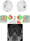
Figure 1
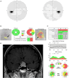
Figure 1
The sensitivity of OCT to detect GCC loss even before visual fields became abnormal for a patient with pituitary adenoma. (Top) Humphrey visual field 24-2 without changes detected in this patient with pituitary macro adenoma. (Middle) Ganglion Cell Analysis of Spectral-domain OCT (Cirrus) showing binasal thinning in the GCC layer despite normal visual fields. (Bottom) MRI T1 coronal view with contrast showing pituitary macro adenoma.
Figure 2
Figure 2
Humphrey visual field 24-2 and OCT with ganglion cell analysis pre- and post-pituitary tumor surgery. (A) Humphrey visual field 30-2 showing bitemporal hemianopia in a patient with a pituitary tumor and chiasmal compression. (B) Ganglion Cell Analysis of Spectral domain-OCT (Cirrus) shows binasal thinning of the ganglion cell layer with relative overall preservation, indicating a good prognosis for visual recovery following surgical excision of a pituitary tumor. (C) Follow-up visual field at one month after surgical decompression of the tumor showing complete recovery of the visual field defect. (D) Ganglion cell analysis at 1 month showing persistence of the binasal ganglion cell loss and the overall preservation of the ganglion cell layer thickness.
Figure 3
Figure 3
Humphrey visual field 24-2 and OCT with ganglion cell analysis and retinal nerve fiber layer thickness of a patient with sellar and suprasellar meningioma. (A) Normal visual field test. (B) Evidence of ganglion cell layer loss in the left eye, especially nasally. (C) Thinning of the retinal nerve fiber layer in both eyes. (D) MRI shows meningioma of the sellar and suprasellar region with homogenous enhancement on T1 with contrast.
Figure 4
Figure 4
MRI T1 pre-contrast (top) and post-contrast (bottom) sagittal view of pituitary adenoma.
Figure 5
Figure 5
(A) MRI T1 with (top) and without (bottom) contrast coronal view of a giant craniopharyngioma with sellar and suprasellar solid enhancing parts; A large cystic component extends into the frontal horn of the right lateral ventricle. (B) A mid sagittal plane CT head of the same patient with evidence of sellar widening and peripheral calcification.
Figure 6
Figure 6
A midsagittal cut T1 with contrast demonstrating a sellar meningioma that has a clear dural tail anteriorly extending to the cribriform plate and normal adenophysis clearly demarcated intra sellarly.

References

    1. Subhawong TK, Fishman EK, Swart JE, Carrino JA, Attar S, Fayad LM. Soft-tissue masses and masslike conditions: what does CT add to diagnosis and management? AJR Am J Roentgenol (2010) 194(6):1559–67. doi: 10.2214/AJR.09.3736 - DOI - PMC - PubMed
    1. Ogra S, Nichols AD, Stylli S, Kaye AH, Savino PJ, Danesh-Meyer HV. Visual acuity and pattern of visual field loss at presentation in pituitary adenoma. J Clin Neurosci (2014) 21(5):735–40. doi: 10.1016/j.jocn.2014.01.005 - DOI - PubMed
    1. Rebolleda G, Diez-Alvarez L, Casado A, Sánchez-Sánchez C, de Dompablo E, González-López JJ, et al. . OCT: New perspectives in neuro-ophthalmology. Saudi J Ophthalmol (2015) 29(1):9–25. doi: 10.1016/j.sjopt.2014.09.016 - DOI - PMC - PubMed
    1. Vuong LN, Hedges TR, 3rd. Ganglion cell layer complex measurements in compressive optic neuropathy. Curr Opin Ophthalmol (2017) 28(6):573–8. doi: 10.1097/ICU.0000000000000428 - DOI - PubMed
    1. Tieger MG, Hedges TR, 3rd, Ho J, Erlich-Malona NK, Vuong LN, Athappilly GK, et al. . Ganglion cell complex loss in chiasmal compression by brain tumors. J Neuroophthalmol (2017) 37(1):7–12. doi: 10.1097/WNO.0000000000000424 - DOI - PMC - PubMed

LinkOut - more resources