. 2024 Jun 25:15:1405597.
doi: 10.3389/fimmu.2024.1405597. eCollection 2024.

Proangiogenic properties of complement protein C1q can contribute to endometriosis

Affiliations

Proangiogenic properties of complement protein C1q can contribute to endometriosis

Chiara Agostinis et al. Front Immunol. .

Abstract

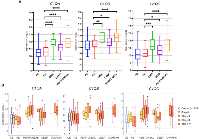
Endometriosis (EM) is defined as the engraftment and proliferation of functional endometrial-like tissue outside the uterine cavity, leading to a chronic inflammatory condition. While the precise etiology of EM remains elusive, recent studies have highlighted the crucial involvement of a dysregulated immune system. The complement system is one of the predominantly altered immune pathways in EM. Owing to its involvement in the process of angiogenesis, here, we have examined the possible role of the first recognition molecule of the complement classical pathway, C1q. C1q plays seminal roles in several physiological and pathological processes independent of complement activation, including tumor growth, placentation, wound healing, and angiogenesis. Gene expression analysis using the publicly available data revealed that C1q is expressed at higher levels in EM lesions compared to their healthy counterparts. Immunohistochemical analysis confirmed the presence of C1q protein, being localized around the blood vessels in the EM lesions. CD68+ macrophages are the likely producer of C1q in the EM lesions since cultured EM cells did not produce C1q in vitro. To explore the underlying reasons for increased C1q expression in EM, we focused on its established pro-angiogenic role. Employing various angiogenesis assays on primary endothelial endometriotic cells, such as migration, proliferation, and tube formation assays, we observed a robust proangiogenic effect induced by C1q on endothelial cells in the context of EM. C1q promoted angiogenesis in endothelial cells isolated from EM lesions (as well as healthy ovary that is also rich in C1q). Interestingly, endothelial cells from EM lesions seem to overexpress the receptor for the globular heads of C1q (gC1qR), a putative C1q receptor. Experiments with siRNA to silence gC1qR resulted in diminished capacity of C1q to perform its angiogenic functions, suggesting that C1q is likely to engage gC1qR in the pathophysiology of EM. gC1qR can be a potential therapeutic target in EM patients that will disrupt C1q-mediated proangiogenic activities in EM.

Keywords: C1q; angiogenesis; complement system; endometriosis; endothelial cells; gC1qR; ovary.


Conflict of interest statement

The authors declare that the research was conducted in the absence of any commercial or financial relationships that could be construed as a potential conflict of interest. The author(s) declared that they were an editorial board member of Frontiers, at the time of submission. This had no impact on the peer review process and the final decision.

Figures

Figure 1
Figure 1
Gene expression analysis of C1q in EM lesions based on EndometDB. (A) Histograms representing C1QA, C1QB, and C1QC mRNA expression in control endometrium (CE), patient endometrium (PE), and in different EM lesions (peritoneal; deep; and ovarian, OMA). Gene expression profiling (GEP) analysis, based on data extracted from GEO (GSE141549), revealed a significantly higher expression of all three C1q genes in EM lesions as compared to CE. (B) Analysis of C1q gene expression in EM patients clustered into different disease stages (stage I-IV). *p < 0.05, **p < 0.01, ***p < 0.001, ****p < 0.0001 (Mann-Whitney U Test).
Figure 2
Figure 2
C1q is abundantly present in endometriotic lesions and in healthy ovary. Representative microphotographs showing the presence of C1q in ovarian endometriotic lesions (A, B, E), patients’ eutopic endometrium (C), and healthy ovary (D). AEC (red) chromogen was used to visualize the binding of rabbit anti-human C1q antibody. Red arrows indicate vessels (A), while yellow arrows indicate isolated cells scattered in EM stroma which resulted due to positive staining for C1q (B). (F, G) Representative microphotographs showing the presence of C4d (F) or C1q (G) in serial sections of endometriotic lesions. C1q is present in the endometriotic lesion; however, the classical pathway is feebly activated. AEC (red) chromogen was used to visualize the binding of secondary antibodies. Nuclei were counterstained in blue with Harris Hematoxylin. Magnification, 10x (A, C-E); 20x (B). Scale bars, 50 µm (A, B); 100 µm (C-G).
Figure 3
Figure 3
Double immunofluorescence microscopy for C1q in EM lesions. Representative images showing double staining for C1q (red) and vWF (A, D, E), CD68 (B), or CD34 (C) (green) in endometriotic lesions. After deparaffinization, tissue sections were incubated overnight with anti-human C1q and anti-human vWF, CD68, or CD34 primary antibodies, followed by incubation with anti-rabbit Cy3 and anti-mouse Alexa Fluor™ 488 secondary antibodies. Cell nuclei were stained with DAPI. Scale bars, 50 µm.
Figure 4
Figure 4
C1QA, C1QB, and C1QC gene expression in endometriotic lesions and primary isolated endometriotic cells. (A) Graphical representation of primary EM cell isolation procedure. (B) Characterization of endometriotic cells (EMCs) isolated from EM ovary cysts by immunofluorescence. Around 10% of EMCs resulted positively stained for cytokeratin (CK)8/18, and 100% were positive for mucin-1 (MUC-1) and vimentin. Cell nuclei were stained with DAPI. (C) After total RNA extraction and retro-transcription, C1q gene expression was analyzed by performing RT-qPCR. Peripheral blood mononuclear cells (PBMCs) were used as positive control. GAPDH was used as the housekeeping gene. Scatter plots were generated with the software GraphPad Prism 8.4.3. (D, E) The clustering of EM patients in stages (I-III or IV) or for adenomyosis presence respectively highlighted a significant difference in terms of C1q expression, with higher C1q levels in the most severe group (D), and in adenomyosis-positive EM patients (E). C1q expression was evaluated by examining the collective mean of individual C1QA, C1QB, and C1QC genes. Data are expressed as box-plots (median, interquartile range). *p < 0.05 (unpaired two-tailed t-test).
Figure 5
Figure 5
Binding of C1q to EECs, OVECs, or HUVECs. (A) Different endothelial cells [ECs; i.e., ECs isolated from healthy ovary (OVECs), n = 3; human umbilical vein ECs (HUVECs), n = 3; and endometriotic ECs (EECs)] grown to confluence on 96-well tissue culture plates were incubated with 10 µg/mL of purified C1q at different time points (0, 15, 30, 60, or 120 minutes) at room temperature. The binding of C1q was revealed by whole-cell ELISA assay. The data are presented as mean ± SD of three separate experiments. (B) Schematic representation of functional assays for the evaluation of C1q proangiogenic properties by migration, scratch, proliferation, and tube formation assays. Image created with BioRender.com, as an adaptation from Laschke et al. (33). *p<0.05; **p<0.01; ****p<0.0001.
Figure 6
Figure 6
C1q promotes angiogenesis in EECs and OVECs. (A) Migration assays were performed in a trans-well system using endothelial cells (ECs) isolated from endometriotic cysts (EECs), healthy ovary (OVECs), or human umbilical vein (HUVECs). Cells were stained with FAST DiI™, seeded in FluoroBlok™ Inserts (1.5x10⁵ cells/insert), and the lower chamber was loaded with C1q (10 μg/mL) or VEGF (20 ng/mL), as chemoattractant stimuli. After 24h, fluorescence was read via INFINITE 200 Fluorescence Plate Reader. Data are expressed as mean ± standard deviation (SD). *p < 0.05; **p < 0.01. (B) Wound healing assays were performed using EECs, OVECs, and HUVECs. Cells (5x10⁴/well) were grown until 60–70% of confluence in a 24-well plate. After scratching the middle of endothelial monolayer, cells were stimulated with C1q (10 μg/mL) or VEGF (20 ng/mL). Images of the wound fields were captured after 18h, allowing calculation of percentage wound closure. Data are expressed as mean ± SD. *p < 0.05; **p < 0.01. (C) Tube formation assays were performed in EECs, OVECs, and HUVECs. Cells (5x10⁴) were seeded onto Matrigel® in CultureSlides, and stimulated with C1q (10 μg/mL) or VEGF (20 ng/mL). After 18h, using TiEsseLab BDS 600 microscope, the capillary-like structures formed by ECs were manually counted, comparing the different conditions. Data are expressed as mean ± SD. *p < 0.05. (D) Proliferation assays were performed using EECs, OVECs, and HUVECs. Cells (7x10³/well) were seeded in a 96-well plate, and stimulated with C1q (10 μg/mL) or VEGF (20 ng/mL) for 24h. MTS was then added in each well, and cell proliferation was measured using PowerWave Select X Microplate Reader. Data are expressed as mean ± SD. REST, resting cells.
Figure 7
Figure 7
gC1qR is expressed in EECs and OVECs and can modulate C1q-induced proangiogenic behaviour. (A, B) Representative images displaying gC1qR positive staining (green) in EECs and OVECs, comparing permeabilized and not permeabilized cells. Cell nuclei were stained with DAPI (blue). (C, D) Surface biotinylation assay for the detection of gC1qR fraction present on the cell surface of EECs (n = 3) and OVECs (n = 3). Cells were treated with Sulfo-NHS-biotin reagent, biotinylated cell surface proteins were isolated upon binding to a Streptavidin-coated resin, and separated on a 10% SDS-PAGE. Membrane was then probed with anti-gC1qR antibody and anti-rabbit IRDye 800CW secondary antibody by Western blot analysis. Signal intensity was detected using an Odyssey CLx near-infrared scanner (LI-COR Biosciences, Lincoln, NE, USA). Image acquisition, processing, and data analysis were performed with Image Studio 5.2 (LI-COR Biosciences). β-actin was used to normalize the results. Data are expressed as mean ± standard deviation (SD). EECs displayed a higher amount of gC1qR compared to OVECs, considering both total protein and cell surface fraction; *p < 0.05. B, biotinylated; MW, molecular weights; NB, not biotinylated; PD, pull down. (E) Wound healing assay was performed using HUVECs (n = 3) after transfection with siC1QBP (gC1qR gene) or siCTRL for 48h. After scratching the middle of endothelial monolayer, cells were stimulated with C1q (10 μg/mL) or VEGF (20 ng/mL). Images of the wound fields were captured at 0 and 8h, percentage wound closure was calculated. The percentage of wound healing was considered as relative to siCTRL in resting conditions (100% of wound healing). Data are expressed as mean ± SD; *p < 0.05.

References

    1. Giudice LC, Kao LC. Endometriosis. Lancet. (2004) 364:1789–99. doi: 10.1016/S0140-6736(04)17403-5 - DOI - PubMed
    1. Moradi Y, Shams-Beyranvand M, Khateri S, Gharahjeh S, Tehrani S, Varse F, et al. A systematic review on the prevalence of endometriosis in women. Indian J Med Res. (2021) 154:446–54. doi: 10.4103/ijmr.IJMR_817_18 - DOI - PMC - PubMed
    1. Feng J, Zhang S, Chen J, Zhu J, Yang J. Global burden of endometriosis in 204 countries and territories from 1990 to 2019. CEOG. (2022) 49(10):235. doi: 10.31083/j.ceog4910235 - DOI
    1. Parasar P, Ozcan P, Terry KL. Endometriosis: epidemiology, diagnosis and clinical management. Curr Obstet Gynecol Rep. (2017) 6:34–41. doi: 10.1007/s13669-017-0187-1 - DOI - PMC - PubMed
    1. Imperiale L, Nisolle M, Noël JC, Fastrez M. Three types of endometriosis: pathogenesis, diagnosis and treatment. State Art J Clin Med. (2023) 12(3):994. doi: 10.3390/jcm12030994 - DOI - PMC - PubMed

MeSH terms

LinkOut - more resources