. 2021 Oct 20:1:755058.
doi: 10.3389/fopht.2021.755058. eCollection 2021.

Choroidal Vascularity Index in CHM Carriers

Affiliations

Choroidal Vascularity Index in CHM Carriers

Dario Pasquale Mucciolo et al. Front Ophthalmol (Lausanne). .

Abstract

Purpose: To assess the choroidal structure using the Choroidal Vascularity Index (CVI) and analyse choroidal changes in choroideremia (CHM) carriers.

Material and methods: Female CHM carriers, genetically characterized, and a control group were recruited at the Eye Clinic of Careggi Teaching Hospital, Florence. The patients underwent a complete ophthalmic evaluation and retinal imaging. In particular, the Stromal Area (SA), Luminal Area (LA), Total Choroidal Area (TCA), CVI, and Subfoveal Choroidal Thickness (SFCT) were calculated for each eye using Optical Coherence Tomography (OCT) examinations.

Results: Twelve eyes of 6 CHM carriers and 14 eyes of 7 age-matched controls were analysed. The mean SFCT was 270.9 ± 54.3μm in carriers and 281.4 ± 36.8μm in controls (p = 0.564); LA was 0.99 ± 0.25mm2 and 1.01 ± 0.13mm2 (p = 0.172); SA was 0.53 ± 0.09mm2 and 0.59 ± 0.07mm2 (p = 0.075), and TCA was 1.53 ± 0.34mm2 and 1.69 ± 0.19mm2 respectively (p = 0.146). Mean CVI measured 64.03 ± 3.98% in the CHM carriers and 65.25 ± 2.55% in the controls (p = 0.360).

Conclusions: The CVI and CVI-related parameters (SA, LA, and TCA) do not differ between CHM female carriers and controls. These findings reveal a preserved choroidal vasculature in eyes with RPE impairment and support the primary role of RPE in the pathogenesis of CHM disease.

Keywords: CHM; CVI; OCT; carriers; choroid; choroideremia.


Conflict of interest statement

The authors declare that the research was conducted in the absence of any commercial or financial relationships that could be construed as a potential conflict of interest.

Figures
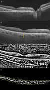
Figure 1
Figure 1
(A) Long horizontal OCT B-scan at the fovea of a control. (B) 9 mm Horizontal OCT B-scan at the fovea. The orange line indicates the SFCT. (C) The image was binarized using Niblack’s auto-local threshold to calculate the CVI; the binarization was performed using public domain software “Image J” through an automated function. The dark pixels represent the luminal area and the white pixels the stromal area. (D) After uploading the images on Image J, a polygon tool was used to select the total choroid area (TCA-area between the yellow lines), with the RPE representing the anterior boundary of the TCA and the scleral-choroidal interface as the posterior boundary of the TCA, across the entire length of the scan.
Figure 2
Figure 2
(A) Ultra-Widefield (UWF) color fundus imaging and (B) Fundus Autofluorescence (FAF) of the right eye of a 42-year-old CHM female carrier. UWF imaging shows large and irregular hypo-pigmented areas with yellowish deposits at the macula. (C) Long horizontal OCT B-scan at the fovea showing a preserved External Limiting Membrane (ELM) and slight irregularities of the Ellipsoid Zone (EZ) and RPE-Bruch’s membrane complex. (D) 9 mm horizontal OCT scan. The orange line indicates the SFCT. (E) The image was binarized using Niblack’s auto-local threshold to calculate the CVI; the binarization was performed using public domain software “Image J” trough an automated function. The dark pixels represent the luminal area and the white pixels the stromal area. (F) The CVI was calculated as the ratio between the Luminal Area (LA) and the total choroidal area (TCA). After uploading the images on Image J, a polygon tool was used to select the total choroid area (TCA-area between the yellow lines), with the RPE representing the anterior boundary of the TCA and the scleral-choroidal interface as the posterior boundary of the TCA, across the entire length of the scan.
Figure 3
Figure 3
(A) Ultra-Widefield (UWF) color fundus imaging and (B) Fundus Autofluorescence (FAF) of the right eye of a 51-year-old female CHM carrier. FAF shows a normal appearance of the macula with multiple hypo-autofluorescent areas at the posterior pole and at the mid-periphery. (C) Long horizontal B-scan OCT passing through the fovea showing atrophy of the outer retinal layers (interdigitation zone, ellipsoid zone, outer nuclear layer) and RPE layer at the peripapillary area. (D) 9 mm horizontal B-scan OCT. The orange line indicates the SFCT. (E) The image was binarized using Niblack’s auto-local threshold in order to calculate the CVI. The binarization was performed using public domain software “Image J” through an automated function. The dark pixels represent the luminal area and the white pixels the stromal area. (F) The CVI was calculated as the ratio between the luminal area (LA) and the total choroidal area (TCA). After uploading the images on Image J, a polygon tool was used to select the total choroid area (TCA-area between the yellow lines), with the RPE as the anterior boundary of the TCA and the scleral-choroidal interface as the posterior boundary of the TCA, across the entire length of the scan.

References

    1. Van den Hurk JA, Schwartz M, van Bokhoven H, van de Pol TJ, Bogerd L, Pinckers AJ, et al. . Molecular Basis of Choroideremia (CHM): Mutations Involving the Rab Escort Protein-1 (REP-1) Gene. Hum Mutat (1997) 9(2):110–7. doi: 10.1002/(SICI)1098-1004(1997)9:2<110::AID-HUMU2>3.0.CO;2-D - DOI - PubMed
    1. Heon E, Alabduljalil T, McGuigan DB, III, Cideciyan AV, Li S, Chen S, et al. . Visual Function and Central Retinal Structure in Choroideremia. Invest Ophthalmol Vis Sci (2016) 57(9):OCT377–87. doi: 10.1167/iovs.15-18421 - DOI - PubMed
    1. Murro V, Mucciolo DP, Passerini I, Palchetti S, Sodi A, Virgili G, et al. . Retinal Dystrophy and Subretinal Drusenoid Deposits in Female Choroideremia Carriers. Graefes Arch Clin Exp Ophthalmol Albrecht Von Graefes Arch Klin Exp Ophthalmol (2017) 255(11):2099–111. doi: 10.1007/s00417-017-3751-5 - DOI - PubMed
    1. Agrawal R, Ding J, Sen P, Rousselot A, Chan A, Nivison-Smith L, et al. . Exploring Choroidal Angioarchitecture in Health and Disease Using Choroidal Vascularity Index. Prog Retin Eye Res (2020) 77:100829. doi: 10.1016/j.preteyeres.2020.100829 - DOI - PubMed
    1. Iovino C, Au A, Hilely A, Violanti S, Peiretti E, Gorin MB, et al. . Evaluation of the Choroid in Eyes With Retinitis Pigmentosa and Cystoid Macular Edema. Invest Ophthalmol Vis Sci (2019) 60(15):5000–6. doi: 10.1167/iovs.19-27300 - DOI - PubMed

LinkOut - more resources