Review
. 2024 Jun 9;16(6):e61996.
doi: 10.7759/cureus.61996. eCollection 2024 Jun.

Selectins in Biology and Human Disease: Opportunity in E-selectin Antagonism

Affiliations
Review

Selectins in Biology and Human Disease: Opportunity in E-selectin Antagonism

John M Peterson et al. Cureus. .

Abstract

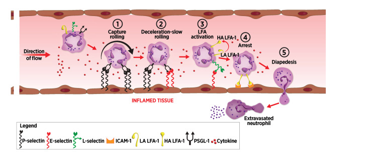
Selectins are cell adhesion proteins discovered in the 1980s. As C-type lectins, selectins contain an essential calcium ion in the ligand-binding pocket and recognize the isomeric tetrasaccharides sialyl Lewisx (sLex) and sialyl Lewisa (sLea). Three selectins, E-selectin, P-selectin, and L-selectin, play distinct, complementary roles in inflammation, hematopoiesis, and tumor biology. They have been implicated in the pathology of diverse inflammatory disorders, and several selectin antagonists have been tested clinically. E-selectin plays a unique role in leukocyte activation, making it an attractive target for intervention, for example, in sickle cell disease (SCD). This review summarizes selectin biology and pathology, structure and ligand binding, and selectin antagonists that have reached clinical testing with an emphasis on E-selectin.

Keywords: acute lung injury; adhesion; antagonism; inflammation; leukocyte activation; selectin; sickle cell disease; thrombosis; vaso-occlusion.


Conflict of interest statement

Conflicts of interest: In compliance with the ICMJE uniform disclosure form, all authors declare the following: Payment/services info: All authors have declared that no financial support was received from any organization for the submitted work. Financial relationships: John M. Peterson declare(s) employment, a patent, stock/stock options and travel from GlycoMimetics, Inc. Edwin P. Rock declare(s) employment, stock/stock options and travel from GlycoMimetics, Inc. John L. Magnani declare(s) personal fees, employment and stock/stock options from GlycoMimetics, Inc. Theodore A. Smith declare(s) employment, a patent and stock/stock options from GlycoMimetics, Inc. Intellectual property info: All authors are currently or were recently employees of GlycoMimetics Inc. As such, we have been involved in the discovery and development of the E-selectin antagonists uproleselan and GMI-1687, as well as the pan-selectin antagonist rivipansel, which are described in this manuscript. Other relationships: All authors have declared that there are no other relationships or activities that could appear to have influenced the submitted work.

Figures

Figure 1
Figure 1. Selectin genes and protein domain structure
(a) Selectin gene cluster on chromosome 1q. (b) Selectin protein structure. The N-terminal CRD is labeled "lectin" at the right side of the figure, followed by an EGF domain (oval) and varying numbers of SCR domains (rectangles). Intracellular domains are represented by green rectangles, and glycosylation sites are represented by tridents. CRD: carbohydrate-recognition domain, EGF: epidermal growth factor, SCR: short consensus repeat Image credits: Dr. Drew Provan using Adobe Illustrator
Figure 2
Figure 2. Selectin binding ligand structures
(a) Structures of sialyl Lewisx and sialyl Lewisa​​​​​​​: the GlcNAc residue is highlighted in red to indicate the isomeric nature of the structure. (b) Structure of synthetic P-selectin ligand SGP-3, comprising the terminal 19 amino acids of PSGL-1, including three sulfated tyrosine residues (sY5, sY7, and sY10) (highlighted in red), an O-linked saccharide at T16 containing a sialyl Lewisx​​​​​​​ (highlighted in red) moiety, and an enterokinase cleavage sequence from R20 to K28 [16]. PSGL-1: P-selectin glycoprotein ligand-1 Image credits: Dr. Drew Provan using Adobe Illustrator
Figure 3
Figure 3. Inflammatory cascade leading to cell migration
(1) Inflammation activates local endothelium to express P-selectin that initiates tethering and rolling of circulating leukocytes along the endothelial wall. (2) Inflammatory cytokines induce de novo synthesis and surface expression of E-selectin that also captures circulating neutrophils. (3) E-selectin binds to sLex residues expressed by L-selectin on captured neutrophils, inducing conformational changes of LFA-1 from an LA to an intermediate affinity, then an HA state. (4) LFA-1 in its HA conformation binds to ICAM-1 expressed by endothelial cells, leading to firm arrest. (5) Following firm arrest, leukocytes extravasate into inflamed tissue by the process of diapedesis. sLex: sialyl Lewisx, LFA-1: leukocyte function-associated antigen-1, LA: low affinity, HA: high affinity, ICAM-1: intercellular adhesion molecule-1, PSGL-1: P-selectin glycoprotein ligand-1 Image credits: Dr. Drew Provan using Adobe Illustrator
Figure 4
Figure 4. Neutrophil extravasation and acute lung injury or pneumonia
In severe lung inflammation, activated endothelial surface selectin expression leads to circulating neutrophil capture, extravasation into alveoli, and activation leading to alveolar fluid accumulation and impaired gas exchange. To eliminate redundancy, the binding cascade here omits PSGL-1 interactions with P-selectin and E-selectin, as well as E-selectin interactions with L-selectin. PSGL-1: P-selectin glycoprotein ligand-1, NETS: neutrophil extracellular traps, HA: high affinity, LFA-1: leukocyte function-associated antigen-1, ICAM-1: intercellular adhesion molecule-1 Image credits: Dr. Drew Provan using Adobe Illustrator
Figure 5
Figure 5. Vaso-occlusion in sickle cell disease
(1) Inflammatory triggers, such as hypoxia and hemolysis products, activate endothelium to express P-selectin, which captures circulating leukocytes. (2) E-selectin is expressed following de novo synthesis, capturing more leukocytes. (3) E-selectin binding to sLex on L-selectin (not shown in the figure), followed by the activation of LFA-1, induces endothelial capture of inflammatory cells, as well as secondary capture of platelets and red blood cells, leading to vascular occlusion. To eliminate redundancy, this binding cascade omits PSGL-1 interactions with P-selectin and E-selectin, as well as E-selectin interactions with L-selectin. sLex: sialyl Lewisx, LFA-1: leukocyte function-associated antigen-1, PSGL-1: P-selectin glycoprotein ligand-1, LA: low affinity, HA: high affinity Image credits: Dr. Drew Provan using Adobe Illustrator
Figure 6
Figure 6. Thrombosis
(1) Endothelium is activated via hypoxia or inflammatory triggers leading to the expression of P-selectin, E-selectin, and vWF. These proteins capture circulating platelets, leukocytes, and TF+MV. (2) Activation of bound leukocytes leads to the expression of tissue factors on their surface. Captured platelets also release tissue factor. (3) Tissue factor then activates the clotting cascade, leading to thrombus formation, including rbc. vWF: von Willebrand factor, TF+MV: tissue factor expressing microvesicles, rbc: red blood cells, PSGL-1: P-selectin glycoprotein ligand-1 Image credits: Dr. Drew Provan using Adobe Illustrator
Figure 7
Figure 7. Selectin family X-ray crystal structures
Tertiary structures of E-selectin, P-selectin, and L-selectin CRD and EGF-like domains from X-ray crystallography. RCSB Protein Data Bank reference numbers are shown. CRD: carbohydrate-recognition domain, EGF: epidermal growth factor, RCSB: Research Collaboratory for Structural Bioinformatics Image credits: Dr. Drew Provan using Pymol
Figure 8
Figure 8. Bound and unbound selectin structures
(a) Crystal structures of CRD and EGF-like domains of E-selectin unbound and bound to sLex. (b) Crystal structures of P-selectin CRD and EGF-like domains unbound and bound to the PSGL-1 derived sulfoglycopeptide, SGP-3. RCSB Protein Data Bank reference numbers are shown. CRD: carbohydrate-recognition domain, EGF: epidermal growth factor, sLex: sialyl Lewisx, PSGL-1: P-selectin glycoprotein ligand-1, RCSB: Research Collaboratory for Structural Bioinformatics Image credits: Dr. Drew Provan using Pymol
Figure 9
Figure 9. E-selectin bound to sLex (4CSY)
(a) Interactions between E-selectin and the fucose residue of sLex. (b) Interactions between E-selectin and the galactose and NeuAc residues of sLex. sLex: sialyl Lewisx Image credits: Dr. Drew Provan using Pymol
Figure 10
Figure 10. P-selectin bound to PSGL-1 derived glycosulfopeptide SGP-3
(a) Interactions between P-selectin and the fucose residue of SGP-3. (b) Interactions between P-selectin and SGP-3 galactose and NeuAc residues. (c) Interactions between P-selectin and SGP-3 sulfotyrosine residues 7 and 10. Image credits: Dr. Drew Provan using Pymol
Figure 11
Figure 11. Structures of selectin antagonists that have been reported
Image credits: Dr. Drew Provan using Adobe Illustrator
Figure 12
Figure 12. Docking of uproleselan into E-selectin crystal structure 4CSY
(a) Predicted interactions in the fucose region of uproleselan. (b) Predicted interactions in the GalNAc-cyclohexylmethyl lactic acid region of uproleselan [109]. Image credits: Dr. Drew Provan using Pymol

Similar articles

Cited by

References

    1. Specific precipitating activity of plant agglutinins (lectins) Boyd WC, Shapleigh E. Science. 1954;119:419. - PubMed
    1. Lectins: cell-agglutinating and sugar-specific proteins. Sharon N, Lis H. Science. 1972;177:949–959. - PubMed
    1. GMP-140, a platelet alpha-granule membrane protein, is also synthesized by vascular endothelial cells and is localized in Weibel-Palade bodies. McEver RP, Beckstead JH, Moore KL, Marshall-Carlson L, Bainton DF. J Clin Invest. 1989;84:92–99. - PMC - PubMed
    1. Identification of an inducible endothelial-leukocyte adhesion molecule. Bevilacqua MP, Pober JS, Mendrick DL, Cotran RS, Gimbrone MA Jr. Proc Natl Acad Sci U S A. 1987;84:9238–9242. - PMC - PubMed
    1. A cell-surface molecule involved in organ-specific homing of lymphocytes. Gallatin WM, Weissman IL, Butcher EC. Nature. 1983;304:30–34. - PubMed

LinkOut - more resources