. 2024 Jun 25:3:1336887.
doi: 10.3389/fnimg.2024.1336887. eCollection 2024.

Dynamic off-resonance correction improves functional image analysis in fMRI of awake behaving non-human primates

Affiliations

Dynamic off-resonance correction improves functional image analysis in fMRI of awake behaving non-human primates

Mo Shahdloo et al. Front Neuroimaging. .

Abstract

Introduction: Use of functional MRI in awake non-human primate (NHPs) has recently increased. Scanning animals while awake makes data collection possible in the absence of anesthetic modulation and with an extended range of possible experimental designs. Robust awake NHP imaging however is challenging due to the strong artifacts caused by time-varying off-resonance changes introduced by the animal's body motion. In this study, we sought to thoroughly investigate the effect of a newly proposed dynamic off-resonance correction method on brain activation estimates using extended awake NHP data.

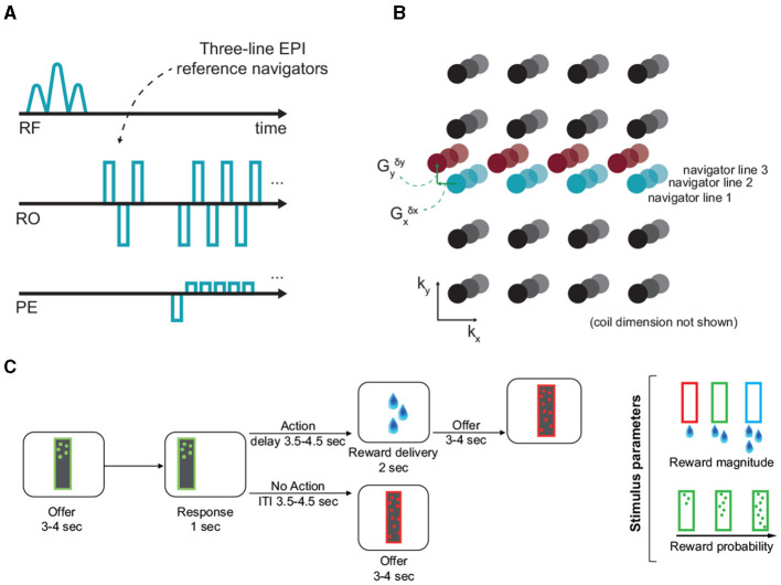
Methods: We correct for dynamic B0 changes in reconstruction of highly accelerated simultaneous multi-slice EPI acquisitions by estimating and correcting for dynamic field perturbations. Functional MRI data were collected in four male rhesus monkeys performing a decision-making task in the scanner, and analyses of improvements in sensitivity and reliability were performed compared to conventional image reconstruction.

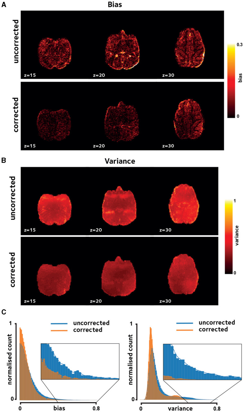
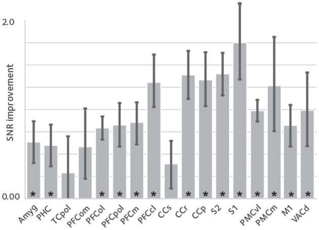
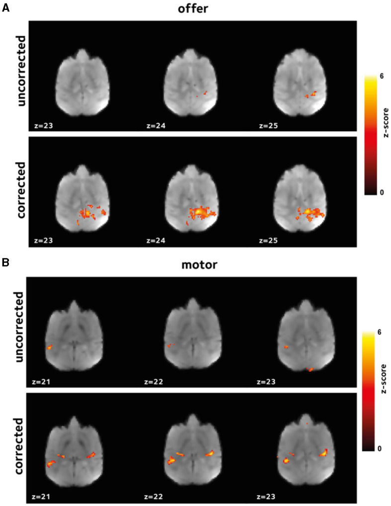
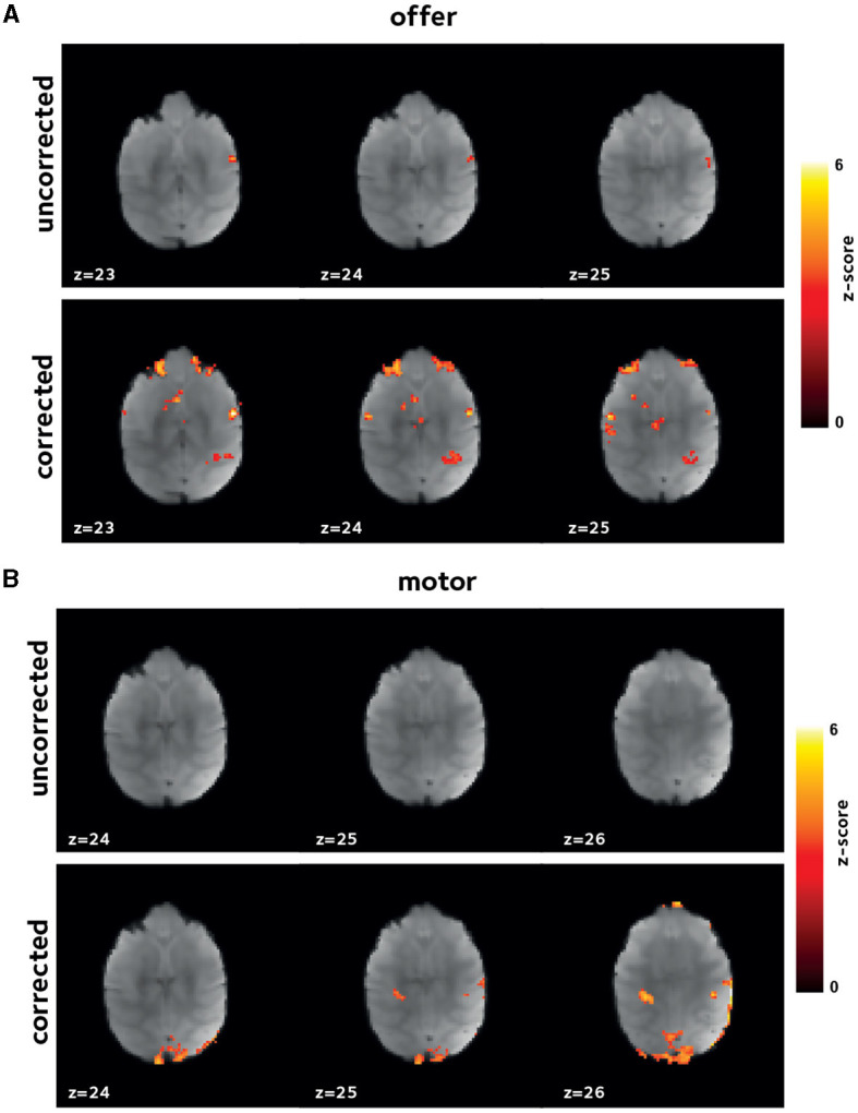
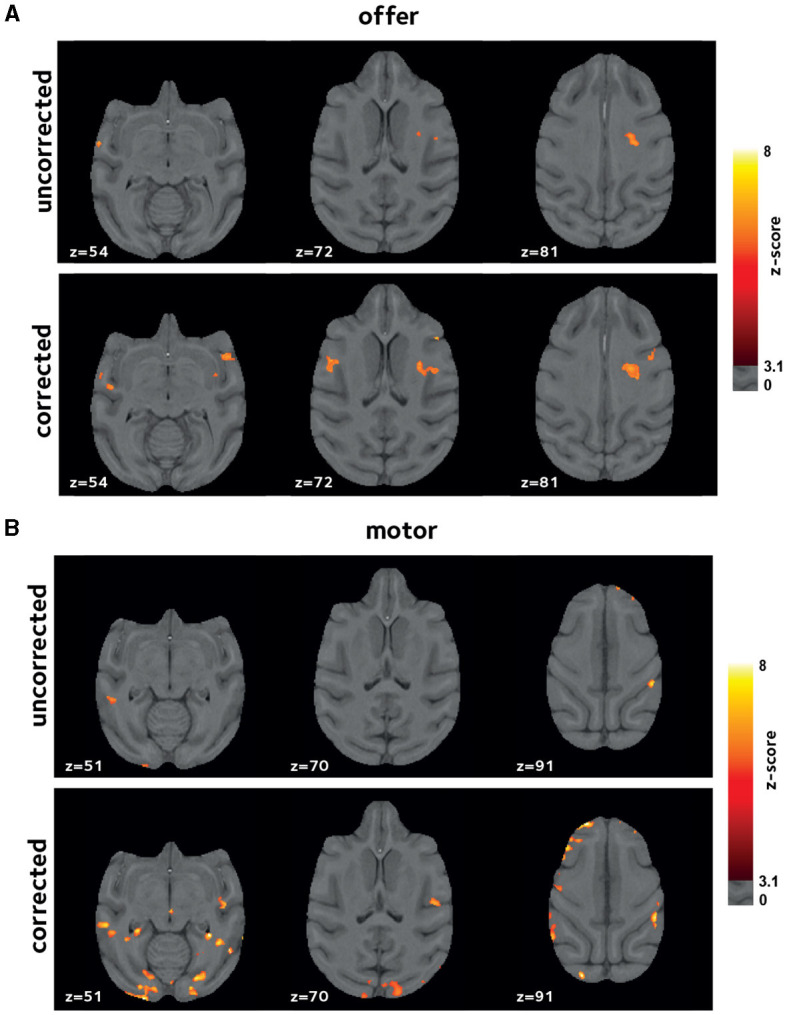
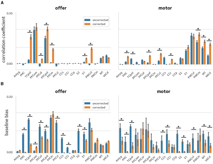
Results: Applying the correction resulted in reduced bias and improved temporal stability in the reconstructed time-series data. We found increased sensitivity to functional activation at the individual and group levels, as well as improved reliability of statistical parameter estimates.

Conclusions: Our results show significant improvements in image fidelity using our proposed correction strategy, as well as greatly enhanced and more reliable activation estimates in GLM analyses.

Keywords: fMRI; non-human primate (NHP); off-resonance artifacts; raw data correction; simultaneous multi-slice.


Conflict of interest statement

The authors declare that the research was conducted in the absence of any commercial or financial relationships that could be construed as a potential conflict of interest.

Figures

Figure 1
Figure 1
Off-resonance correction, and the experimental paradigm. (A, B) EPI reference navigators in each frame are compared to a reference frame to estimate the dynamic linear off-resonance. These estimates are then used to correct the imaging data (Shahdloo et al., 2022). (C) This off-resonance correction method was validated in a decision-making task in awake NHPs, where the animals decide to act based on the number and color of dots appearing on the screen, respond by touching a pad, and receive a liquid reward based on the response. The experimental paradigm involves a wide range of body motion, as well as irregular events.
Figure 2
Figure 2
Reconstruction fidelity. Improvements in image quality were assessed using (A) reconstruction bias and (B) temporal variation, shown for a representative session. Off-resonance correction reduces the reconstruction bias and variance across the brain. (C) Histogram of these measures taken over brain voxels and pooled across sessions in the same representative monkey verifies the observed reduction in bias and variance.
Figure 3
Figure 3
tSNR enhancement. To assess the effect of image quality improvements on the fMRI time series, temporal signal to noise ratio (tSNR) was compared between reconstructions, in anatomical ROIs [mean ± sem; asterisks denote statistical significance at q(FDR) <0.05]. tSNR is significantly improved in most of the studied ROIs that cover the whole brain.
Figure 4
Figure 4
Improvement in first level GLM analysis. GLM models were fit to the data in each session to identify voxels that were activated (A) by offer, and (B) by the motor_response variables. Thresholded z-scores in a representative session are shown. Functional signal quality is improved using the off-resonance correction, as reflected by the larger number of voxels passing the threshold.
Figure 5
Figure 5
Improvement in first level GLM analysis. Thresholded z-score maps in a representative session from a second animal showing activated voxels (A) by offer, and (B) by the motor_response variables. The improvements in functional signal quality are consistent across animals.
Figure 6
Figure 6
Improvements in second-level analysis. A second-level mixed-effects analysis was performed taking scanning sessions as random effect. Thresholded z-score maps are shown indicating activated voxels (A) by offer, and (B) by the motor_response variables. The off-resonance correction significantly improves the results of the higher-level GLM analysis.
Figure 7
Figure 7
Reliability of activation estimates. To quantify the improvements in reliability of the estimated activations, a cross-validation procedure was used were the sessions pooled across animals were randomly divided in two and models were fit in each sub-group separately. Reliability was quantified by taking the (A) correlation, and (B) baseline bias between the estimates in the two sub-groups [asterisks denote statistical significance at q(FDR) <0.05]. The off-resonance correction significantly improves the reliability of activation estimates across the brain.

References

    1. Andersen R. A., Aflalo T., Kellis S. (2019). From thought to action: the brain-machine interface in posterior parietal cortex. Proc. Natl. Acad. Sci. U. S. A. 116, 26274–26279. 10.1073/pnas.1902276116 - DOI - PMC - PubMed
    1. Autio J. A., Glasser M. F., Ose T., Donahue C. J., Bastiani M., Ohno M., et al. . (2020). Towards HCP-Style macaque connectomes: 24-Channel 3T multi-array coil, MRI sequences and preprocessing. NeuroImage 215:116800. 10.1016/j.neuroimage.2020.116800 - DOI - PMC - PubMed
    1. Autio J. A., Zhu Q., Li X., Glasser M. F., Schwiedrzik C. M., Fair D. A., et al. . (2021). Minimal specifications for non-human primate MRI: challenges in standardizing and harmonizing data collection. NeuroImage 236:118082. 10.1016/j.neuroimage.2021.118082 - DOI - PMC - PubMed
    1. Dymerska B., Poser B. A., Bogner W., Visser E., Eckstein K., Cardoso P., et al. . (2016). Correcting dynamic distortions in 7T echo planar imaging using a jittered echo time sequence. Magn. Reson. Med. 76, 1388–1399. 10.1002/mrm.26018 - DOI - PMC - PubMed
    1. Feinberg D. A., Moeller S., Smith S. M., Auerbach E., Ramanna S., Glasser M. F., et al. . (2010). Multiplexed echo planar imaging for sub-second whole brain fMRI and fast diffusion imaging. PLoS ONE 5:e15710. 10.1371/journal.pone.0015710 - DOI - PMC - PubMed

LinkOut - more resources