. 2024 Jul 1;4(7):1881-1893.
doi: 10.1158/2767-9764.CRC-24-0081.

Inhibition of Notch4 Using Novel Neutralizing Antibodies Reduces Tumor Growth in Murine Cancer Models by Targeting the Tumor Endothelium

Affiliations

Inhibition of Notch4 Using Novel Neutralizing Antibodies Reduces Tumor Growth in Murine Cancer Models by Targeting the Tumor Endothelium

Jason W-L Eng et al. Cancer Res Commun. .

Abstract

Endothelial Notch signaling is critical for tumor angiogenesis. Notch1 blockade can interfere with tumor vessel function but causes tissue hypoxia and gastrointestinal toxicity. Notch4 is primarily expressed in endothelial cells, where it may promote angiogenesis; however, effective therapeutic targeting of Notch4 has not been successful. We developed highly specific Notch4-blocking antibodies, 6-3-A6 and humanized E7011, allowing therapeutic targeting of Notch4 to be assessed in tumor models. Notch4 was expressed in tumor endothelial cells in multiple cancer models, and endothelial expression was associated with response to E7011/6-3-A6. Anti-Notch4 treatment significantly delayed tumor growth in mouse models of breast, skin, and lung cancers. Enhanced tumor inhibition occurred when anti-Notch4 treatment was used in combination with chemotherapeutics. Endothelial transcriptomic analysis of murine breast tumors treated with 6-3-A6 identified significant changes in pathways of vascular function but caused only modest change in canonical Notch signaling. Analysis of early and late treatment timepoints revealed significant differences in vessel area and perfusion in response to anti-Notch4 treatment. We conclude that targeting Notch4 improves tumor growth control through endothelial intrinsic mechanisms.

Significance: A first-in-class anti-Notch4 agent, E7011, demonstrates strong antitumor effects in murine tumor models including breast carcinoma. Endothelial Notch4 blockade reduces perfusion and vessel area.


Conflict of interest statement

Y. Kato reports a patent to US9969812B2 issued. Y. Adachi reports personal fees from Eisai Co., Ltd. during the conduct of the study, as well as a patent to US9969812B2 issued. Y. Sakamoto reports a patent to US9969812B2 issued. Y. Nakazawa reports personal fees from Eisai Co., Ltd. during the conduct of the study, as well as a patent to US9969812B2 issued. S. Tachino reports a patent to US9969812B2 issued. K. Ito reports a patent to US9969812B2 issued. T. Abe reports a patent to US9969812B2 issued. H. Ogasawara reports a patent to US9969812B2 issued. Y. Ozawa reports a patent to US9969812B2 issued. T. Imai reports grants and personal fees from Eisai Co., Ltd. outside the submitted work. Y. Funahashi reports personal fees from Eisai Co., Ltd. during the conduct of the study. J. Matsui reports a patent to US9527921B2 licensed. J. Kitajewski reports grants from Eisai Co., Ltd. during the conduct of the study, as well as a patent, US9969812B2, issued to Eisai Co., Ltd. related to this work. No other disclosures were reported.

Figures

Figure 1
Figure 1
Development and validation of 6-3-A6 and E7011 as Notch4 inhibitors. A, Schematic of the immunizing epitope used to generate clone 6-3-A6. EGF, EGF repeats; Igk, immunoglobulin κ chain; SEAP, secreted embryonic alkaline phosphatase. B, Binding assay of the fully humanized E7011 and 6-3-A6 fused to human Fc for immobilization. Affinity to recombinant human Notch4 was calculated with surface plasmon resonance. C, Affinity of 6-3-A6 and E7011 to Raji cell lines generated to overexpress full-length human Notch1, Notch2, Notch3, and Notch4 (hNotch1 to 4, respectively). Binding was determined by MFI with flow cytometry analysis (n = 3). D, Luciferase reporter assay using a Notch4-ECD/Notch1-ICD-Gal4 fusion protein expressed in bEnd.3-immortalized ECs. Luminescence was measured with increasing concentrations of added E7011 or 6-3-A6. Statistical analysis performed with one-way ANOVA with the Dunnett multiple comparison test, plotted as average ± SEM; **, P < 0.01; ****, P < 0.0001.
Figure 2
Figure 2
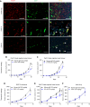
Treatment of human tumor xenografts with 6-3-A6 delays growth of tumors which express endothelial Notch4. A, IF staining of Notch4 (red) and CD31+ tumor vessels (green) in human tumor xenografts. Arrowheads indicate Notch4-expressing vessels (Scale bar = 200 μm). B and C, Tumor growth curves of subcutaneously implanted tumors from the indicated cell lines. B, MDA-MB-231 xenografts treated intravenously with 6-3-A6 (n = 5). C, Calu-6 human NSCLC xenograft tumors treated intravenously with 6-3-A6. All treatments started 12 hours after tumor implantation. Statistical analysis performed with two-way ANOVA for tumor growth curves, plotted as average ± SEM; *, P < 0.05.
Figure 3
Figure 3
6-3-A6 treatment alone or in combination with other chemotherapeutic agents delays tumor growth in human tumor xenografts. A, IF staining of Notch4 (red) and CD31+ tumor vessels (green) in additional human tumor xenografts. B, SEKI melanoma human xenografts treated with 6-3-A6 alone, i.v. bevacizumab alone, or lenvatinib alone (n = 5). C, DU145 prostate xenografts treated with i.v. 6-3-A6, i.v. paclitaxel, or combination (n = 4 mice/group). All treatments started 12 hours after tumor implantation. Statistical analysis performed with two-way ANOVA for tumor growth curves, plotted as average ± SEM; *, P < 0.05; **, P < 0.005; ***, P < 0.001.
Figure 4
Figure 4
Treatment of tumors with 6-3-A6/E7011 delays tumor growth in syngeneic tumor models which express endothelial Notch4. A, IF staining of murine Lewis lung carcinoma, murine Py8119 breast carcinoma, and murine B16F10 melanoma with Notch4 and CD31. Arrowheads indicate Notch4-expressing vessels (scale bar = 200 μm). B and C, Preventative strategy using Py8119 tumor cells orthotopically implanted into the mammary fat pad and treated with B, E7011 (n = 18 human IgG, n = 17 E7011) or C, 6-3-A6 (n = 4) twice weekly starting 12 hours following tumor implantation. D and F, Tumor growth curve of subcutaneously implanted D, Lewis lung (n = 4) and E, B16F10 (n = 7) treated intravenously with E7011 (25 mg/kg twice weekly) and orthotopically implanted F, Py8119 murine breast carcinomas (n = 5) treated intravenously with 6-3-A6 (25 mg/kg twice weekly) once the average tumor volume of each cohort had reached approximately 30 mm3. Statistical analysis performed with two-way ANOVA for tumor growth curves, plotted as average ± SEM; *, P < 0.05; ***, P < 0.001. TNBC, triple-negative breast cancer.
Figure 5
Figure 5
Notch4 is expressed on tumor ECs, and treatment alters the tumor vessel transcriptional profile. A, Representative flow plots of gating for CD31+ cells in Py8119 and B16F10 tumors. B, Histograms comparing E7011 and human IgG binding to CD31+ and CD31 cell populations from dissociated Py8119 and B16F10 tumors (n = 3). Left, E7011 samples were concatenated and merged into a single representative histogram. Right, Graph of concatenated MFI from Py8119 and B16F10 samples bound with E7011 or human IgG. C, Heatmaps of significantly upregulated and downregulated differentially expressed genes isolated from IP polysomes from tumor ECs treated with 6-3-A6 or control IgG (n = 3). Boxes indicate genes implicated in GPCR control of angiogenesis (red), endothelial nitric oxide regulation (blue), and inflammatory/immune response (green). D, Reactome pathway analysis of upregulated and downregulated pathways was performed using g:Profiler, and significance threshold by Benjamini–Hochberg FDR was set at 0.05. A, Fold cutoff of −log10(Padj) > 2 was used to select the displayed pathways. Statistical analysis performed with the t test for perfusion studies, plotted as average ± SEM; *, P < 0.05; **, P < 0.005. Heatmap data presented as Z-scores, which integrate both fold change and significance.
Figure 6
Figure 6
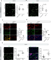
Anti-Notch4 treatment alters vessel perfusion and angiogenesis. A and B, Assessment of vessel coverage area by CD31+ staining in A, B16F10 melanoma treated for 4 days with two doses (days 1 and 3) of 6-3-A6 (n = 3) and B, Py8119 tumors treated for 10 days with four doses (days 1, 3, 6, and 9) of 6-3-A6 (n = 6). Right, Representative images; left, quantification of colocalization. (Scale bar = 100 μm). C and D, Assessment of tumor perfusion in syngeneic C, B16F10 melanoma (n = 3) and D, Py8119 tumors (n = 6) using intravenously injected fluorescent tomato lectin–Alex Fluor 488. Right, Representative images costained with CD31 (red); left, quantification of colocalization. (Scale bar = 100 μm). E and F, IF staining of E, CD31+ tumor vessels in orthotopically implanted Py8119 tumors treated for 4 weeks with E7011 (n = 4), and F, vessel perfusion by fluorescent tomato lectin–Alexa Fluor 647 labeling with CD31 in Py8119 tumors treated for an extended duration (n = 5). Right, Representative images; left, quantification of the staining or colocalization. (Scale bar = 200 μm). Statistical analysis by the t test, plotted as average ± SEM; *, P < 0.05.

References

    1. Bolós V, Grego-Bessa J, de la Pompa JL. Notch signaling in development and cancer. Endocr Rev 2007;28:339–63. - PubMed
    1. Uyttendaele H, Marazzi G, Wu G, Yan Q, Sassoon D, Kitajewski J. Notch4/int-3, a mammary proto-oncogene, is an endothelial cell-specific mammalian Notch gene. Development 1996;122:2251–9. - PubMed
    1. Sargis T, Youn S-W, Thakkar K, Naiche LA, Paik NY, Pajcini KV, et al. . Notch1 and Notch4 core binding domain peptibodies exhibit distinct ligand-binding and anti-angiogenic properties. Angiogenesis 2023;26:249–63. - PMC - PubMed
    1. Sprinzak D, Blacklow SC. Biophysics of notch signaling. Annu Rev Biophys 2021;50:157–89. - PMC - PubMed
    1. Naiche LA, Villa SR, Kitajewski JK. Endothelial cell fate determination: a top notch job in vascular decision-making. Cold Spring Harb Perspect Med 2022;12:a041183. - PMC - PubMed

Publication types

MeSH terms