

Review
doi: 10.1107/S2059798324005588.
Epub 2024 Jul 10.
A snapshot love story: what serial crystallography has done and will do for us
Affiliations
- PMID: 38984902
- PMCID: PMC11301758
- DOI: 10.1107/S2059798324005588
Item in Clipboard
Review
A snapshot love story: what serial crystallography has done and will do for us
Acta Crystallogr D Struct Biol.
.
Abstract
Serial crystallography, born from groundbreaking experiments at the Linac Coherent Light Source in 2009, has evolved into a pivotal technique in structural biology. Initially pioneered at X-ray free-electron laser facilities, it has now expanded to synchrotron-radiation facilities globally, with dedicated experimental stations enhancing its accessibility. This review gives an overview of current developments in serial crystallography, emphasizing recent results in time-resolved crystallography, and discussing challenges and shortcomings.
Keywords: serial crystallography; structural dynamics; time-resolved.
open access.
Figures

Diagram of a serial crystallography experiment using either an X-ray free-electron laser (XFEL) or synchrotron radiation. Protein crystals are delivered to the X-ray beam in random orientations. The intense X-rays interact with the crystals, producing diffraction patterns that are recorded by a detector. Every crystal is only probed once. Time-resolved experiments can be conducted either by light activation using a laser or by chemical activation. In the latter case the reactant is mixed with the protein crystals at a defined time delay before probing the crystals with X-rays. (Created with BioRender.com.)
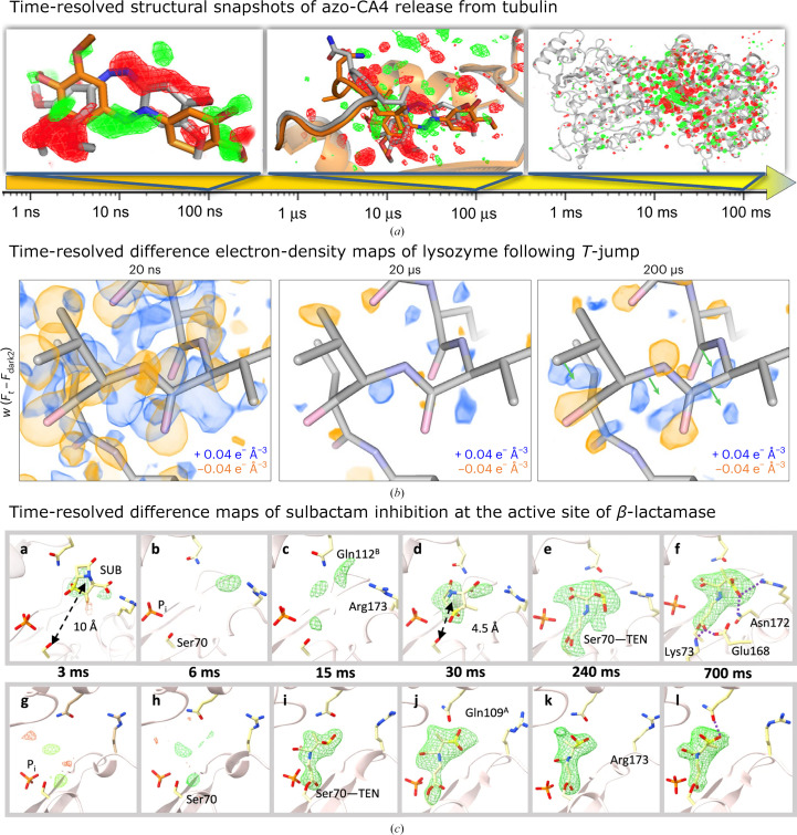
Examples of results from time-resolved SX using three different methods for reaction initiation. (a) Time-resolved structural snapshots of the release of azo-CA4 from tubulin (Wranik, Weinert et al., 2023 ▸). The time arrow depicts the investigated time regime. The panels from left to right show the isomorphous difference maps obtained at 100 ns with changes centered on the ligand, 100 µs with changes centered on the binding pocket and 100 ms with conformational changes propagating throughout the protein. All panels show isomorphous difference maps in red (negative) and green (positive) at 3σ. The structure in the given time range (colored orange) is compared with that in the previous time range (colored gray). (b) shows the time-resolved difference electron-density (DED) maps of lysozyme following a temperature jump (T-jump; Wolff et al., 2023 ▸). Weighted DED maps are depicted for each time point around residues 97–100 of lysozyme visualized at an absolute contour level of ±0.04 e− Å−3. Positive DED is displayed as a blue map and the negative DED map is shown in orange. The green arrows depicted at the 200 µs time step indicate proposed coordinate motions derived from analysis of the DED. (c) Difference electron-density maps around the active center of both subunits (subunit A, top row; subunit B, lower row) of a lactamase at different time points of a mix-and-inject time-resolved experiment with the lactamase inhibitor sulbactam (Malla et al., 2023 ▸). Omit maps are shown in all subpanels except for subpanels d, f, j and l, which show polder maps (contour levels ±3σ). SUB, sulbactam; TEN, trans-enamine. Images and captions were taken and adapted from (a) Wranik, Weinert et al. (2023 ▸), (b) Wolff et al. (2023 ▸) and (c) Malla et al. (2023 ▸), licensed under a Creative Commons Attribution 4.0 International License (https://creativecommons.org/licenses/by/4.0/ ).

High-throughput compatibility of various sample-delivery methods for SSX/SFX compared with rotational macromolecular crystallography (MX) and how these scale with detector developments towards higher frame rates. The assumptions made were that 10 000 indexed frames are required for an SSX/SFX data set and that 1800 images need to be recorded for a data set from rotational MX. Furthermore, based on the literature (Bosman et al., 2024; Zielinski et al., 2022 ▸), we assumed a 100% indexing rate (indexable images per recorded images) for fixed-target sample delivery and tape drive/microfluidics. For liquid jets indexing rates of greater than 80% have been reported (Williamson et al., 2023 ▸), but showing two cases, based on either a 10% or 50% indexing rate, seemed to be more realistic for most experiments. For rotational MX and fixed targets, we assumed the time it takes a robotic arm to exchange the sample to be 20 s. In the case of rotational MX we factored in another 10 s for crystal centring for each data set. For the tape drive/microfluidics we assumed 2 h downtime per day for the exchange of sample/tape/microfluidics. For all jets and tape drive/microfluidics in the 100 kHz detector case, we assumed 6 h of downtime per day. The number of data sets per day is plotted on a logarithmic scale. As can be seen, multi-compartment fixed targets and high-indexing jets are excellent in terms of high-throughput capability up to a 10 kHz frame rate. Single-compartment fixed-target chips and rotational MX are already not now competitive when detectors that are already available, such as JUNGFRAU or PILATUS4, are being used. Note that for SSX/SFX experiments further developments are required in order to unlock the full potential in terms of high-throughput capability. (Created with BioRender.com.)

Development of the number of new PDB entries per year from SFX/SSX experiments (blue line) and the number of publications mentioning ‘serial crystallography’ since 2010 (pink line). The number of publications is based on a query using Google Scholar. For the number of PDB entries the deposition date, rather than the release date, was used.
Similar articles
-
Use of fixed targets for serial crystallography.Methods Enzymol. 2024;709:29-55. doi: 10.1016/bs.mie.2024.10.002. Epub 2024 Oct 19. Methods Enzymol. 2024. PMID: 39608947
-
Macromolecular crystallography and biology at the Linac Coherent Light Source.J Synchrotron Radiat. 2025 May 1;32(Pt 3):548-566. doi: 10.1107/S1600577525002735. Epub 2025 Apr 23. J Synchrotron Radiat. 2025. PMID: 40266725 Free PMC article.
-
Opportunities and challenges for time-resolved studies of protein structural dynamics at X-ray free-electron lasers.Philos Trans R Soc Lond B Biol Sci. 2014 Jul 17;369(1647):20130318. doi: 10.1098/rstb.2013.0318. Philos Trans R Soc Lond B Biol Sci. 2014. PMID: 24914150 Free PMC article. Review.
-
Dynamic Structural Biology Experiments at XFEL or Synchrotron Sources.Methods Mol Biol. 2021;2305:203-228. doi: 10.1007/978-1-0716-1406-8_11. Methods Mol Biol. 2021. PMID: 33950392 Review.
-
Crystal structure of a bacterial photoactivated adenylate cyclase determined by serial femtosecond and serial synchrotron crystallography.IUCrJ. 2024 Nov 1;11(Pt 6):991-1006. doi: 10.1107/S2052252524010170. IUCrJ. 2024. PMID: 39470573 Free PMC article.
Cited by
-
Present and future structural biology activities at DESY and the European XFEL.J Synchrotron Radiat. 2025 Mar 1;32(Pt 2):474-485. doi: 10.1107/S1600577525000669. Epub 2025 Feb 18. J Synchrotron Radiat. 2025. PMID: 39964790 Free PMC article.
-
Microcrystals in structural biology: small samples, big insights.IUCrJ. 2025 May 1;12(Pt 3):259-261. doi: 10.1107/S2052252525003653. IUCrJ. 2025. PMID: 40293197 Free PMC article.
-
Advancing time-resolved structural biology: latest strategies in cryo-EM and X-ray crystallography.Nat Methods. 2025 Jul;22(7):1420-1435. doi: 10.1038/s41592-025-02659-6. Epub 2025 May 1. Nat Methods. 2025. PMID: 40312512 Review.
-
Scalable fabrication of an array-type fixed-target device for automated room temperature X-ray protein crystallography.Sci Rep. 2025 Jan 2;15(1):334. doi: 10.1038/s41598-024-83341-3. Sci Rep. 2025. PMID: 39747265 Free PMC article.
References
-
- Austin, R. H., Beeson, K. W., Eisenstein, L., Frauenfelder, H. & Gunsalus, I. C. (1975). Biochemistry, 14, 5355–5373. - PubMed
-
- Barends, T. R., Foucar, L., Ardevol, A., Nass, K., Aquila, A., Botha, S., Doak, R. B., Falahati, K., Hartmann, E., Hilpert, M., Heinz, M., Hoffmann, M. C., Köfinger, J., Koglin, J. E., Kovacsova, G., Liang, M., Milathianaki, D., Lemke, H. T., Reinstein, J., Roome, C. M., Shoeman, R. L., Williams, G. J., Burghardt, I., Hummer, G., Boutet, S. & Schlichting, I. (2015). Science, 350, 445–450. - PubMed
-
- Barends, T. R. M., Gorel, A., Bhattacharyya, S., Schirò, G., Bacellar, C., Cirelli, C., Colletier, J.-P., Foucar, L., Grünbein, M. L., Hartmann, E., Hilpert, M., Holton, J. M., Johnson, P. J. M., Kloos, M., Knopp, G., Marekha, B., Nass, K., Nass Kovacs, G., Ozerov, D., Stricker, M., Weik, M., Doak, R. B., Shoeman, R. L., Milne, C. J., Huix-Rotllant, M., Cammarata, M. & Schlichting, I. (2024). Nature, 626, 905–911. - PMC - PubMed
Publication types
MeSH terms
Substances
LinkOut - more resources
Full Text Sources
