. 2024 Jul 22;63(29):13191-13196.
doi: 10.1021/acs.inorgchem.4c01991. Epub 2024 Jul 10.

Active Site Characterization of a Campylobacter jejuni Nitrate Reductase Variant Provides Insight into the Enzyme Mechanism

Affiliations

Active Site Characterization of a Campylobacter jejuni Nitrate Reductase Variant Provides Insight into the Enzyme Mechanism

Jing Yang et al. Inorg Chem. .

Abstract

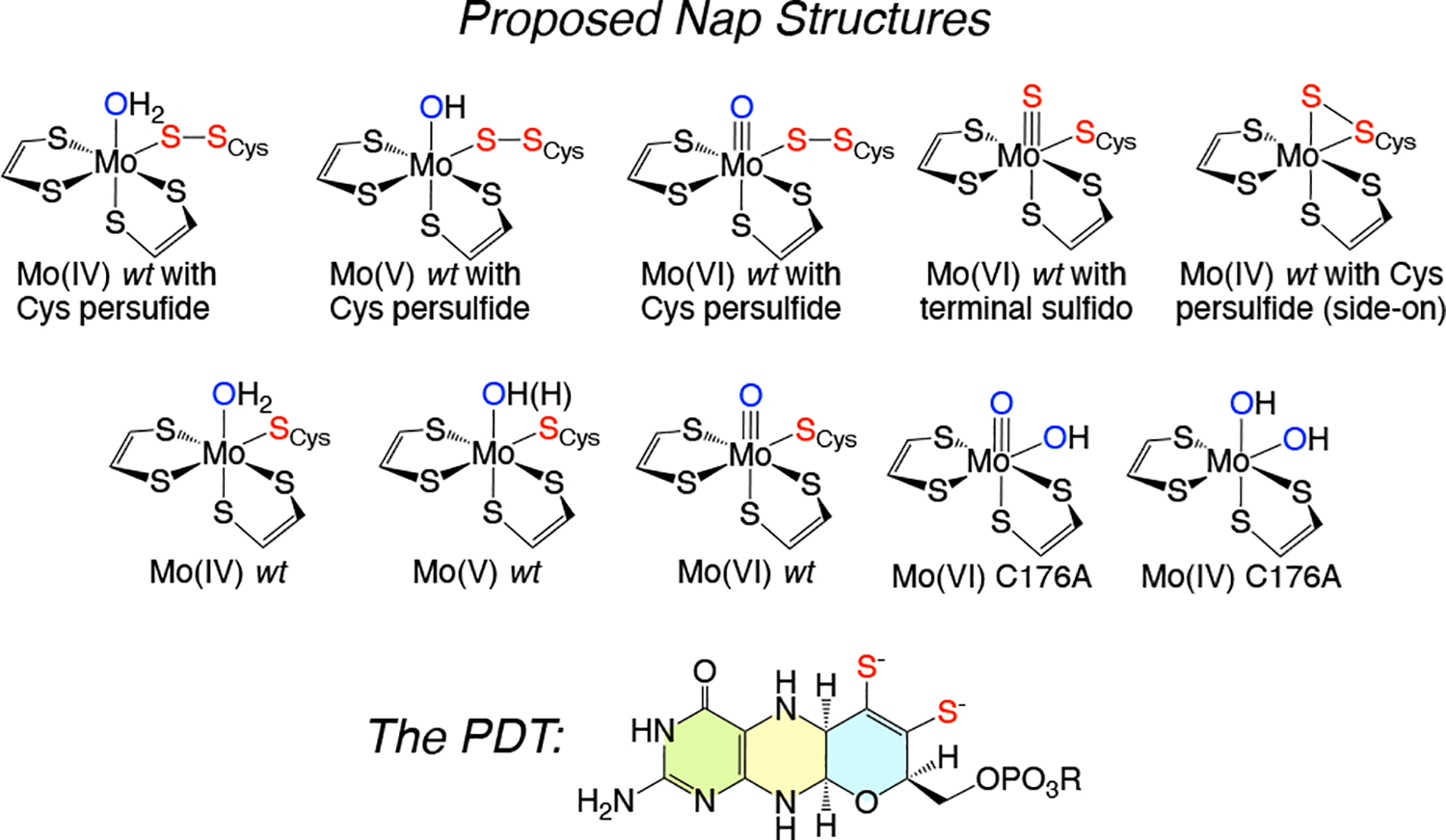
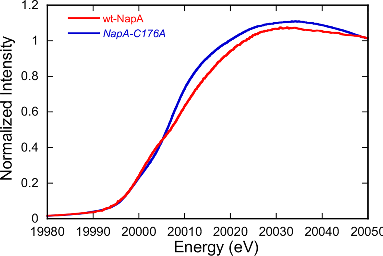
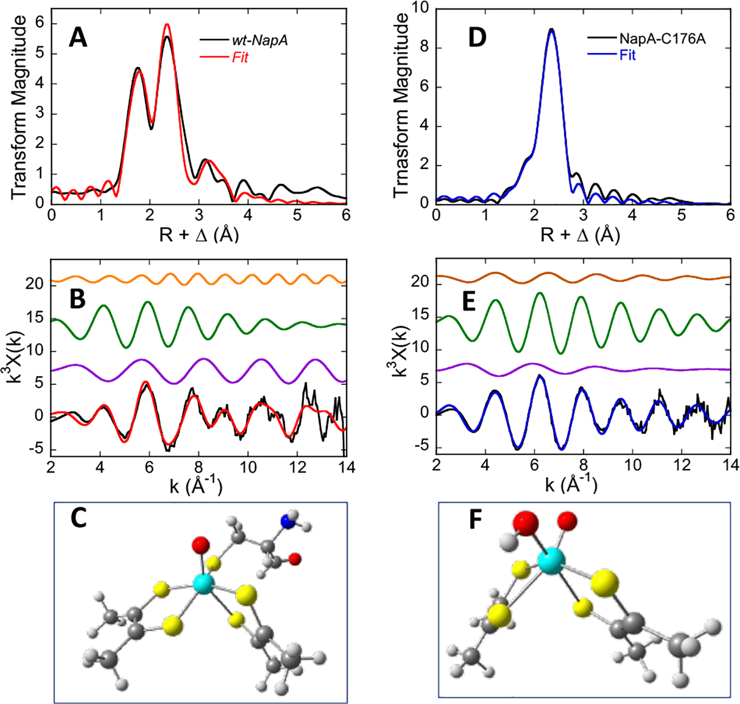
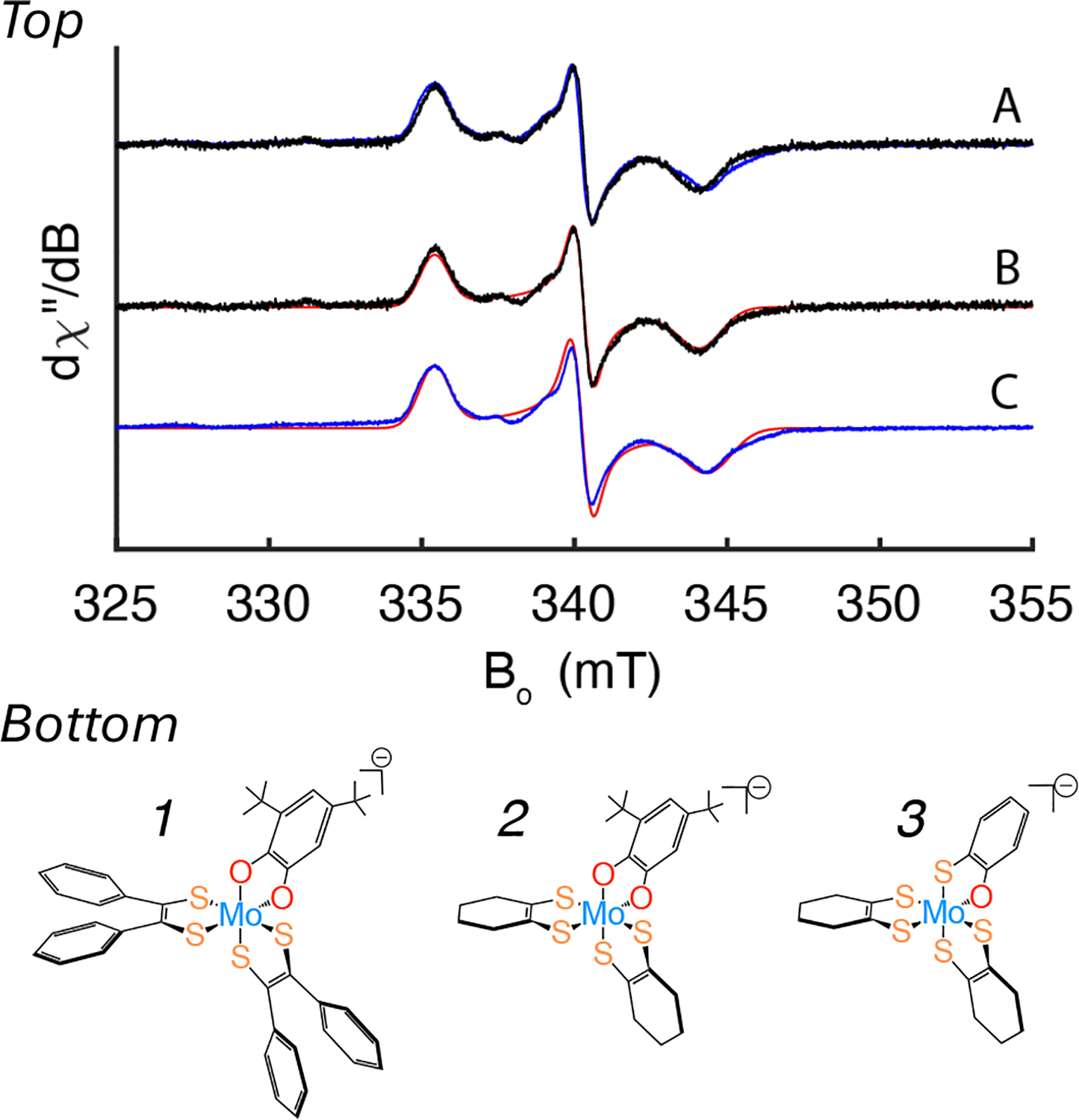
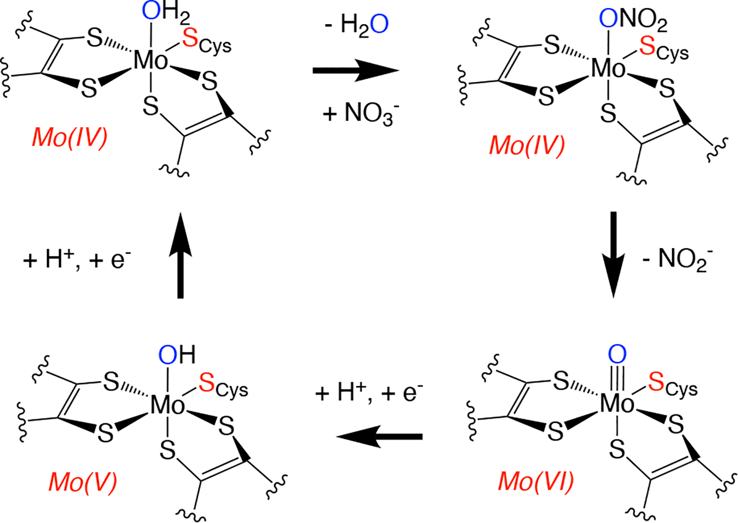
Mo K-edge X-ray absorption spectroscopy (XAS) is used to probe the structure of wild-type Campylobacter jejuni nitrate reductase NapA and the C176A variant. The results of extended X-ray absorption fine structure (EXAFS) experiments on wt NapA support an oxidized Mo(VI) hexacoordinate active site coordinated by a single terminal oxo donor, four sulfur atoms from two separate pyranopterin dithiolene ligands, and an additional S atom from a conserved cysteine amino acid residue. We found no evidence of a terminal sulfido ligand in wt NapA. EXAFS analysis shows the C176A active site to be a 6-coordinate structure, and this is supported by EPR studies on C176A and small molecule analogs of Mo(V) enzyme forms. The SCys is replaced by a hydroxide or water ligand in C176A, and we find no evidence of a coordinated sulfhydryl (SH) ligand. Kinetic studies show that this variant has completely lost its catalytic activity toward nitrate. Taken together, the results support a critical role for the conserved C176 in catalysis and an oxygen atom transfer mechanism for the catalytic reduction of nitrate to nitrite that does not employ a terminal sulfido ligand in the catalytic cycle.


Figures

Figure 1.
Figure 1.
Top: Selected structures that have been previously proposed for sulfido-based nitrate reductase enzyme forms. Middle: Selected structures proposed for nonsulfido-based NapA enzyme forms. Bottom generalized structure of the reduced PDT ligand.
Figure 2.
Figure 2.
Mo K-edge XANES spectra for nitrate-treated wt-Cj NapA and its as isolated NapA-C176A variant. The rising edge energies, defined as the inflection point of the first derivative, are observed at 20014.0 eV for oxidized wt-NapA and 20011.4 eV for NapA-C176A variant.
Figure 3.
Figure 3.
Mo K-edge EXAFS data for oxidized wt-NapA (A and C) and the NapA-C176A variant (B and D). The experimental data are shown in black, and the best fits to the data are in red for wt-NapA and blue for NapA-C176A. Four Mo-C scatters from the dithiolene C=C carbon atoms of the two PDT ligands have been included in the fit for wt-NapA, yielding Mo-C vectors at an average distance of 3.339 Å. The observed oscillations arise from terminal oxo, S, C (second coordination sphere), and OH scatterers, and are colored in purple, green, orange, and brown, respectively. The computational models used as initial guesses in the fitting procedure are given in C and F. The XAS data for the wt protein was obtained by incubating the enzyme with nitrate.
Figure 4.
Figure 4.
Top: X-band EPR spectra of the as-isolated NapA C176A variant in H2O (Black) and D2O (Blue) (50 mM HEPES pH 7.4) with simulations of the spectra in red. Enzyme concentrations are approximately 200 μM. The spectra in H2O and D2O buffer are virtually identical (A) and show no evidence for strongly coupled protons. EPR spin quantitation yields ~ 10–20% of the variant being in the Mo5+ oxidation state (see SI). The g-values determined from spectral simulations are g1,2,3 = 2.0170, 1.9876, 1.9644 in H2O sample (B) and g1,2,3 = 2.0178, 1.9888, 1.9645 in D2O (C). Bottom: Bond line drawings for small molecule analogs of the Mo5+ forms of C176A NapA (1-2) and wt-NapA (3).
Figure 5.
Figure 5.
Proposed NapA oxygen atom transfer mechanism based on the results of this study.

Similar articles

Cited by

References

    1. Cerqueira N; Fernandes PA; Gonzalez PJ; Moura JJG; Ramos MJ The Sulfur Shift: An Activation Mechanism for Periplasmic Nitrate Reductase and Formate Dehydrogenase. Inorg. Chem 2013, 52 (19), 10766–10772. DOI: 10.1021/ic3028034. - DOI - PubMed
    1. Sparacino-Watkins C; Stolz JF; Basu P Nitrate and periplasmic nitrate reductases. Chem. Soc. Rev 2014, 43 (2), 676–706. DOI: 10.1039/c3cs60249d. - DOI - PMC - PubMed
    1. Cerqueira N; Gonzalez PJ; Fernandes PA; Moura JJG; Ramos MJ Periplasmic Nitrate Reductase and Formate Dehydrogenase: Similar Molecular Architectures with Very Different Enzymatic Activities. Acc. Chem. Res 2015, 48 (11), 2875–2884. DOI: 10.1021/acs.accounts.5b00333. - DOI - PubMed
    1. Hille R; Hall J; Basu P The Mononuclear Molybdenum Enzymes. Chemical Reviews 2014, 114 (7), 3963–4038. DOI: 10.1021/cr400443z. - DOI - PMC - PubMed
    1. Hille R; Schulzke C; Kirk ML Molybdenum and Tungsten Enzymes. In RSC Metallobiology Series, Garner CD, Sun H, Wedd A, Ciurli SL, Eds.; The Royal Society of Chemistry: Cambridge, UK, 2017.

MeSH terms

Substances

LinkOut - more resources