Review
. 2025 Jan;50(1):290-304.
doi: 10.1007/s00261-024-04491-6. Epub 2024 Jul 10.

Urologic prosthetics: an imaging review of short- and long-term complications

Affiliations
Review

Urologic prosthetics: an imaging review of short- and long-term complications

Jared Raikin et al. Abdom Radiol (NY). 2025 Jan.

Abstract

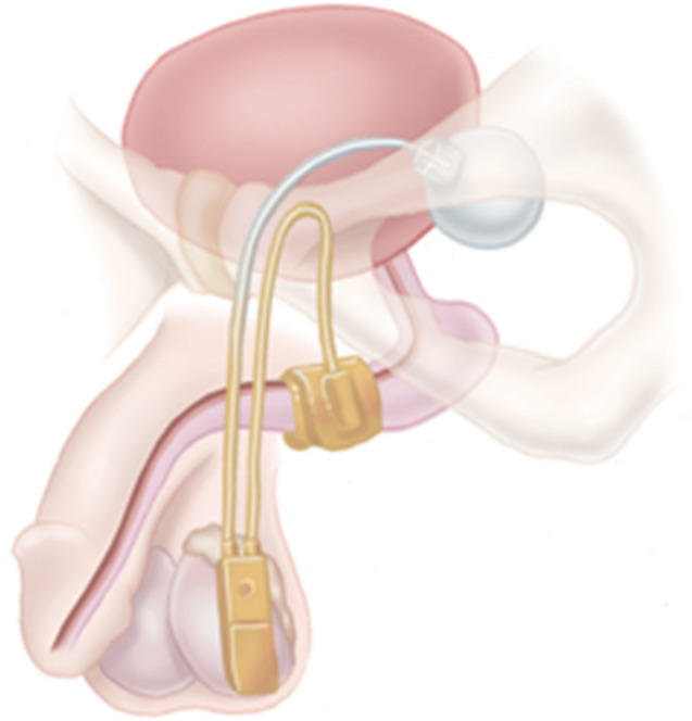
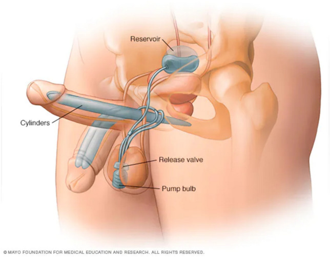
Purpose: Urologic prosthetics offer significant quality of life enhancements for patients with stress urinary incontinence and erectile dysfunction. Artificial urinary sphincter and penile prosthesis are the most commonly used prosthetics for these patients. Radiographic imaging offers important insight, guiding treatment when patients present with complications. Herein, we pictorialize normal radiographic findings and complications alike.

Methods: We reviewed our IRB-approved prosthetics database, highlighting patients with prosthetic complications with available imaging. We collected imaging from patients without complications for baseline reference.

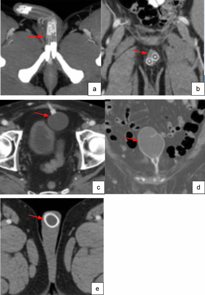
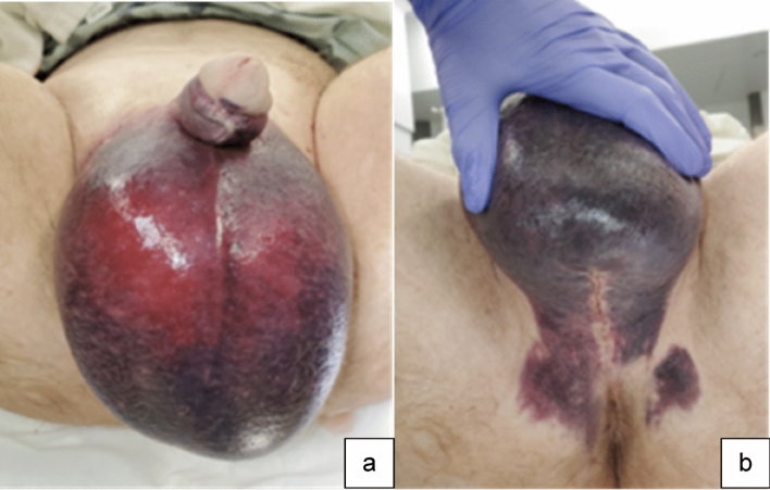
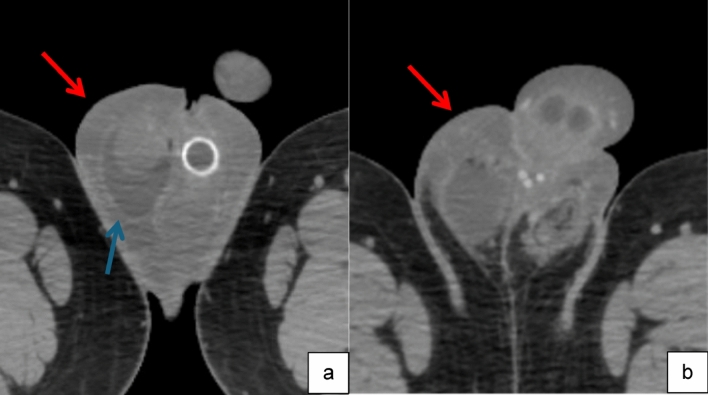
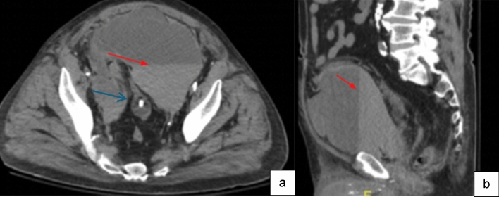
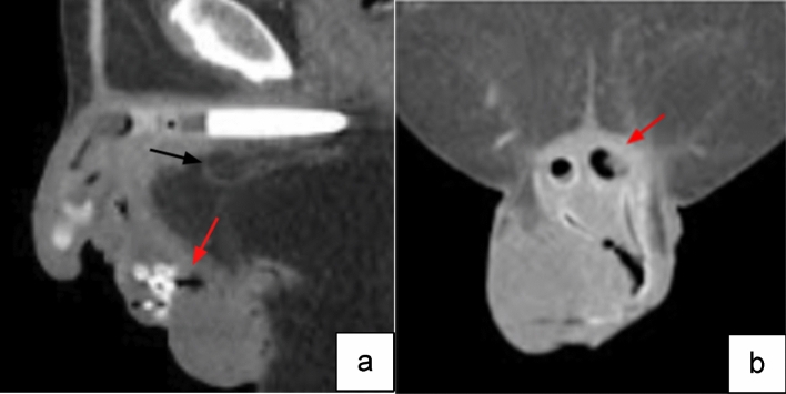
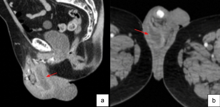
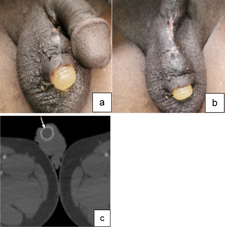
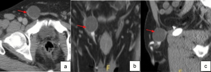
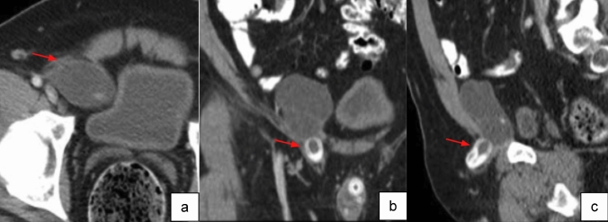
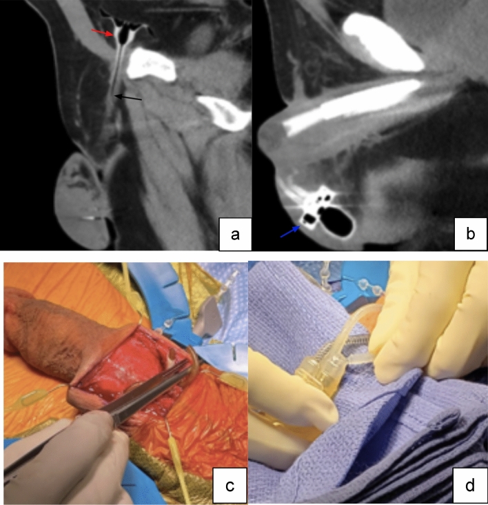
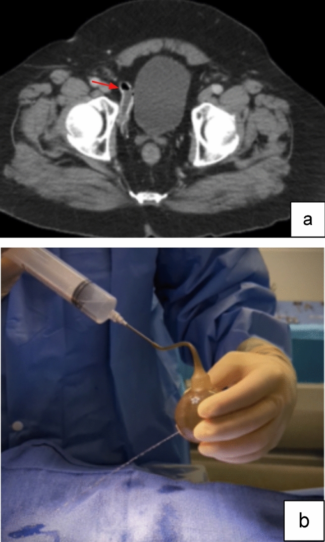
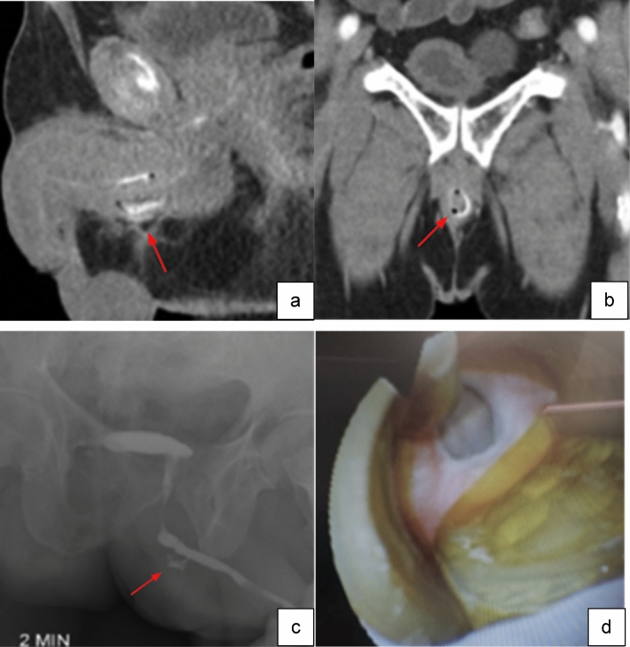
Results: The radiographic appearance of orthotopic genitourinary prosthetics and a review of short- and long-term complications including hematoma, infection, malpositioning, leak and erosion are pictorialized.

Conclusion: Radiologic imaging serves as a vital complement to history and physical examination, aiding in the identification of complications and potentially streamlining surgical preparations. It is important for radiologists to familiarize themselves with standard prosthetic nomenclature, normal positioning and appearance, along with imaging findings of common complications.

Keywords: Complications; Genitourinary; Prosthetics; Review; Urology.


Conflict of interest statement

Declarations. Competing interests: The authors have not disclosed any competing interests.

Figures

Fig. 1
Fig. 1
Image of artificial urinary sphincter AMS 800 includes infrapubic PRB, urethral cuff, and scrotal pump [15]. Image adapted from Boston Scientific
Fig. 2
Fig. 2
Coronal (a), and sagittal (b) non-contrast CT images of normal AUS pump location in the lateral scrotum (red arrow). Coronal (c), sagittal (d) and axial (e) non-contrast CT images of normal urethral cuff around the bulbar urethra (red arrows)
Fig. 3
Fig. 3
Image of a 3-piece inflatable penile prosthesis with a reservoir, 2 cylinders, and a pump [19]. Image adapted from Mayo Clinic
Fig. 4
Fig. 4
Axial (a) and coronal (b) CT images with IV contrast of normal three-piece PP paired inflatable cylinders in corpora cavernosa (red arrows). Axial (c) and coronal (d) CT images of normal PP abdominal placement of reservoir (red arrows). Axial (e) CT image of normal PP pump location in the midline inferior scrotum (red arrow)
Fig. 5
Fig. 5
Clinical photo demonstrating scrotal (a) and perineal (b) enlargement and skin discoloration due to postoperative scrotal hematoma
Fig. 6
Fig. 6
Axial images inferior (a) and superior (b) from CT pelvis with IV contrast show diffuse penoscrotal skin thickening and subcutaneous edema (red arrow) without discrete hematoma or rim-enhancing fluid collection. Small right hydrocele is incidentally noted (blue arrow). Portion of a PP pump is visible
Fig. 7
Fig. 7
Axial (a) and sagittal (b) non-contrast CT images, showing large fluid collection in the pelvis with fluid–fluid level, representing layering hematocrit level (red arrows), and compatible with postoperative hematoma. This collection produced significant regional mass effect, displacing the urinary bladder, decompressed around the foley catheter, posterior in the pelvis (blue arrow)
Fig. 8
Fig. 8
Sagittal (a) and coronal (b) CT images demonstrate gas (red arrows) within the scrotum and corpora cavernosa components of the PP with surrounding fat stranding (black arrows) compatible with infection
Fig. 9
Fig. 9
Sagittal (a) and axial (b) CT images with IV contrast show an intrascrotal peripherally enhancing fluid collection (red arrows), consistent with an abscess
Fig. 10
Fig. 10
Photos show skin breakdown with the penile pump prosthesis protruding through the skin of the scrotum (a, b). Axial CT image with IV contrast (c) shows absence of skin barrier overlying the pump (white arrow)
Fig. 11
Fig. 11
Axial (a), coronal (b) and sagittal (c) CT images with IV contrast show reservoir of IPP in the inguinal canal (red arrows)
Fig. 12
Fig. 12
Axial (a), coronal (b) and sagittal (c) CT images with IV contrast show IPP reservoir partially extending into the inguinal canal (red arrows)
Fig. 13
Fig. 13
Sagittal (a) and axial (b) CT images without contrast demonstrate a compressed PP reservoir containing air (red arrow), fractured free-floating tubing (black arrow), and air within the scrotal pump (blue arrow). Intraoperative photos of penoscrotal incision at time of repair demonstrate multiple fractures of prosthetic connection tubing (c, d)
Fig. 14
Fig. 14
Axial CT image with IV contrast (a) shows a collapsed reservoir with an air-fluid level, compatible with system leak (red arrow). Intraoperative photo demonstrates pinpoint fluid leak from reservoir (b)
Fig. 15
Fig. 15
Sagittal (a) and coronal (b) CT images with IV contrast demonstrate foci of gas and fat stranding at the base of the penis concerning for cuff erosion (red arrows). Portable radiograph after retrograde urethrogram (c) shows contrast extravasation at anterior penile urethra in the region of foci of gas on CT (red arrow). Intraoperative cystoscopic view (d) of circumferential artificial urinary sphincter erosion

Similar articles

References

    1. Rodriguez KM, Pastuszak AW (2017) A history of penile implants. Transl Androl Urol. 6(Suppl 5):S851-s857. doi:10.21037/tau.2017.04.02 - PMC - PubMed
    1. Cordon BH, Singla N, Singla AK (2016) Artificial urinary sphincters for male stress urinary incontinence: current perspectives. Med Devices (Auckl) 9:175-83. doi:10.2147/mder.S93637 - PMC - PubMed
    1. Grewal S, Vetter J, Brandes SB, Strope SA (2014) A population-based analysis of contemporary rates of reoperation for penile prosthesis procedures. Urology 84(1):112-6. doi:10.1016/j.urology. - PMC - PubMed
    1. Baas W, O'Connor B, Welliver C, et al (2020) Worldwide trends in penile implantation surgery: data from over 63,000 implants. Transl Androl Urol 9(1):31-37. doi:10.21037/tau.2019.09.26 - PMC - PubMed
    1. Luber KM (2004) The definition, prevalence, and risk factors for stress urinary incontinence. Rev. in urology 6(Suppl 3):S3. - PMC - PubMed

Publication types

MeSH terms

LinkOut - more resources