Review
. 2024 Jul 17;481(14):903-922.
doi: 10.1042/BCJ20210352.

The C-terminal sequences of Bcl-2 family proteins mediate interactions that regulate cell death

Affiliations
Review

The C-terminal sequences of Bcl-2 family proteins mediate interactions that regulate cell death

Dang Nguyen et al. Biochem J. .

Abstract

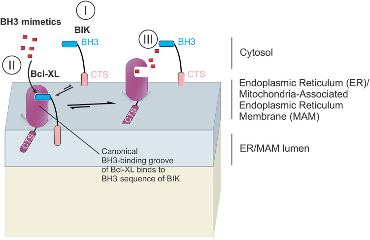
Programmed cell death via the both intrinsic and extrinsic pathways is regulated by interactions of the Bcl-2 family protein members that determine whether the cell commits to apoptosis via mitochondrial outer membrane permeabilization (MOMP). Recently the conserved C-terminal sequences (CTSs) that mediate localization of Bcl-2 family proteins to intracellular membranes, have been shown to have additional protein-protein binding functions that contribute to the functions of these proteins in regulating MOMP. Here we review the pivotal role of CTSs in Bcl-2 family interactions including: (1) homotypic interactions between the pro-apoptotic executioner proteins that cause MOMP, (2) heterotypic interactions between pro-apoptotic and anti-apoptotic proteins that prevent MOMP, and (3) heterotypic interactions between the pro-apoptotic executioner proteins and the pro-apoptotic direct activator proteins that promote MOMP.

Keywords: BH3-only proteins; Bax; Bcl-2; apoptosis; protein–protein interactions; transmembrane domain.


Conflict of interest statement

The authors declare that there are no competing interests associated with the manuscript.

Figures

Figure 1.
Figure 1.. The CTSs of Bax and Bak regulate their pore-forming functions.
(A) The CTS and BH3 of Bim are required for Bim to bind Bax in the cytosol [15] (l-a) and on the MOM (l-b). It is unknown where the Bim CTS binds on Bax. Binding of Bim BH3 to the trigger site of Bax induces conformational changes that displace the CTS of Bax from the canonical BH3-binding groove enabling Bax to bind to the MOM (II) and expose the BH3 of Bax [78] (III). As a result, the canonical BH3-binding groove of Bax is accessible for binding a Bim BH3 sequence. This leads to further conformational changes in Bax (core-latch domain separation [71]) (V) enabling Bax to dimerize with the BH3 of one Bax inserted into the canonical BH3-binding groove of another Bax (BH3-in-groove model [96]) (VI). The Bax CTSs can interact with each other to form an additional dimerization interface either in the Bax BH3- in-groove dimer itself [40] (Vl-a) or between monomers from two different BH3-in-groove Bax dimers [41] (Vl-b) to form higher-order Bax oligomers. Finally, Bax oligomers form large ring-like pores on the MOM releasing pro-apoptotic factors such as cytochrome c and SMAC/Diablo from the intermembrane space into the cytosol [21,97] (VII). It remains unclear that binding to the trigger site is always required and in which circumstances BH3 sequences may compete directly with CTS binding to activate Bax. (B) The Bak CTS is required for Bak to interact with Voltage-dependent Anion Channel 2 (VDAC2) which keeps Bak in its inactive state [44] (I). Caspase 8 cleaves Bid to generate (cleaved) cBid in which the two fragments (p7 and p15) are held together by hydrophobic interactions [98,99] (II). The truncated C-terminal p15 fragment of cBid (tBid) inserts into the MOM where it binds to Bak displacing it from VDAC2 thereby activating Bak [100] (III). Activated Bak forms dimers via both the BH3 and CTS sequences where BH3 of one Bak is inserted into the canonical BH3-binding groove of another Bak [101] and the Bak CTSs dimerize [42] (IV). Similar to Bax, it is possible that Bak CTSs dimerize Bak from different Bak dimers (IV-b) as well as Bak from within the same BH3-in-groove dimer (IV-a). Interactions between Bak CTSs of different Bak dimers (IV-b) would enable formation of high-order oligomers that permeabilize the MOM [102] (V).
Figure 2.
Figure 2.. The CTS of Bax binds to the CTS of Bcl-2.
The Bax CTS interacts with the CTS of Bcl-2. Upon activation, Bax binds to the MOM but becomes inhibited by binding to the anti-apoptotic protein Bcl-2, and by analogy Bcl-XL (not shown here), such that the exposed Bax BH3 becomes sequestered in the canonical BH3-binding groove of Bcl-2 [114] (I). The CTSs of Bax and of Bcl-2 bind and form an additional interface in this heterodimer [18]. Upon treatment with BH3-mimetics like ABT-263, targeting the BH3-binding groove of Bcl-XL/Bcl-2, activated Bax might be released (II) from the anti-apoptotic proteins to bind to another MOM- bound Bax (III), thereby forming Bax dimers (IV) and oligomers which permeabilize the MOM (V). It is currently unclear whether BH3-mimetics directly displace activated Bax from Bcl-2 or prevent the formation of new Bax-Bcl-2 heterodimeric complexes. For simplicity, only one pathway of Bax dimerization and oligomerization is shown. Both potential pathways for Bax oligomerization are shown in Figure 1.
Figure 3.
Figure 3.. The CTS of Bim contributes to binding to anti-apoptotic proteins and Bax.
(A) Bim BH3 and CTS sequences strongly bind to Bcl-XL in solution and at the MOM (thick arrows) ensuring that Bcl-XL outcompetes Bax for binding to Bim [14] (l-a). The Bim BH3 binds to the canonical BH3-binding groove of Bcl-XL [126], whereas the binding site for Bim CTS on Bcl-XL is unknown. The two binding sequences create a double-bolt lock that prevents BH3-mimetics from displacing Bim from the anti-apoptotic proteins. Bim that is not bound to Bcl-XL can bind to and activate Bax (l-b) as described in Figure 1. (B) In contrast to Bim, BH3-only protein Bid requires prior activation by caspase-8 mediated cleavage to form cBid (I). The p15 fragment of cBid (tBid) binds to Bcl-XL on the MOM via a BH3-dependent mechanism (II). Therefore, unlike Bim, tBid can be displaced by BH3-mimetics from the anti-apoptotic proteins [14] (III). The displaced tBid can then bind to Bax (or Bak) (IV) inducing conformational changes that expose Bax BH3 (V). This results in Bax dimerization (VI) and oligomerization (VII) on the MOM. For simplicity, only one Bax oligomerization pathway is shown.
Figure 4.
Figure 4.. The CTS of BIK is a canonical tail anchor sequence that localizes BIK to the ER or the MAM but does not confer resistance of BIK to displacement by BH3-mimetics from Bcl-XL.
BIK is inserted into the ER or MAM via the canonical tail anchor CTS [123,136] (I). BIK binds to Bcl-XL solely via the BIK Bh3 sequence binding to the canonical BH3-binding groove of Bcl-XL (II) and Bcl-2 (not shown) and does not bind to Bax or Bak. BH3-mimetics compete with the BIK BH3 sequence for the binding site on Bcl-XLthereby displacing BIK [127] (III).

Similar articles

Cited by

References

    1. Green, D.R. and Tait, S.W.G. (2010) Mitochondria and cell death: outer membrane permeabilization and beyond. Nat. Rev. Mol. Cell Biol. 11, 621–632 10.1038/nrm2952 - DOI - PubMed
    1. Shamas-Din, A., Kale, J., Leber, B. and Andrews, D.W. (2013) Mechanisms of action of Bcl-2 family proteins. Cold Spring Harb. Perspect. Biol. 5, a008714 10.1101/cshperspect.a008714 - DOI - PMC - PubMed
    1. Singh, R., Letai, A. and Sarosiek, K. (2019) Regulation of apoptosis in health and disease: the balancing act of BCL-2 family proteins. Nat. Rev. Mol. Cell Biol. 20, 175–193 10.1038/s41580-018-0089-8 - DOI - PMC - PubMed
    1. Kale, J., Liu, Q., Leber, B. and Andrews, D. (2012) Shedding light on apoptosis at subcellular membranes. Cell 151, 1179–1184 10.1016/j.cell.2012.11.013 - DOI - PubMed
    1. Leber, B., Lin, J. and Andrews, D.W. (2007) Embedded together: the life and death consequences of interaction of the Bcl-2 family with membranes. Apoptosis 12, 897–911 10.1007/s10495-007-0746-4 - DOI - PMC - PubMed

MeSH terms

Substances