Spinal dementia: Don't miss it, it's treatable
- PMID: 38985320
- PMCID: PMC11424741
- DOI: 10.1007/s00234-024-03425-9
Spinal dementia: Don't miss it, it's treatable
Abstract
Background & purpose: Around 5% of dementia patients have a treatable cause. To estimate the prevalence of two rare diseases, in which the treatable cause is at the spinal level.
Methods: A radiology information system was searched using the terms CT myelography and the operation and classification system (OPS) code 3-241. The clinical charts of these patients were reviewed to identify patients with a significant cognitive decline.
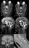
Results: Among 205 patients with spontaneous intracranial hypotension (SIH) and proven CSF leaks we identified five patients with a so-called frontotemporal brain sagging syndrome: Four of those had CSF venous fistulas and significantly improved by occluding them either by surgery or transvenous embolization. Another 11 patients had infratentorial hemosiderosis and hearing problems and ataxia as guiding symptoms. Some cognitive decline was present in at least two of them. Ten patients had ventral dural tears in the thoracic spine and one patient a lateral dural tear at C2/3 respectively. Eight patients showed some improvement after surgery.
Discussion: It is mandatory to study the (thoracic) spine in cognitively impaired patients with brain sagging and/ or infratentorial hemosiderosis on MRI. We propose the term spinal dementia to draw attention to this region, which in turn is evaluated with dynamic digital subtraction and CT myelography.
Keywords: Dementia; Frontotemporal brain sagging syndrome; Infratentorial hemosiderosis – CT myelography; Spontaneous intracranial hypotension.
© 2024. The Author(s).
Conflict of interest statement
The authors declare that they have no conflict of interest.
Figures


Similar articles
-
Spontaneous intracranial hypotension: diagnostic and therapeutic workup.Neuroradiology. 2021 Nov;63(11):1765-1772. doi: 10.1007/s00234-021-02766-z. Epub 2021 Jul 23. Neuroradiology. 2021. PMID: 34297176 Free PMC article. Review.
-
Prevalence of hyperdense paraspinal vein sign in patients with spontaneous intracranial hypotension without dural CSF leak on standard CT myelography.Diagn Interv Radiol. 2018 Jan-Feb;24(1):54-59. doi: 10.5152/dir.2017.17220. Diagn Interv Radiol. 2018. PMID: 29217497 Free PMC article.
-
Spontaneous spinal cerebrospinal fluid-venous fistulas in patients with orthostatic headaches and normal conventional brain and spine imaging.Headache. 2021 Feb;61(2):387-391. doi: 10.1111/head.14048. Epub 2021 Jan 23. Headache. 2021. PMID: 33484155
-
Spontaneous Intracranial Hypotension: A Systematic Imaging Approach for CSF Leak Localization and Management Based on MRI and Digital Subtraction Myelography.AJNR Am J Neuroradiol. 2019 Apr;40(4):745-753. doi: 10.3174/ajnr.A6016. Epub 2019 Mar 28. AJNR Am J Neuroradiol. 2019. PMID: 30923083 Free PMC article.
-
Spinal Cerebrospinal Fluid Leak Localization with Digital Subtraction Myelography: Tips, Tricks, and Pitfalls.Radiol Clin North Am. 2024 Mar;62(2):321-332. doi: 10.1016/j.rcl.2023.10.004. Epub 2023 Nov 11. Radiol Clin North Am. 2024. PMID: 38272624 Review.
Cited by
-
Neuraxial biomechanics, fluid dynamics, and myodural regulation: rethinking management of hypermobility and CNS disorders.Front Neurol. 2024 Dec 10;15:1479545. doi: 10.3389/fneur.2024.1479545. eCollection 2024. Front Neurol. 2024. PMID: 39719977 Free PMC article. Review.
-
Mild cognitive impairment in spontaneous intracranial hypotension and its rapid reversal by repair of a spinal cerebrospinal fluid leak.Headache. 2025 Feb;65(2):382-388. doi: 10.1111/head.14882. Epub 2024 Dec 15. Headache. 2025. PMID: 39676275 Free PMC article.
-
Reply: Dementia in spontaneous intracranial hypotension: look at the spine.Neuroradiology. 2025 Jan;67(1):3-5. doi: 10.1007/s00234-024-03529-2. Epub 2024 Dec 23. Neuroradiology. 2025. PMID: 39714482 Free PMC article. No abstract available.
-
[Brain sagging dementia-A rare potentially reversible cause of dementia].Nervenarzt. 2025 Mar;96(2):192-196. doi: 10.1007/s00115-025-01803-z. Epub 2025 Jan 22. Nervenarzt. 2025. PMID: 39841179 Free PMC article. German. No abstract available.
-
Dementia in spontaneous intracranial hypotension: a call for accurate terminology.Neuroradiology. 2025 Jan;67(1):1-2. doi: 10.1007/s00234-024-03530-9. Epub 2024 Dec 18. Neuroradiology. 2025. PMID: 39692763 No abstract available.
References
-
- Jessen F, Bohr L, Kruse C, Dodel R (2023) The German S3 guidelines on dementia. Nervenarzt 94:609–613 - PubMed
-
- Gifford DR, Holloway RG, Vickrey BG (2000) Systematic review of clinical prediction rules for neuroimaging in the evaluation of dementia. Arch Intern Med 160:2855–2862 - PubMed
-
- Schievink WI, Moser FG, Maya MM (2014) CSF-venous fistula in spontaneous intracranial hypotension. Neurology 83:472–473 - PubMed
-
- Hong M, Shah GV, Adams KM et al (2002) Spontaneous intracranial hypotension causing reversible frontotemporal dementia. Neurology 58:1285–1287 - PubMed
MeSH terms
LinkOut - more resources
Full Text Sources
Medical
Miscellaneous

