. 2024 Aug 1;35(8):ar114.
doi: 10.1091/mbc.E23-11-0428. Epub 2024 Jul 10.

Pulmonary matrix-derived hydrogels from patients with idiopathic pulmonary fibrosis induce a proinflammatory state in lung fibroblasts in vitro

Affiliations

Pulmonary matrix-derived hydrogels from patients with idiopathic pulmonary fibrosis induce a proinflammatory state in lung fibroblasts in vitro

Jorge G Fernandez Davila et al. Mol Biol Cell. .

Abstract

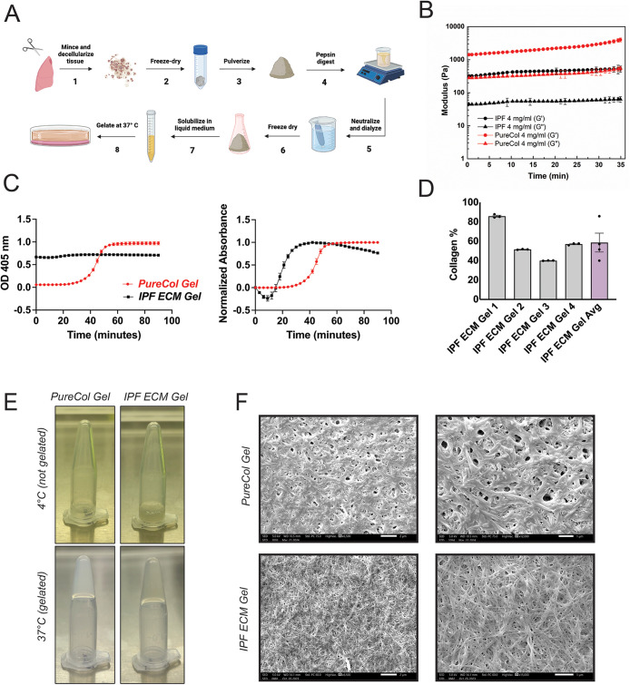
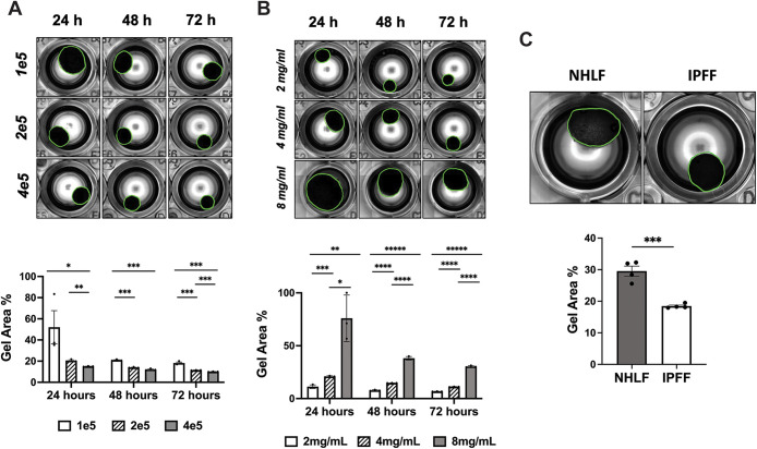
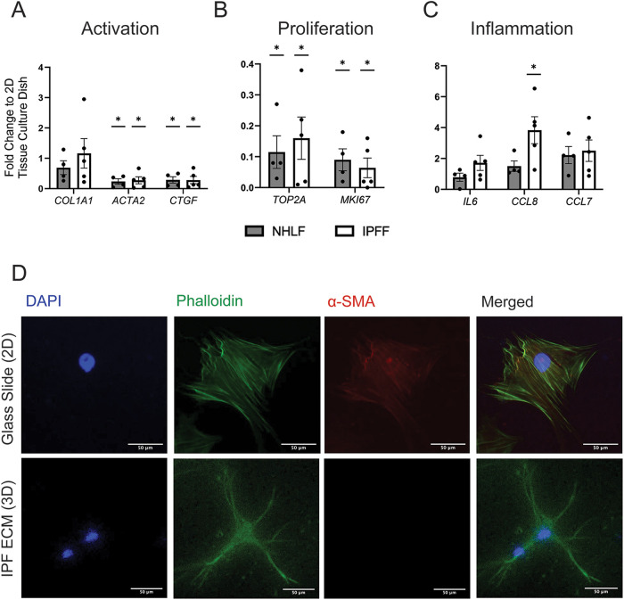
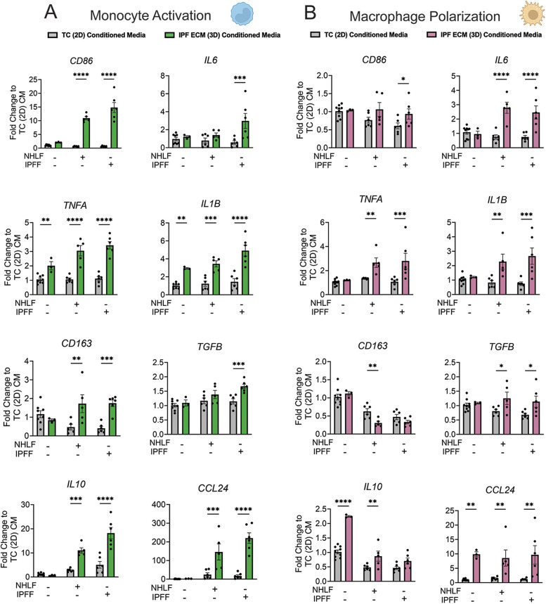
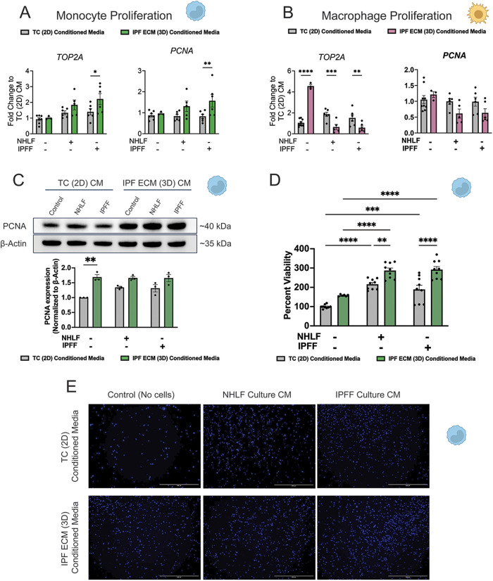
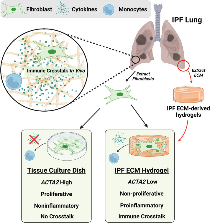
Idiopathic pulmonary fibrosis (IPF), one of the most common forms of interstitial lung disease, is a poorly understood, chronic, and often fatal fibroproliferative condition with only two FDA-approved medications. Understanding the pathobiology of the fibroblast in IPF is critical to evaluating and discovering novel therapeutics. Using a decellularized lung matrix derived from patients with IPF, we generate three-dimensional hydrogels as in vitro models of lung physiology and characterize the phenotype of fibroblasts seeded into the hydrogels. When cultured in IPF extracellular matrix hydrogels, IPF fibroblasts display differential contractility compared with their normal counterparts, lose the classical myofibroblast marker α-smooth muscle actin, and increase expression of proinflammatory cytokines compared with fibroblasts seeded two-dimensionally on tissue culture dishes. We validate this proinflammatory state in fibroblast-conditioned media studies with monocytes and monocyte-derived macrophages. These findings add to a growing understanding of the lung microenvironment effect on fibroblast phenotypes, shed light on the potential role of fibroblasts as immune signaling hubs during lung fibrosis, and suggest intervention in fibroblast-immune cell cross-talk as a possible novel therapeutic avenue.


Figures

FIGURE 1:
FIGURE 1:
IPF ECM hydrogel preparation and characterization in comparison to PureCol gel. (A) Biorender schematic demonstrating the preparation of our IPF ECM hydrogel in eight steps from decellularization of tissue to gelation of hydrogel. (B) Rheological time sweep performances of hydrogels at 4 mg/ml. (C) Gelation curves of IPF ECM and PureCol hydrogels at 4 mg/ml (n = 3) at an absorbance of 405 nm and normalized absorbance. (D) SIRCOL soluble collagen detection assay in IPF ECM pregel solutions (n = 3). (D) Images of IPF ECM and PureCol pregel solutions at 4 mg/ml in a microcentrifuge tube before and after gelation. (E) FESEM images of IPF ECM and PureCol hydrogels at 4 mg/ml, scale bar on left images: 2 µm and right images: 1 µm.
FIGURE 2:
FIGURE 2:
Contraction rate of IPF ECM hydrogels. (A) Top: IPF hydrogel (4 mg/ml ECM protein concentration) seeded with varying fibroblast densities: 1e5, 2e5, and 4e5 cells/well. Bottom: Bar graph demonstrating a significant increase at all timepoints in hydrogel contraction as cell density increases. (B) Top: IPF hydrogel at varying ECM protein concentrations: 2 mg/mL, 4 mg/mL, and 8 mg/mL with a fibroblast density of 2e5 cells/well. Bottom: Bar graph demonstrating a significant decrease of hydrogel contractions at all timepoints as the concentration of IPF ECM in the hydrogel increases. (C) Top: IPF hydrogel seeded with NHLF and IPFF at a density of 2e5 and imaged after 24 h. Bottom: Significantly increased contraction of IPFF in comparison to NHLF in IPF ECM hydrogels after 24 h (n = 2). *: p < 0.05, **: p < 0.005, ***: p < 0.0005, ****: p < 0.00005, *****: p < 0.000005.
FIGURE 3:
FIGURE 3:
Gene expression of IPFF and NHLF fibroblasts seeded in IPF hydrogel and tissue culture plastic: (A) Gene expression of fibroblast activation markers: Col1a1, ACTA2, and CTGF. (B) Gene expression of ligands associated with inflammation: IL6, CCL8, and CCL7 (C) Gene expression of markers associated with cell proliferation: TOP2A and MKI67. *: p < 0.05. (D) Fluorescence microscopy images of IPF fibroblasts stained for DAPI, phalloidin and α-SMA seeded on a 2D glass slide or embedded in the IPF ECM hydrogel. Scale bar: 50 µm.
FIGURE 4:
FIGURE 4:
Proinflammatory gene expression of fibroblasts in fibrotic ECM hydrogel. (A) Microarray data highlighting gene expression changes of IPF fibroblasts immediately prior to culture and after 3 weeks. (B) STRING analysis of protein interactions compiled from genes in the bottom block of the heat map in Figure 4A. (C) Bar graphs demonstrating fold change of gene expression (CCL20, CXCL2, CXCL3, CXCL13, CXCL14, CXCR4) from STRING diagram in Figure 4B comparing NHLF and IPFF seeded in IPF ECM hydrogels against the average of NHLF expression. IL6, CCL7, and CCL8 are not part of the genes in the STRING diagram but were identified as inflammatory genes in Figure 3B. *: p < 0.05.
FIGURE 5:
FIGURE 5:
Gene expression in monocytes after fibroblast conditioned media (CM) challenge. (A) Gene expression fold change of proinflammatory genes (CD86, IL6, TNFA, IL1B) and proresolution genes (CD163, TGFB, CCL24, IL10) in U937 monocytes. (B) Gene expression of the same gene panel in U937-derived macrophages. *: p < 0.05, **: p < 0.005, ***: p < 0.0005, ****: p<0.00005.
FIGURE 6:
FIGURE 6:
Proliferation of monocytes after 48-h fibroblast conditioned media challenge. (A and B) Gene expression fold change in U937 monocytes and U937-derived macrophage proliferation markers: TOP2A and PCNA. (C) Immunoblotting image of PCNA and β-Actin on U937 lysates exposed to fibroblast conditioned media (CM) for 48 h. PCNA protein expression normalized to β-Actin of immunoblot in Figure 6C is shown as a bar graph. (D) The percent viability of U937 monocytes exposed to fibroblast-conditioned media (CCK-8 assay) is shown as a bar graph; a complete media was used as a control. *: p < 0.05, **: p < 0.005, ***: p < 0.0005, ****: p < 0.00005. (E) Fluorescent microscopy images of live U937 monocytes (NucBlue stain) exposed to fibroblast conditioned media. Scale bar: 400 µm.
FIGURE 7:
FIGURE 7:
Immune-fibroblast cross-talk in the IPF lung is a poorly understood aspect of disease biology that is difficult to model in vitro. Fibroblasts isolated from the IPF lungs and seeded into IPF ECM hydrogels maintain high levels of cytokine expression compared with fibroblasts seeded on tissue culture plastic. These cytokines are potent activators of both monocytes and monocyte-derived macrophages.

Similar articles

Cited by

References

    1. Adams TS, Schupp JC, Poli S, Ayaub EA, Neumark N, Ahangari F, Chu SG, Raby BA, DeIuliis G, Januszyk M, et al. (2020). Single-cell RNA-seq reveals ectopic and aberrant lung-resident cell populations in idiopathic pulmonary fibrosis. Sci Adv 6, eaba1983. - PMC - PubMed
    1. Arora S, Dev K, Agarwal B, Das P, Syed MA (2018). Macrophages: Their role, activation and polarization in pulmonary diseases. Immunobiology 223, 383–396. - PMC - PubMed
    1. Arora PD, Narani N, McCulloch CAG (1999). The compliance of collagen gels regulates transforming growth factor-β induction of α-smooth muscle actin in fibroblasts. Am J Pathol 154, 871–882. - PMC - PubMed
    1. Betensley A, Sharif R, Karamichos D (2016). A systematic review of the role of dysfunctional wound healing in the pathogenesis and treatment of idiopathic pulmonary fibrosis. J Clin Med 6, 2. - PMC - PubMed
    1. Booth AJ, Hadley R, Cornett AM, Dreffs AA, Matthes SA, Tsui JL, Weiss K, Horowitz JC, Fiore VF, Barker TH, et al. (2012). Acellular normal and fibrotic human lung matrices as a culture system for in vitro investigation. Am J Respir Crit Care Med 186, 866–876. - PMC - PubMed

LinkOut - more resources