. 2024 Oct 3;144(14):1521-1531.
doi: 10.1182/blood.2024023887.

RBCs regulate platelet function and hemostasis under shear conditions through biophysical and biochemical means

Affiliations

RBCs regulate platelet function and hemostasis under shear conditions through biophysical and biochemical means

Debbie Jiang et al. Blood. .

Abstract

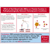
Red blood cells (RBCs) have been hypothesized to support hemostasis by facilitating platelet margination and releasing platelet-activating factors such as adenosine 5'-diphosphate (ADP). Significant knowledge gaps remain regarding how RBCs influence platelet function, especially in (patho)physiologically relevant hemodynamic conditions. Here, we present results showing how RBCs affect platelet function and hemostasis in conditions of anemia, thrombocytopenia, and pancytopenia and how the biochemical and biophysical properties of RBCs regulate platelet function at the blood and vessel wall interface and in the fluid phase under flow conditions. We found that RBCs promoted platelet deposition to collagen under flow conditions in moderate (50 × 103/μL) but not severe (10 × 103/μL) thrombocytopenia in vitro. Reduction in hematocrit by 45% increased bleeding in mice with hemolytic anemia. In contrast, bleeding diathesis was observed in mice with a 90% but not with a 60% reduction in platelet counts. RBC transfusion improved hemostasis by enhancing fibrin clot formation at the site of vascular injury in mice with severe pancytopenia induced by total body irradiation. Altering membrane deformability changed the ability of RBCs to promote shear-induced platelet aggregation. RBC-derived ADP contributed to platelet activation and aggregation in vitro under pathologically high shear stresses, as observed in patients supported by left ventricular assist devices. These findings demonstrate that RBCs support platelet function and hemostasis through multiple mechanisms, both at the blood and vessel wall interface and in the fluidic phase of circulation.


Conflict of interest statement

Conflict-of-interest disclosure: T.B.G. claims no relevant conflicts of interest but reports being a member of the medical advisory board for Platelet Disorder Support Association; receiving consultation fees from Sanofi Corporation, Sobi Corporation, Alpine Immune Science: Bioproducts Laboratory, Cellphire Corporation, Novartis, and Amgen; is a member of the data safety monitoring board of Palisade Bio and Novartis; and receives honoraria from Amgen Inc and Sobi Corporation. The remaining authors declare no competing financial interests.

Figures
None
Graphical abstract
Figure 1.
Figure 1.
RBCs increased platelet thrombus formation on collagen under flow conditions. Human blood reconstituted to varying hematocrits (HCTs) and platelet counts was perfused over a collagen-coated surface using a flow perfusion chamber at a shear stress of 2 Pa. The proportion of surface area covered by platelet thrombus was calculated and is expressed as mean thrombus surface percentage ± standard deviation. (A-C) Representative images of platelet thrombus formed on immobilized collagen after 5-minute perfusion (scale bar, 50 μm) and collective data from multiple independent experiments with different platelet counts and HCTs (n = 49-50 per experimental condition; Student t test). (D) Representative images of thrombus surface coverage (scale bar, 50 μm) after 10-minute perfusion of blood reconstituted to a platelet concentration of 10 × 103/μL and varying HCT (n = 50-59 per experimental condition; 1-way analysis of variance [ANOVA] and Tukey honestly standard deviation).
Figure 2.
Figure 2.
Influence of RBCs and platelet concentration on tail bleeding time. For the hemolytic anemia model, mice were evaluated 24 hours after being treated with PHZ (n = 13) or an equal volume of buffer control (n = 15) for hematocrit (A), platelet count (B), reticulated platelet percentage (C), and tail bleeding time (D) (t test). For the ITP model, mice were evaluated for hematocrit (E), platelet count (F), and tail bleeding time (G) 3 hours after IV injection of a monoclonal (n = 6) or polyclonal (n = 10) anti-GP1bα antibody (1-way ANOVA).
Figure 3.
Figure 3.
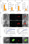
Hemostasis at the site of laser-induced injury. Mice subjected to 6.5 Gy total body irradiation had significantly reduced hematocrit (A), platelet count (B), white blood cell (WBC) count (C), and neutrophil count (D) (n = 6, paired t test). (E) Top panels: Representative images of platelet aggregation identified by Dylight 488 anti-GPIbβ antibody (green) and fibrin clot formation detected by Alexa Fluor 647 anti-fibrin antibody (red) in response to a penetrating vascular injury induced by pulse laser in cremaster arterioles of irradiated mice with or without RBC transfusion (bar = 50 μm); bottom panels: time-dependent changes in the formation of platelet and fibrin clots quantified using mean fluorescence intensity (MFI) of antibody-bound fibrin (left) and platelets (right) from 11 independent experiments (6 transfused with RBCs and 5 with cell-free plasma; 2-way ANOVA, P < .001) (bottom). (F) Representative images of platelet and fibrin hemostatic plugs at the site of an identical laser injury to nonirradiated C57BL/6J mice (bar = 50 μm).
Figure 4.
Figure 4.
Facilitation of SIPA and its regulation by membrane cholesterol. (A) SIPA observed after WB and PRP were exposed to different shear rates for 5 minutes at 37°C (n = 6 per condition; 2-way ANOVA). (B) Viscosities of WB and PRP under different shear rates (n = 88; 46% male; Mann-Whitney rank sum test). (C) SIPA in WB and PRP exposed different shear stresses for 5 minutes at 37°C (n = 5-26 per condition; 2-way ANOVA). (D) SIPA of platelets reconstituted with fresh, partially fixed (0.03% glutaraldehyde at 5 minutes), and completely fixed (0.1% glutaraldehyde at 1 hour) RBCs (n = 4 per condition; 1-way ANOVA, P = .0016). (E) MβCD (10 mM) treated and untreated RBCs were drawn through microfluidic channels under negative pressure (2 psi; bar = 20 μm) and their movements were recorded at 2000 fps. RBC deformability was quantified by Taylor deformation (top panel: deformation distributions at the entrance, middle, and exit of the constriction, representative of 5 independent runs). (F) Shear-induced aggregation of platelets reconstituted with MβCD-treated RBCs (n = 6-8 per condition; 1-way ANOVA, P < .0001). fps, frames per second.
Figure 5.
Figure 5.
Synergistic activities of shear stress and RBC-derived ADP. (A) Levels of CD62p+ platelets in response to 10 Pa shear stress exposure after reconstitution with fRBCs or sRBCs; n = 3 per condition; t test). (B) ADP released from washed RBCs subjected to increasing levels of shear stress (n = 5-7 per condition; 1-way ANOVA). The white bar indicates the amount of ADP from lysed RBCs at 40% hematocrit (n = 5). (C) ADP-induced aggregation of resting platelets and those primed with shear stress (10 Pa for 5 minutes at 37°C; n = 7-8 per condition; 2-way ANOVA); a representative aggregation curve in response to 2 μM ADP (left). (D) SIPA in response to 8 Pa when PRP was reconstituted with unwashed, washed, and apyrase-treated RBCs exposed to high shear stress (n = 8-12 per condition; 1-way ANOVA). (E) Collagen-induced aggregation of resting platelets and those primed with shear stress (10 Pa for 5 minutes at 37°C; n = 7-8 per condition; 2-way ANOVA) with representative curve in response to 2 μg/mL collagen on the left. (F) Plasma levels of RBC-derived EVs (CD235+ EVs) of patients at baseline and after LVAD implantation (n = 12; paired t test). BL, baseline; fRBC, fresh RBC; sRBC, cold-stored RBC.

Comment in

References

    1. Livio M, Marchesi D, Remuzzi G, Mecca G, Remuzzi G, de Gaetano G. Uraemic bleeding: role of anaemia and beneficial effect of red cell transfusions. Lancet. 1982;2(8306):1013–1015. - PubMed
    1. Valeri CR, Cassidy G, Pivacek LE, et al. Anemia-induced increase in the bleeding time: implications for treatment of nonsurgical blood loss. Transfusion (Paris) 2001;41(8):977–983. - PubMed
    1. Crowley JP, Metzger JB, Robert Valeri C. The volume of blood shed during the bleeding time correlates with the peripheral venous hematocrit. Am J Clin Pathol. 1997;108(5):579–584. - PubMed
    1. Ho CH. The hemostatic effect of packed red cell transfusion in patients with anemia. Transfusion (Paris) 1998;38(11-12):1011–1014. - PubMed
    1. Hung Ho C. Increase of red blood cells can shorten the bleeding time in patients with iron deficiency anemia. Blood. 1998;91(3):1094. - PubMed

Publication types

Substances