. 2024 Jul 12;10(28):eado1453.
doi: 10.1126/sciadv.ado1453. Epub 2024 Jul 10.

Targeting a microbiota Wolbachian aminoacyl-tRNA synthetase to block its pathogenic host

Affiliations

Targeting a microbiota Wolbachian aminoacyl-tRNA synthetase to block its pathogenic host

Guillaume Hoffmann et al. Sci Adv. .

Abstract

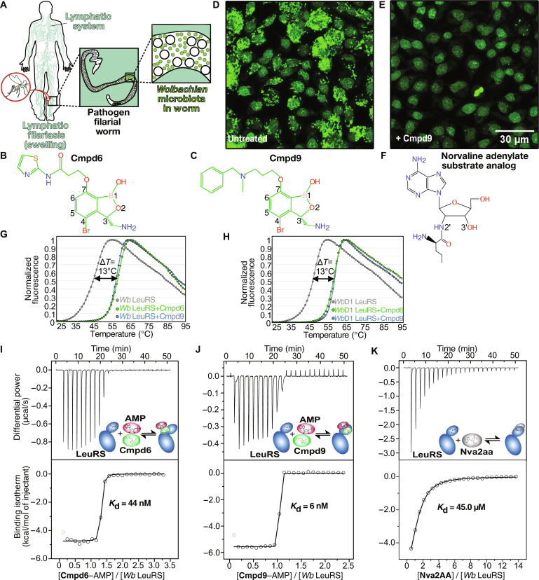
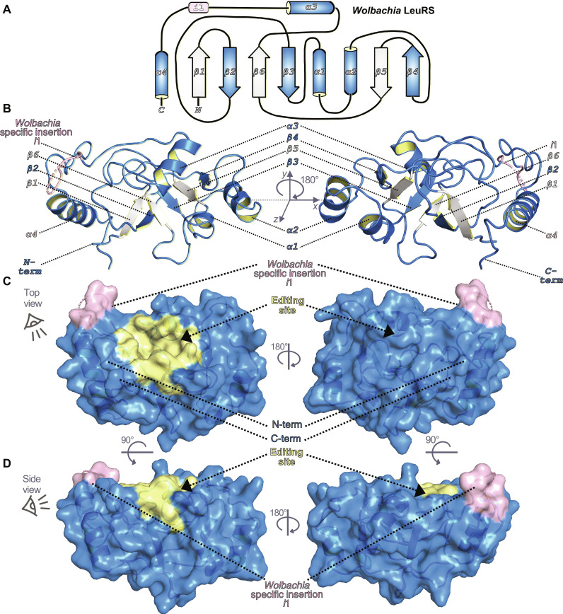
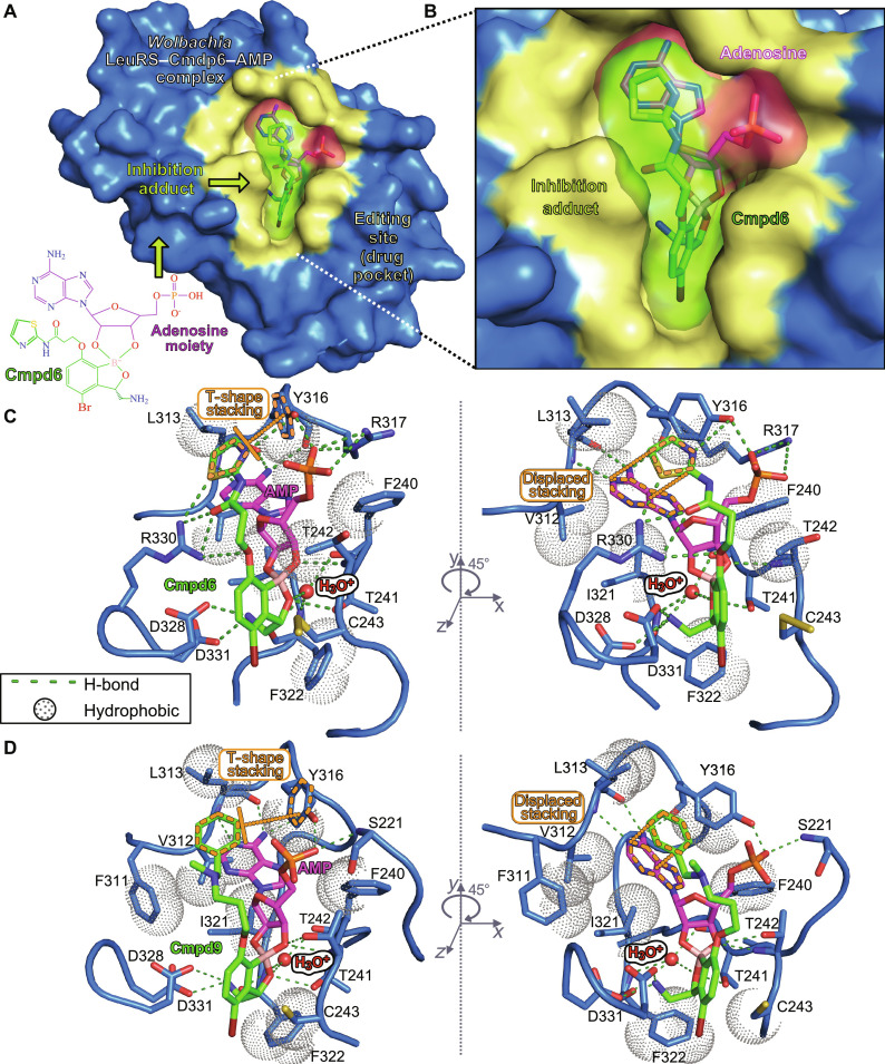
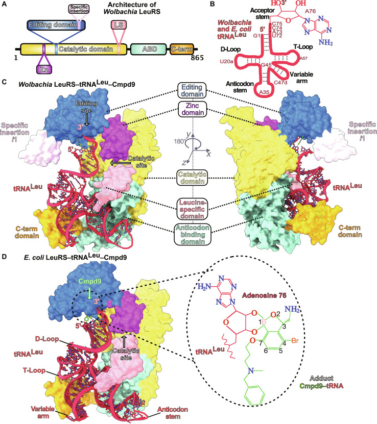
The interplay between humans and their microbiome is crucial for various physiological processes, including nutrient absorption, immune defense, and maintaining homeostasis. Microbiome alterations can directly contribute to diseases or heighten their likelihood. This relationship extends beyond humans; microbiota play vital roles in other organisms, including eukaryotic pathogens causing severe diseases. Notably, Wolbachia, a bacterial microbiota, is essential for parasitic worms responsible for lymphatic filariasis and onchocerciasis, devastating human illnesses. Given the lack of rapid cures for these infections and the limitations of current treatments, new drugs are imperative. Here, we disrupt Wolbachia's symbiosis with pathogens using boron-based compounds targeting an unprecedented Wolbachia enzyme, leucyl-tRNA synthetase (LeuRS), effectively inhibiting its growth. Through a compound demonstrating anti-Wolbachia efficacy in infected cells, we use biophysical experiments and x-ray crystallography to elucidate the mechanism behind Wolbachia LeuRS inhibition. We reveal that these compounds form adenosine-based adducts inhibiting protein synthesis. Overall, our study underscores the potential of disrupting key microbiota to control infections.


Figures

Fig. 1.
Fig. 1.. Benzoxaborole compounds target microbiota Wolbachian LeuRS.
(A) Transmitted by mosquito bites, filarial worm pathogens can reach the lymphatic nodes where they cause severe chronic pain and swelling of tissues. (B, C, and F) Chemical structures of Cmpd6, Cmpd9, and editing substrate analog of norvaline (Nva), respectively. (D) C6/36 insect cells (A. albopictus) stably infected with W. pipientis (wAlbB) were analyzed by fluorescence and high-content image analysis. Single-cell image analysis shows the nucleus (fluorescent large shapes) in the center surrounded by colonizing Wolbachia (small fluorescent dots) in the cytoplasm. (E) Reduction of Wolbachia load in host cells treated with Cmpd9 at 9 μM for 7 days. (G and H) Thermal-shift assays showing the stabilization of Wolbachia LeuRS proteins in the presence of the inhibitors Cmpd6 and Cmpd9. The experiments were performed in triplicates for both the wild-type Wolbachia LeuRS and the construct Wolbachia LeuRS D1 in the presence of AMP. (I, to K) ITC binding experiments showed one order of magnitude higher affinity of Cmpd6/9 in the presence of AMP compared to norvaline posttransfer analog (physiological editing substrate of LeuRS). DSF and ITC experiments were performed in biological and technical triplicates; N = 3.
Fig. 2.
Fig. 2.. Crystal structure of apo Wolbachia LeuRS.
(A) 2-D architecture of the Wolbachia LeuRS editing domain showing an iterative α-β secondary structure organization. For clarity, α helices are colored blue and β sheets are intermittently colored white/blue. The Wolbachia LeuRS specific insertion i1 is colored pink. (B) Crystal structure of Wolbachia LeuRS D1 editing domain shown in cartoon representation with key secondary elements colored as in (A). (C and D) Semi-transparent surface representations of the Wolbachia LeuRS crystal structure showing a top view rotated by 180° around the y axis and a side view rotated by 45° around the x axis, respectively. The editing site (drug-binding site) is shown in yellow and the insertion i1 is shown in light pink.
Fig. 3.
Fig. 3.. Crystal structures of the Wolbachia LeuRS complexes with AMP-compound adducts.
(A) Overall structure of the Wolbachia LeuRS complex with the adduct AMP-Cmpd6 bound into the editing site. Van der Waals surface representation in blue was used for the protein, with the drug-binding site in yellow. (B) Zoomed-in view showing the inhibition adduct in surface and stick representations, with the AMP group in pink and Cmpd6 in green. (C and D) Main interactions established by the adenosine-drug inhibition adducts formed by Cmpd6 and Cmpd9, respectively. Hydrogen bonds are shown as green dashed lines and hydrophobic interactions are shown as dotted spheres. Key protein residues are shown as blue sticks, the hydroxonium ion is shown as a red sphere, and the compound-AMP adducts are in the same color code as in (B). π-π aromatic stackings of the thiazole/phenyl rings of Cmpd6/9 are highlighted in orange dashed lines. For clarity, panels are rotated by 45° around the y axis.
Fig. 4.
Fig. 4.. Structures of full-length LeuRS complexes with tRNALeu adenosine-compound adducts.
(A) Domain architecture of Wolbachia LeuRS protein. (B) Cloverleaf-like secondary structures of Wolbachia and E. coli tRNAsLeu with highlighted main regions. Key tRNALeu bases are numbered with the last base of the acceptor stem (Ade76) shown as sticks. (C) Wolbachian LeuRS-tRNALeu complex with Cmpd9 built by using as templates the crystal structures of the Wolbachia LeuRS editing domain and of the full-length E. coli LeuRS-tRNALeu-Cmpd9. The LeuRS protein is shown in surface representation, and the different domains are in the same color code as in (A). An insertion that is unique to Wolbachia LeuRS, named specific insertion i1, is shows as light-pink surface. The tRNALeu is shown in sticks/ribbon representation and Cmpd9 is shown as green sticks. (D) Crystal structure of the E. coli LeuRS-tRNALeu-Cmpd9 complex determined at 2.1 Å resolution. Protein and tRNALeu are shown in the same color code as Wolbachia LeuRS complexes. The structure of Cmpd9 is shown as green sticks. The zoomed-in view shows the adduct formed by the tRNALeu terminal adenosine with the oxaborole group of Cmpd9.

References

    1. Honda K., Littman D. R., The microbiota in adaptive immune homeostasis and disease. Nature 535, 75–84 (2016). - PubMed
    1. Garrett W. S., Cancer and the microbiota. Science 348, 80–86 (2015). - PMC - PubMed
    1. Hertig M., Wolbach S. B., Studies on rickettsia-like micro-organisms in insects. J. Med. Res. 44, 329–374.7 (1924). - PMC - PubMed
    1. Kaur R., Shropshire J. D., Cross K. L., Leigh B., Mansueto A. J., Stewart V., Bordenstein S. R., Bordenstein S. R., Living in the endosymbiotic world of Wolbachia: A centennial review. Cell Host Microbe 29, 879–893 (2021). - PMC - PubMed
    1. Johnston K. L., Hong W. D., Turner J. D., O’Neill P. M., Ward S. A., Taylor M. J., Anti-Wolbachia drugs for filariasis. Trends Parasitol. 37, 1068–1081 (2021). - PubMed

Publication types

LinkOut - more resources