. 2024 Jul 23;96(29):11716-11724.
doi: 10.1021/acs.analchem.4c00660. Epub 2024 Jul 10.

Affinity-Resolved Size Exclusion Chromatography Coupled to Mass Spectrometry: A Novel Tool to Study the Attribute-and-Function Relationship in Therapeutic Monoclonal Antibodies

Affiliations

Affinity-Resolved Size Exclusion Chromatography Coupled to Mass Spectrometry: A Novel Tool to Study the Attribute-and-Function Relationship in Therapeutic Monoclonal Antibodies

Yuetian Yan et al. Anal Chem. .

Abstract

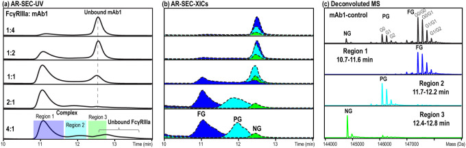
Assessment of critical quality attributes (CQAs) is an important aspect during the development of therapeutic monoclonal antibodies (mAbs). Attributes that affect either the target binding or Fc receptor engagement may have direct impacts on the drug safety and efficacy and thus are considered as CQAs. Native size exclusion chromatography (SEC)-based competitive binding assay has recently been reported and demonstrated significant benefits compared to conventional approaches for CQA identification, owing to its faster turn-around and higher multiplexity. Expanding on the similar concept, we report the development of a novel affinity-resolved size exclusion chromatography-mass spectrometry (AR-SEC-MS) method for rapid CQA evaluation in therapeutic mAbs. This method features wide applicability, fast turn-around, high multiplexity, and easy implementation. Using the well-studied Fc gamma receptor III-A (FcγRIIIa) and Fc interaction as a model system, the effectiveness of this method in studying the attribute-and-function relationship was demonstrated. Further, two case studies were detailed to showcase the application of this method in assessing CQAs related to antibody target binding, which included unusual N-linked glycosylation in a bispecific antibody and Met oxidation in a monospecific antibody, both occurring within the complementarity-determining regions (CDRs).


Conflict of interest statement

The authors declare the following competing financial interest(s): Y.Y., T.X., X.H., W.P., S.W., and N.L. are full-time employees and shareholders of Regeneron Pharmaceuticals Inc.

Figures

Figure 1
Figure 1
General workflow for affinity-resolved SEC-MS.
Figure 2
Figure 2
Affinity-resolved SEC-MS to study the FcγRIIIa (ectodomain, 38–43 kDa) binding of mAb1. (a) SEC-UV traces from analyses of FcγRIIIa and mAb1 mixtures prepared at ratios from the insufficient level (1:4) to excess level (4:1) of FcγRIIIa. (b) XICs of fully glycosylated (FG), partially glycosylated (PG), and nonglycosylated (NG) mAb1 from SEC-PCD-MS analyses of each mixture sample. The XICs were reconstructed using the most abundant charge state of each species. (c) Deconvoluted mass spectrum of mAb1 from native SEC-MS analysis in the absence of FcγRIIIa (1st panel, black); deconvoluted mass spectra of mAb1 species eluting between 10.7 and 11.6 min (2nd panel, blue), 11.7 and 12.2 min (3rd panel, cyan), and 12.4 and 12.8 min (4th panel, green) from SEC-PCD-MS analysis of the 4:1 mixture sample. The corresponding raw mass spectra are shown in Figure S3.
Figure 3
Figure 3
Affinity-resolved SEC-MS analysis to study the FcγRIIIa binding of mAb2. (a) SEC-UV traces from analyses of FcγRIIIa and mAb2 mixtures prepared at ratios from the insufficient level (1:4) to excess level (2:1) of FcγRIIIa. (b) XICs of mAb2 containing G0/G0F (yellow), G0/G1F (orange), G0/G2F (red), G0F/G0F (blue), G0F/G1F (cyan), and G1F/G1F (or G0F/G2F, green) glycoforms from SEC-PCD-MS analyses of each mixture sample. The XICs were constructed using the most abundant charge state of each species. (c) Deconvoluted mass spectrum of mAb2 from native SEC-MS analysis in the absence of FcγRIIIa (1st panel); deconvoluted mass spectra of mAb2 species eluting between 10.7 and 11.4 min (2nd panel) and 11.4 and 12.3 min (3rd panel) from SEC-PCD-MS analysis of the 2:1 mixture sample.
Figure 4
Figure 4
(a) Native SEC-UV/MS analysis of mAb3 after IdeS digestion illustrating the chromatographic separation of the unmodified and CDR N-glycosylated F(ab′)2 species and their corresponding deconvoluted mass spectra (inset). (b) SEC-UV traces from affinity-resolved SEC-MS analysis using Ag3 (ectodomain of Her2, M.W. = 86–92 kDa) and F(ab′)2 mixtures prepared at ratios from the insufficient level (1:2) to excess level (3:1) of Ag3. (c) XICs of the unmodified F(ab′)2 species from SEC-PCD-MS analyses of each mixture sample showing its distribution in the complex and unbound forms. (d) XICs of the N-glycosylated F(ab′)2 (with G2FS2) from SEC-PCD-MS analyses of each mixture sample showing its distribution in the complex and unbound forms. The XICs were reconstructed using the most abundant charge state of each species.
Figure 5
Figure 5
(a) Native SEC-UV/MS analysis of mAb4 after IdeS digestion, limited reduction, and alkylation showing the elution profiles of Fab and Fc by UV (solid line) and XICs (dotted and filled lines) and their corresponding deconvoluted mass spectra (inset). (b) SEC-UV traces from affinity-resolved SEC-MS analysis using Ag4 (soluble C5, M.W. = 192–198 kDa) and Fab mixtures prepared at ratios from the insufficient level (1:2) to excess level (5:2) of Ag4. (c) XICs of LC from SEC-PCD-MS analyses of each mixture sample showing its distribution in the complex and unbound forms. (d) XICs of the unmodified Fd from SEC-PCD-MS analyses of each mixture sample showing its distribution in the complex and unbound forms. (e) XICs of the oxidized Fd from SEC-PCD-MS analyses of each mixture sample showing a single unbound peak. All XICs were generated using the most abundant charge state of each corresponding species.

References

    1. Ambrogelly A.; Gozo S.; Katiyar A.; Dellatore S.; Kune Y.; Bhat R.; Sun J.; Li N.; Wang D.; Nowak C.; et al. Analytical comparability study of recombinant monoclonal antibody therapeutics. MAbs 2018, 10 (4), 513–538. 10.1080/19420862.2018.1438797. - DOI - PMC - PubMed
    1. Jefferis R. Posttranslational Modifications and the Immunogenicity of Biotherapeutics. J. Immunol. Res. 2016, 2016, 535827210.1155/2016/5358272. - DOI - PMC - PubMed
    1. Jenkins N.; Murphy L.; Tyther R. Post-translational modifications of recombinant proteins: significance for biopharmaceuticals. Mol. Biotechnol 2008, 39 (2), 113–118. 10.1007/s12033-008-9049-4. - DOI - PubMed
    1. Wang Q.; Chung C. Y.; Chough S.; Betenbaugh M. J. Antibody glycoengineering strategies in mammalian cells. Biotechnol. Bioeng. 2018, 115 (6), 1378–1393. 10.1002/bit.26567. - DOI - PubMed
    1. Wang W.; Singh S.; Zeng D. L.; King K.; Nema S. Antibody structure, instability, and formulation. J. Pharm. Sci. 2007, 96 (1), 1–26. 10.1002/jps.20727. - DOI - PubMed

Publication types

MeSH terms