. 2024 Jul 10;15(1):5789.
doi: 10.1038/s41467-024-50080-y.

HLTF disrupts Cas9-DNA post-cleavage complexes to allow DNA break processing

Affiliations

HLTF disrupts Cas9-DNA post-cleavage complexes to allow DNA break processing

Giordano Reginato et al. Nat Commun. .

Abstract

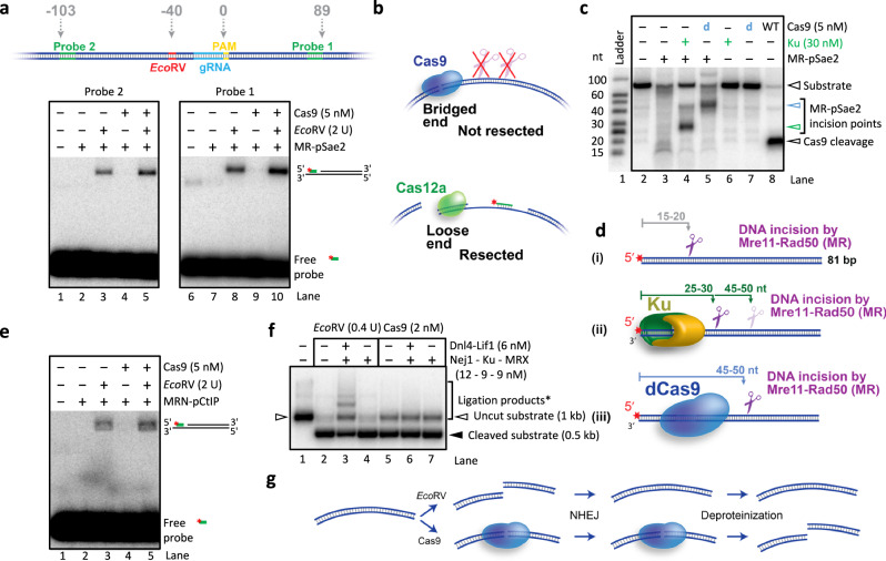
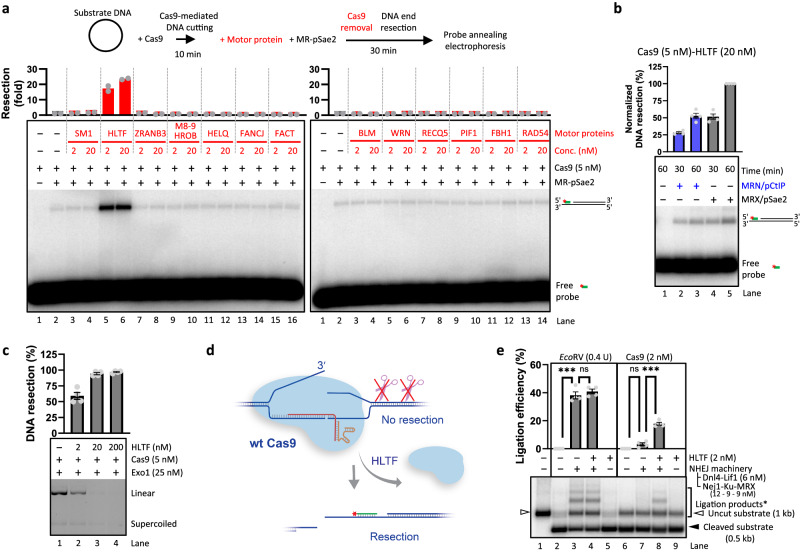
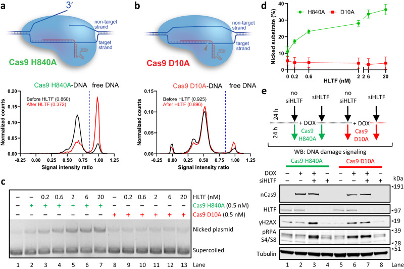
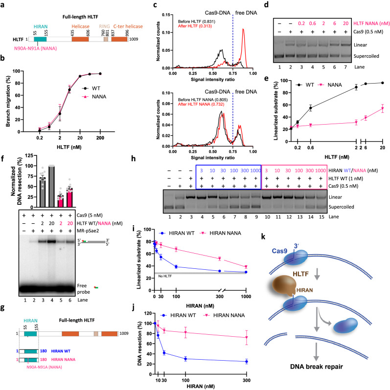
The outcome of CRISPR-Cas-mediated genome modifications is dependent on DNA double-strand break (DSB) processing and repair pathway choice. Homology-directed repair (HDR) of protein-blocked DSBs requires DNA end resection that is initiated by the endonuclease activity of the MRE11 complex. Using reconstituted reactions, we show that Cas9 breaks are unexpectedly not directly resectable by the MRE11 complex. In contrast, breaks catalyzed by Cas12a are readily processed. Cas9, unlike Cas12a, bridges the broken ends, preventing DSB detection and processing by MRE11. We demonstrate that Cas9 must be dislocated after DNA cleavage to allow DNA end resection and repair. Using single molecule and bulk biochemical assays, we next find that the HLTF translocase directly removes Cas9 from broken ends, which allows DSB processing by DNA end resection or non-homologous end-joining machineries. Mechanistically, the activity of HLTF requires its HIRAN domain and the release of the 3'-end generated by the cleavage of the non-target DNA strand by the Cas9 RuvC domain. Consequently, HLTF removes the H840A but not the D10A Cas9 nickase. The removal of Cas9 H840A by HLTF explains the different cellular impact of the two Cas9 nickase variants in human cells, with potential implications for gene editing.


Conflict of interest statement

The authors declare no competing interests.

Figures

Fig. 1
Fig. 1. Cas9-dependent breaks are invisible to DNA end resection enzymes. See also Supplementary Fig. 1.
a Sequence of a DNA segment within the ~2.4 kbp circular plasmid-based DNA substrate used for the Cas experiments. The sequence of the crRNAs is shown in bold for both Cas9 and Cas12a. The PAM sequence is underlined, and the arrowheads indicate the putative cleavage sites. The sequence in green corresponds to the radioactive probe that anneals to the substrates if resection has taken place. The red asterisk indicates the position of the radioactive label. b Representative agarose gel electrophoresis of the substrates treated with the indicated Cas variants. The separation was performed on a 1% TAE gel in the presence of GelRed. The position of the various products is indicated. DA: Cas9 D10A; HA: Cas9 H840A; d: catalytically inactive Cas9; Fn: Francisella novicida Cas12a; Mb: Moraxella bovoculi Cas12a. A representative of two independent experiments is shown. c Top, a schematic overview of the assay. Bottom, representative annealing DNA end resection assay showing resection by the MRE11 complex (25 nM Mre11-Rad50 with 200 nM phosphorylated Sae2, MR-pSae2) of plasmid-based DNA substrate treated with the indicated Cas variants. The probe anneals 92 bp away from the cleavage site for Cas9 and 106 bp for Cas12a. A representative of two independent experiments is shown. d Representative Exo1-MR-pSae2-mediated resection of the plasmid-based DNA substrate treated with EcoRV, Cas9, or Cas12a, as indicated. DNA was stained with GelRed (Biotium). The DNA products, constituted mostly by mono- and dinucleotides, are not stained effectively by the dye. A representative of two independent experiments is shown. Source data are provided as a Source Data file.
Fig. 2
Fig. 2. A loose DNA end is required for the MRE11 complex activation. See also Supplementary Fig. 2.
a A schematic representation of the assay used to detect Mre11-Rad50 (MR)-mediated resection of a substrate in the presence of dimeric LacI (orange ovals, bound to LacO sequence, in red) acting as a protein block. A radioactively labeled probe (green with a red asterisk indicating the 32P label) was annealed to the substrate if the 5′-terminated strand was successfully resected. b Representative annealing DNA end resection assay on supercoiled, circular nicked, and linear substrates (~2.4 kbp) in the presence of dimeric LacI. In the cartoon, LacI is represented as an orange oval, the probe is represented with a green line and the gray arrowheads represent the position of the nick in the circular nicked substrates (~0.9 kbp away from or adjacent to the LacO for nicked circular 1 and 2, respectively). Phosphorylated Sae2 (pSae2) is a co-factor of the Mre11-Rad50 (MR) complex. We note that all the reactions were incubated with 84 nM LacI (in monomers) before the addition of the resection factors. c Quantitation of experiments such as shown in b. n = 3; error bars, SEM. Resection efficiency was normalized to the signal of the linear substrate, which was set to 100%. d A schematic representation of the assay used to detect MR-mediated resection of open and loop-ended substrates. The red portion of the DNA molecule indicates the position of the LacO site, which binds LacI (not shown). The green line represents the radioactively labeled probe (the red asterisk indicates the 32P label). e Representative annealing assay of MR-mediated resection on open and loop-ended DNA substrates (~2.8 kbp) in the presence of dimeric LacI. f Quantitation of experiments such as shown in e. n = 3; error bars, SEM. Resection efficiency was normalized to the signal of the open-end substrate, which was set to 100%. Source data are provided as a Source Data file.
Fig. 3
Fig. 3. Bridging of Cas9-dependent DSBs prevents their immediate processing. See also Supplementary Fig. 3.
a Comparison of resection on either side of the Cas9 break. Top, a cartoon of the circular plasmid DNA substrate used. The relative distance of the various elements from the boundary between the PAM and the protospacer is indicated in bp. Bottom, representative DNA end resection annealing assays with substrates treated with EcoRV and/or Cas9, as indicated. Resection was monitored using a probe on the PAM-distal (left, Probe 2) or PAM-proximal (right, Probe 1) side of the break. A representative of two independent experiments is shown. b A model explaining the difference in the processing of Cas9 vs Cas12a breaks by MR and pSae2. Cas9 breaks are not resected because they are bridged. Cas12a breaks are not bridged and, therefore, can be resected. Resection on only one side of the break is depicted for simplicity. c Representative MR-pSae2 endonuclease assay with an 81 bp 5′ radioactively labeled oligonucleotide-based substrate in the presence of the Ku complex or catalytically dead Cas9 (dCas9). The position of the various cleavage products is indicated by arrowheads (refer to d for the positions of the DNA incision sites). A representative of two independent experiments is shown. d A cartoon of the assay shown in c. The positions of the observed DNA incision points are indicated by scissors. The higher transparency of the scissors symbol indicates lower DNA cleavage efficiency. The red asterisk shows the position of the 32P label. e Representative DNA end resection annealing assay using human MRN-pCtIP. A representative of two independent experiments is shown. f Representative in vitro non-homologous end-joining (NHEJ) assay of the plasmid-length DNA substrate cleaved with EcoRV or Cas9. The bracket (Ligation products*) refers to lanes 2–7, and indicates DNA ligation products, along with a fraction of uncleaved DNA. A representative of two independent experiments is shown. g Cartoon of the assay shown in f. EcoRV-mediated breaks can be ligated in vitro by the yeast NHEJ machinery, while the ends generated by Cas9 are not ligated. Source data are provided as a Source Data file.
Fig. 4
Fig. 4. HLTF removes Cas9 from DNA post-cleavage complex. See also Supplementary Fig. 4.
a Annealing DNA end resection assays of Cas9-mediated DNA breaks by MR and pSae2 in the presence of various human DNA translocases. Top, a schematic overview of the assay. Middle: quantitation of resection efficiency expressed as fold increase compared to the “no translocase” sample (lane 2), which was set to 1. Averages shown, n = 2. Bottom, representative of two independent experiments. SM1: SMARCAL1; M8-9: phosphorylated MCM8-MCM9; FACT: hSpt16-SSRP1. b Annealing DNA end resection assay of Cas9-mediated DNA breaks by yeast MRX-pSae2 or human MRN-pCtIP in the presence of HLTF. Top, quantitation of resection efficiency normalized to the resection obtained by MRX-pSae2 after 1 h, which was set to 100%. n = 4; error bars, SEM. Bottom, a representative experiment. c Exo1-mediated resection of the plasmid-based DNA substrate cleaved with Cas9 and its dependence on HLTF. Top, quantitation; n = 4; error bars, SEM. Bottom, a representative experiment. DNA was stained with GelRed (Biotium). The DNA products, constituted mostly by mono- and dinucleotides, are not stained effectively by the dye. d A cartoon depicting HLTF-mediated Cas9-removal. HLTF is capable of removing Cas9 from the DNA post-cleavage complex, thus allowing DNA end resection of the DSB. e In vitro non-homologous end-joining of EcoRV or Cas9-dependent DNA breaks, and the effect of HLTF. Top, quantitation of ligation efficiency. n = 4; error bars, SEM. Bottom, a representative experiment. The bracket (Ligation products*) refers to lanes 2–7, and indicates DNA ligation products, along with a proportion of uncleaved DNA. (ns, non significant) p = 0.3221 (lanes 3 and 4) and p = 0.0520 (lanes 6 and 7), (***) p = 0.0006 (lanes 2 and 3) and p = 0.0007 (lanes 7 and 8), two-tailed paired t test. The amount of uncleaved substrate in lanes 2 and 6 was used as background. Source data are provided as a Source Data file.
Fig. 5
Fig. 5. HLTF removes Cas9 allowing it to cut multiple targets. See also Supplementary Fig. 5.
a A schematic of the multi-turnover assay used to determine the mechanism of Cas9-removal by HLTF. b Quantitation of multi-turnover experiments of Cas9 and DNA (1 nM, in molecules) in the presence of increasing concentrations of HLTF as shown in Supplementary Fig. 5a. n = 3; error bars, SEM. c Quantitation of multi-turnover experiments with DNA (1 nM, in molecules), varying concentration of Cas9 (as indicated), and HLTF (20 nM) as shown in Supplementary Fig. 5b. n = 4; error bars, SEM. The values next to each data point represent the fold stimulation of Cas9 cleavage by HLTF compared to the conditions without HLTF (lane 2 in Supplementary Fig. 5b; 16 ± 1% linearization; n = 4; SEM) at the given concentration. We note that the fold stimulation for the 0.5 nM sample (lane 6, Supplementary Fig. 5b) is limited by the amount of substrate used. d A cartoon of the single-molecule fluorescence-based Cas9 dissociation assay. The DNA is visualized by Cy5-labeling, while Cas9 is visualized via ATTO550 labeling of the tracrRNA. A signal intensity ratio value of 0.85 was used as a threshold between Cas9-bound DNA (<0.85) and unbound DNA (>0.85). The formula used for the calculation of the signal intensity ratio (SIR) is presented on the right. e, f Single-molecule fluorescence-based assay monitoring the removal of wild type (e) or catalytically dead (f) Cas9 by HLTF. The distribution of the normalized counts of more than 1000 molecules from two independent experiments is presented. The numbers in parentheses indicate the fraction of bound molecules. Source data are provided as a Source Data file.
Fig. 6
Fig. 6. HLTF acts on 3′-ends released by Cas9 after DNA cleavage. See also Supplementary Fig. 6.
a Single-molecule fluorescence-based assay showing the removal of Cas9 H840A by HLTF. Top, a cartoon of the Cas9 H840A-DNA post-cleavage complex. Cas9 H840A fails to cleave the target strand but is proficient in creating the 3′ ssDNA upon cleavage of the non-target strand. Bottom, the distribution of the normalized counts of >1000 molecules from two independent experiments is presented. The dashed blue line indicates the separation between DNA only (>0.85) and Cas9-bound DNA (<0.85). The numbers in parentheses indicate the fraction of bound molecules. b Single-molecule fluorescence-based assay showing the removal of Cas9 D10A by HLTF. Top, a cartoon of the Cas9 D10A-DNA post-cleavage complex. Cas9 D10A only cleaves the target strand. Bottom, the distribution of the normalized counts of more than 1000 molecules from two independent experiments is presented. The dashed blue line indicates the separation between DNA only (>0.85) and Cas9-bound DNA (<0.85). The numbers in parentheses indicate the fraction of bound molecules. c Representative multi-turnover assay with Cas9 H840A and D10A nickase variants in the presence of increasing concentration of HLTF. d Quantitation of experiments such as shown in c. n = 4; error bars, SEM. e DNA damage response after induction of breaks using a guide targeting the Alu and LINE1 repetitive elements. Top, schematic overview of the cellular assay. Bottom, representative western blots from two independent experiments. Cells were treated 48 h before collection with siRNA against HLTF, as indicated. Expression of the indicated Cas9 nickase was induced with doxycycline (DOX) 24 h before collection, as indicated. pRPA S4/S8: antibody against phosphorylated Ser4 and Ser8 of RPA32. Source data are provided as a Source Data file.
Fig. 7
Fig. 7. The HIRAN domain of HLTF is important for Cas9 removal. See also Supplementary Fig. 7.
a A cartoon representation of the primary structure of HLTF. The position of the HIRAN domain mutations (N90A-N91A, NANA) is indicated. b Quantitation of DNA branch-migration experiments such as shown in Supplementary Fig. 7b, c. n = 4 (WT) and n = 3 (NANA); error bars, SEM. c Single-molecule fluorescence-based assays showing the removal of wild type Cas9 by HLTF WT (top) and NANA mutant (bottom). The distribution of the normalized counts of more than 1000 molecules from two independent experiments is presented. The dashed blue line indicates the separation between DNA only (>0.75) and Cas9-bound DNA (<0.75). d Representative Cas9 multi-turnover assay in the presence of Cas9, 1 nM DNA, and increasing concentration of HLTF NANA mutant. e Quantitation of experiments such as shown in d. n = 3; error bars, SEM. The HLTF WT data is reproduced from Fig. 5b (ATP). f Annealing DNA end resection assay of Cas9 breaks by MR-pSae2 in the presence of HLTF WT or NANA mutant. Top, quantitation. n = 9; error bar, SEM. Bottom, a representative experiment. DNA resection efficiency was normalized to the sample with 20 nM HLTF WT, which was set to 100%. g A cartoon representation of the domain structure of the HLTF protein and the purified HIRAN domain variants (1–180). The position of the NANA HIRAN domain mutations is indicated. h Representative Cas9 multi-turnover assay in the presence of Cas9, 1 nM DNA, HLTF, and increasing concentration of WT and NANA mutant HIRAN domain. i Quantitation of experiments such as shown in h. n = 5; error bars, SEM. The gray line indicates the fraction of linearized substrate in the absence of HLTF. j Quantitation of annealing DNA end resection assays such as shown in Supplementary Fig. 7e. n = 4 (WT) and n = 3 (NANA); error bars, SEM. k A model for HLTF-mediated Cas9 removal from a DNA post-cleavage complex. HLTF recognizes the 3′-end released by Cas9 after DNA cleavage through its HIRAN domain. The motor activity is subsequently engaged and leads to Cas9 displacement allowing DNA repair. Source data are provided as a Source Data file.

References

    1. Cannavo E, Cejka P. Sae2 promotes dsDNA endonuclease activity within Mre11-Rad50-Xrs2 to resect DNA breaks. Nature. 2014;514:122–125. doi: 10.1038/nature13771. - DOI - PubMed
    1. Anand R, Ranjha L, Cannavo E, Cejka P. Phosphorylated CtIP functions as a co-factor of the MRE11-RAD50-NBS1 endonuclease in DNA end resection. Mol. Cell. 2016;64:940–950. doi: 10.1016/j.molcel.2016.10.017. - DOI - PubMed
    1. Sartori AA, et al. Human CtIP promotes DNA end resection. Nature. 2007;450:509–514. doi: 10.1038/nature06337. - DOI - PMC - PubMed
    1. Huertas P, Cortes-Ledesma F, Sartori AA, Aguilera A, Jackson SP. CDK targets Sae2 to control DNA-end resection and homologous recombination. Nature. 2008;455:689–692. doi: 10.1038/nature07215. - DOI - PMC - PubMed
    1. Ranjha L, Howard SM, Cejka P. Main steps in DNA double-strand break repair: an introduction to homologous recombination and related processes. Chromosoma. 2018;127:187–214. doi: 10.1007/s00412-017-0658-1. - DOI - PubMed

MeSH terms

LinkOut - more resources