. 2024 Jul 10;14(1):111.
doi: 10.1038/s41408-024-01089-5.

Immune dysfunction prior to and during vaccination in multiple myeloma: a case study based on COVID-19

Collaborators, Affiliations

Immune dysfunction prior to and during vaccination in multiple myeloma: a case study based on COVID-19

Esperanza Martín-Sánchez et al. Blood Cancer J. .

Abstract

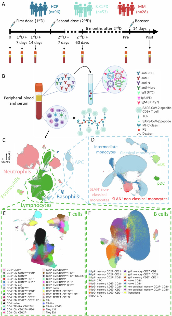
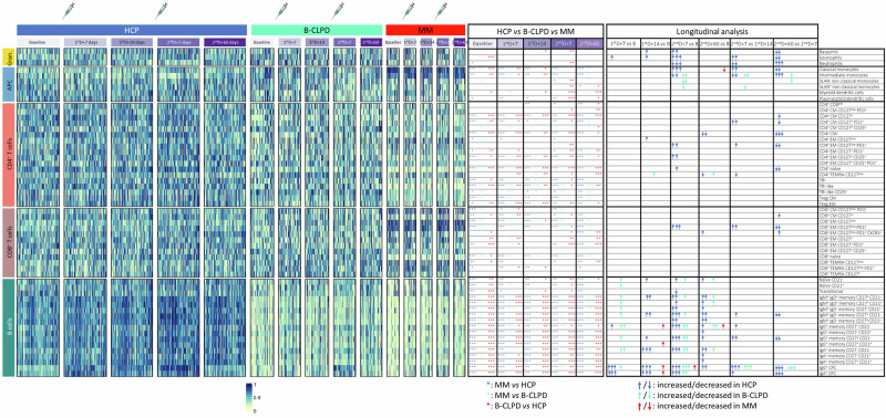
Infection is the leading cause of death in multiple myeloma (MM). However, the cellular composition associated with immune dysfunction is not defined. We analyzed immune profiles in the peripheral blood of patients with MM (n = 28) and B-cell chronic lymphoproliferative disorders (n = 53) vs. health care practitioners (n = 96), using multidimensional and computational flow cytometry. MM patients displayed altered distribution of most cell types (41/56, 73%), particularly within the B-cell (17/17) and T-cell (20/30) compartments. Using COVID-19 as a case study, we compared the immune response to vaccination based on 64,304 data points generated from the analysis of 1099 longitudinal samples. MM patients showed limited B-cell expansion linked to lower anti-RBD and anti-S antibody titers after the first two doses and booster. The percentages of B cells and CD4+ T cells in the blood, as well as the absolute counts of B cells and dendritic cells, predicted vaccine immunogenicity at different time points. In contrast with the humoral response, the percentage and antigen-dependent differentiation of SARS-CoV-2-specific CD8+ T cells was not altered in MM patients. Taken together, this study defined the cellular composition associated with immune dysfunction in MM and provided biomarkers such as the B-cell percentage and absolute count to individualize vaccination calendars.


Conflict of interest statement

The authors declare no competing interests.

Figures

Fig. 1
Fig. 1. Study design.
A Longitudinal analysis of peripheral blood and serum samples during COVID-19 vaccination in a total of 177 individuals: 28 patients with multiple myeloma (MM), 53 patients with a mature B-cell lymphoproliferative disorder (B-CLPD), and 96 health care practitioners (HCP) older than 50. Samples were drawn before vaccination, 7 and 14 days after the first dose, 7 and 60 days after the second dose, as well as before and 14 days after the booster. B In all samples, SARS-CoV-2-specific antibodies and CD8+ T-cells, as well as other non-virus-specific immune cells were analyzed. Antibodies were measured using a CE-IVD serological SARS-CoV-2 multiplex bead-based flow cytometry immunoassay, which allows the simultaneous and quantitative detection of specific IgM, IgG, and IgA antibodies to four SARS-CoV-2 antigens: (1) the receptor-binding domain (RBD) and (2) the stable trimer of the spike (S) glycoprotein; (3) the nucleocapsid (N) protein; and (4) the main viral protease (Mpro). Virus-specific CD8+ T-cells were detected by flow cytometry using a phycoerythrin (PE)-labeled dextran bound to MHC class I proteins presenting five viral peptides of the S-glycoprotein, membrane, ORF3a and N proteins. C Immune profiling was performed using multidimensional and computational flow cytometry that systematically identified a total of 56 immune cell types in peripheral blood, including basophils, eosinophils, neutrophils, antigen-presenting cells, and lymphocytes. D Antigen-presenting cells were sub-clustered into classical, intermediate, SLAN and SLAN+ non-classical monocytes, as well as plasmacytoid and myeloid dendritic cells (pDC and mDC, respectively). E Sub-clustering of T cells into 30 subsets related to antigen-dependent differentiation, along with activation and exhaustion phenotypes in helper and cytotoxic compartments. F Sub-clustering of B cells into 17 subsets related to antigen-dependent differentiation. CM central memory, CPC circulating plasma cells, EM effector memory, TEMRA effector memory T-cells re-expressing CD45RA, Tfh follicular helper T-cells, Treg regulatory T-cells.
Fig. 2
Fig. 2. Immune composition in blood and response to vaccination.
The 56 immune cell types were categorized into five main groups: granulocytes, antigen-presenting cells (APC), CD4 and CD8 T cells, as well as B cells. The percentile-defined frequency in the peripheral blood from 96 health care practitioners (HCP), 53 patients with a mature B-cell lymphoproliferative disorder (B-CLPD) and 28 patients with multiple myeloma (MM) is shown at baseline (B), 7 (1stD + 7) and 14 (1stD + 14) days after the first dose of the vaccine, and 7 (2ndD + 7) and 60 (2ndD + 60) days after the second dose. Each administration is indicated with a syringe. Blue, green, and red asterisks represent significant differences among groups, as indicated. A statistically significant increase or decrease in the frequency of immune cell types across time points in HCP (blue), B-CLPD patients (green), and MM patients (red) are indicated with up or down arrows that are respectively colored. A more detailed and graphical representation of such differences is shown in Supplemental Fig. 1. One symbol (asterisk or arrow), P < 0.05; two symbols, P < 0.01; three symbols, P < 0.001. CM central memory, CPC circulating plasma cells, EM effector memory, TEMRA terminally effector memory CD45RA+, Tfh follicular helper T-cells, Treg regulatory T-cells.
Fig. 3
Fig. 3. Antibody and cellular response to SARS-CoV-2 vaccination.
Indexes of A IgM, B IgA, and C IgG antibodies, as well as D concentration of IgG against the receptor-binding domain (RBD) of the spike-glycoprotein were calculated at baseline, 7 days after the first dose (1stD + 7) as well as at days 7 (2ndD + 7) and 60 (2ndD + 60) after the second dose, in patients with multiple myeloma (MM, n = 28), a mature B-cell lymphoproliferative disorder (B-CLPD, n = 53), and health care practitioners (HCP, n = 96). Each administration is indicated with a syringe and a vertical line. Blue, green, and red asterisks indicate significant differences between the defined time points in HCP, B-CLPD patients, and MM patients, respectively. Black asterisks within tables show significant differences between groups of individuals at each time point. E SARS-CoV-2-specific CD8+ T-cells were identified one week after the second dose of the vaccine using a panel of five dextramers that bound to CD8+ T-cell receptor specific for the top five most immunodominant HLA-A*0201-restricted epitopes of the spike, membrane, ORF3a, and nucleocapsid viral proteins. This was performed in 70 individuals carrying the HLA-A2 allele: 34 HCP, 25 B-CLPD, and 11 MM patients. F Antigen-dependent differentiation of SARS-CoV-2-specific CD8+ T-cells was characterized 7 days after the first (1stD + 7) and second (2ndD + 7) dose of the vaccine using anti-CCR7 and anti-CD45RA antibodies to distinguish between naïve (CCR7+ CD45RA+), central memory (CM, CCR7+ CD45RA-), effector memory (EM, CCR7 CD45RA), and effector memory T-cells re-expressing CD45RA (TEMRA, CCR7 CD45RA+). In all panels, one symbol, P < 0.05; two symbols, P < 0.01; three symbols, P < 0.001; ns non-significant, AI antibody index, AU arbitrary units.
Fig. 4
Fig. 4. Cellular biomarkers predictive of vaccine immunogenicity.
A Odds ratio multivariate analysis with 95% confidence intervals (CI) was included in the logistic regression model. After 10-fold cross-validation, the frequencies of B cells and CD4 T cells measured before vaccination significantly predicted inadequate seroconversion one week after the second dose, defined as below the median observed in patients at this time point (left panel). Area under the curve (AUC) of the prediction probabilities of the model in the training dataset (right panel). B Odds ratio multivariate analysis with 95% CI was included in the logistic regression for validation. Frequency of B cells and CD4 T cells measured before the booster administration significantly predicted inadequate seroconversion 14 days after the booster, defined as below the median of all patients at this time point (left panel). AUC of the prediction probabilities of the model in the validation dataset (right panel). C Association of the presence of none, one or two immune risk-factor predictive of inadequate seroconversion (left panel) with immunoparesis (middle panel) and treatment with anti-CD38 monoclonal antibodies (right panel).

References

    1. Blimark C, Holmberg E, Mellqvist UH, Landgren O, Bjorkholm M, Hultcrantz M, et al. Multiple myeloma and infections: a population-based study on 9253 multiple myeloma patients. Haematologica. 2015;100:107–13. doi: 10.3324/haematol.2014.107714. - DOI - PMC - PubMed
    1. John L, Miah K, Benner A, Mai EK, Kriegsmann K, Hundemer M, et al. Impact of novel agent therapies on immune cell subsets and infectious complications in patients with relapsed/refractory multiple myeloma. Front Oncol. 2023;13:1078725. doi: 10.3389/fonc.2023.1078725. - DOI - PMC - PubMed
    1. Holstein SA, Grant SJ, Wildes TM. Chimeric antigen receptor T-cell and bispecific antibody therapy in multiple myeloma: moving into the future. J Clin Oncol. 2023;41:4416–29. doi: 10.1200/JCO.23.00512. - DOI - PMC - PubMed
    1. Lancman G, Parsa K, Kotlarz K, Avery L, Lurie A, Lieberman-Cribbin A, et al. IVIg use associated with ten-fold reduction of serious infections in multiple myeloma patients treated with anti-BCMA bispecific antibodies. Blood Cancer Discov. 2023;4:440–51. doi: 10.1158/2643-3230.BCD-23-0049. - DOI - PMC - PubMed
    1. Chari A, Samur MK, Martinez-Lopez J, Cook G, Biran N, Yong K, et al. Clinical features associated with COVID-19 outcome in multiple myeloma: first results from the International Myeloma Society data set. Blood. 2020;136:3033–40. doi: 10.1182/blood.2020008150. - DOI - PMC - PubMed

Publication types