. 2024 Jul 11;12(1):40.
doi: 10.1038/s41413-024-00338-4.

Caspase-8 promotes scramblase-mediated phosphatidylserine exposure and fusion of osteoclast precursors

Affiliations

Caspase-8 promotes scramblase-mediated phosphatidylserine exposure and fusion of osteoclast precursors

Brenda Krishnacoumar et al. Bone Res. .

Abstract

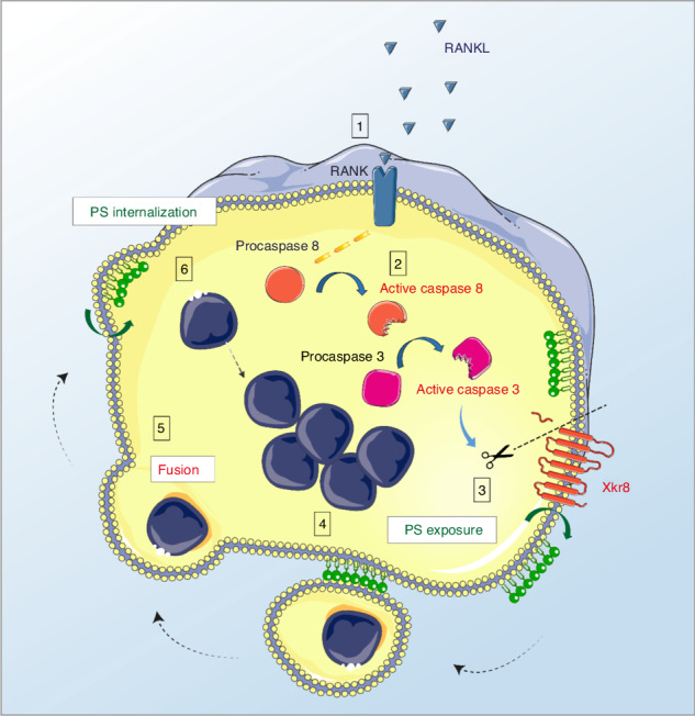
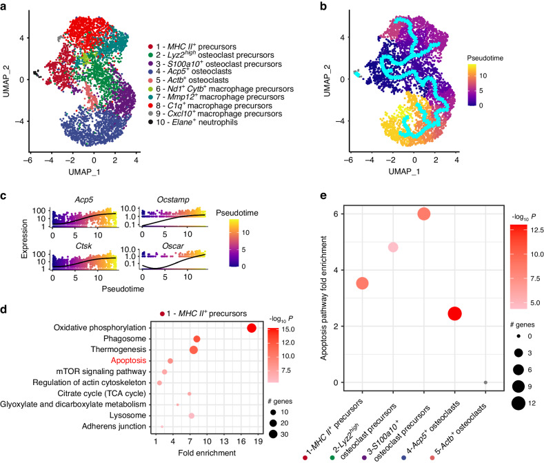
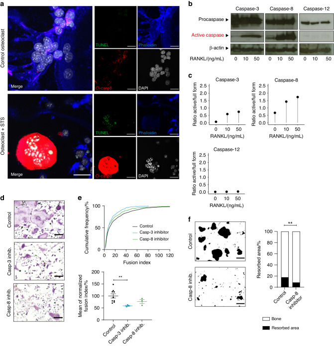
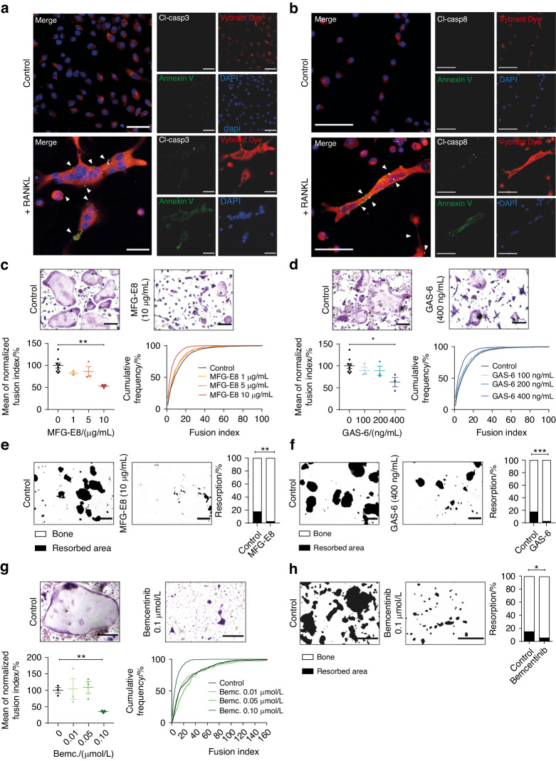
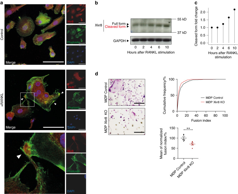
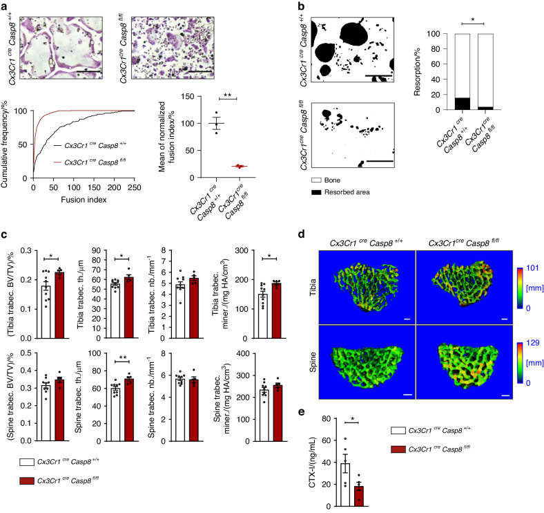
Efficient cellular fusion of mononuclear precursors is the prerequisite for the generation of fully functional multinucleated bone-resorbing osteoclasts. However, the exact molecular factors and mechanisms controlling osteoclast fusion remain incompletely understood. Here we identify RANKL-mediated activation of caspase-8 as early key event during osteoclast fusion. Single cell RNA sequencing-based analyses suggested that activation of parts of the apoptotic machinery accompanied the differentiation of osteoclast precursors into mature multinucleated osteoclasts. A subsequent characterization of osteoclast precursors confirmed that RANKL-mediated activation of caspase-8 promoted the non-apoptotic cleavage and activation of downstream effector caspases that translocated to the plasma membrane where they triggered activation of the phospholipid scramblase Xkr8. Xkr8-mediated exposure of phosphatidylserine, in turn, aided cellular fusion of osteoclast precursors and thereby allowed generation of functional multinucleated osteoclast syncytia and initiation of bone resorption. Pharmacological blockage or genetic deletion of caspase-8 accordingly interfered with fusion of osteoclasts and bone resorption resulting in increased bone mass in mice carrying a conditional deletion of caspase-8 in mononuclear osteoclast precursors. These data identify a novel pathway controlling osteoclast biology and bone turnover with the potential to serve as target for therapeutic intervention during diseases characterized by pathologic osteoclast-mediated bone loss. Proposed model of osteoclast fusion regulated by caspase-8 activation and PS exposure. RANK/RANK-L interaction. Activation of procaspase-8 into caspase-8. Caspase-8 activates caspase-3. Active capase-3 cleaves Xkr8. Local PS exposure is induced. Exposed PS is recognized by the fusion partner. FUSION. PS is re-internalized.


Conflict of interest statement

The authors declare no competing interests.

Figures

None
Proposed model of osteoclast fusion regulated by caspase-8 activation and PS exposure. RANK/RANK-L interaction. Activation of procaspase-8 into caspase-8. Caspase-8 activates caspase-3. Active capase-3 cleaves Xkr8. Local PS exposure is induced. Exposed PS is recognized by the fusion partner. FUSION. PS is re-internalized.
Fig. 1
Fig. 1
ScRNAseq profiling of differentiating osteoclast precursors. a Uniform manifold approximation and projection (UMAP) visualization of 10 clusters in the osteoclast culture system 3 days after initiation of RANKL-induced osteoclastogenesis. b Differentiation trajectory estimated by the cluster-based minimum spanning tree on a UMAP. c Kinetics plot representing relative expression of osteoclastic marker genes Acp5, Ctsk, Ocstamp and Oscar along the osteoclastogenesis trajectory (clusters 1, 2, 3, 4 and 5). d Enrichment analysis of biological processes in cells of the root monocytic osteoclast precursors cluster 1. The significant differentially regulated genes determined with Seurat were used to perform a PathfindR overrepresentation analysis using the KEGG (Kyoto Encyclopedia of Genes and Genomes) protein interaction network database. e Fold Enrichment of the Apoptosis pathway (KEGG mmu04210) in the clusters belonging to the osteoclastogenesis trajectory (clusters 1, 2, 3, 4 and 5)
Fig. 2
Fig. 2
RANKL-induced caspase activity promotes fusion of osteoclast precursors. a Representative confocal laser scanning microscopy images of bone marrow-derived osteoclasts differentiated for 72 h in medium containing 10% L929-conditioned supernatant and 50 ng/mL RANKL either in the presence or absence of 2 μmol/L staurosporine (STS). Cells were fixed and stained for phalloidin (blue), DAPI (white), cleaved caspase-8 (“Cl-casp 8”, red) and TUNEL (green). Scale bar: 30 µm. b Western blot analysis of total cell extracts obtained from bone marrow-derived osteoclasts differentiated for 72 h in medium containing 10% L929-conditioned supernatant and 0, 10, or 50 ng/mL RANKL. Antibodies against the full form (“Procaspase”) or the cleaved active form (“Active caspase”) of caspases 3, 8, and 12 were used. β-actin signal served as loading control. c Quantification of band intensity ratios between cleaved active and full form of the Western Blot shown in b. d, e Bone marrow-derived osteoclasts were differentiated for 72 h in medium containing 10% L929-conditioned supernatant and 10 ng/mL RANKL in the presence of 10 μmol/L caspase-3 inhibitor, 10 μmol/L caspase-8 inhibitor, or vehicle only. Cells were fixed and stained for TRAP. d Representative micrographs of osteoclast size and nuclei per cell. Scale bar: 200 µm. e Quantification of the fusion index defined as the number of nuclei per cell. For every treatment group of the experiment, the cumulative frequency in percent of each occurring fusion index was calculated (top). The average fusion index was then determined, normalized to vehicle-only control and quantified (bottom). Indicated is mean ± SEM. Unpaired, two-tailed Student’s t-test, *P < 0.05; **P < 0.01; ***P < 0.001, n = 3 for each caspase inhibitor-treated group and n = 9 for control group. f Bone marrow-derived osteoclasts were seeded onto bone-coated osteoplates and differentiated for 72 h in medium containing 10% L929-conditioned supernatant and 10 ng/mL RANKL in the presence of 10 μmol/L caspase-8 inhibitor or vehicle only. Left panel: Representative micrographs showing resorbed area (black) vs. remaining bone (white). Scale bar: 200 µm. Right: Quantification of resorbed area vs. remaining bone area in percent. Indicated is the mean. Unpaired, two-tailed Student’s t-test, *P < 0.05; **P < 0.01; ***P < 0.001, n = 4 per group. All images and data are representative of three independent experiments
Fig. 3
Fig. 3
Exposure of phosphatidylserine mediates fusion of osteoclast precursors. a, b Representative confocal laser scanning microscopy images of bone marrow-derived osteoclasts differentiated for 72 h in medium containing 10% L929-conditioned supernatant either in the presence or absence of 50 ng/mL RANKL. Caspase signals are shown in white (arrowheads). Scale bar: 30 µm. c, d Bone marrow-derived osteoclasts were differentiated for 72 h in medium containing 10% L929-conditioned supernatant and 10 ng/mL RANKL in the presence of 1, 5, and 10 µg/mL MFG-E8 or vehicle only c or in the presence of 0, 100, 200, and 400 ng/mL GAS-6 d. Cells were fixed and stained for TRAP. Scale bar: 200 μm. Indicated is mean ± SEM. Unpaired, two-tailed Student’s t-tests, *P < 0.05; **P < 0.01; ***P < 0.001, n = 3 for each tested concentration of MFG-E8 or GAS-6 and n = 9 for control groups. e, f Bone marrow-derived osteoclasts were seeded onto bone-coated osteoplates and differentiated for 72 h in medium containing 10% L929-conditioned supernatant and 10 ng/mL RANKL in the presence of 10 µg/mL MFG-E8 or vehicle only e or in the presence of 0 or 400 ng/mL GAS-6 f. Scale bar: 200 µm. Indicated is the mean. Unpaired, two-tailed Student’s t-test, *P < 0.05; **P < 0.01; ***P < 0.001, n = 4 per group. g Bone marrow-derived osteoclasts were differentiated for 72 h in medium containing 10% L929-conditioned supernatant and 10 ng/mL RANKL in the presence of 0.01, 0.05, and 0.10 μmol/L Bemcentinib or vehicle only. Cells were fixed and stained for TRAP. Scale bar: 200 µm. Indicated is mean ± SEM. Unpaired, two-tailed Student’s t-tests, *P < 0.05; **P < 0.01; ***P < 0.001, n = 3 per group. h Bone marrow-derived osteoclasts were seeded onto bone-coated osteoplates and differentiated for 72 h in medium containing 10% L929-conditioned supernatant and 10 ng/mL RANKL in the presence of 0.1 μmol/L Bemcentinib or vehicle only. Scale bar: 200 µm. Indicated is the mean. Unpaired, two-tailed Student’s t-test, *P < 0.05; **P < 0.01; ***P < 0.001, n = 4 per group. Images and data are representative of three independent experiments except for data shown in e, f, and h, which are representative of two independent experiments
Fig. 4
Fig. 4
RANKL-induced osteoclast fusion is mediated via the phospholipid scramblase Xkr8. a Representative confocal laser scanning microscopy images of human monocyte-derived osteoclasts differentiated for 6 days in medium containing 30 ng/mL hM-CSF, 1 ng/mL hTGF-β, and 3 ng/mL hRANKL (middle and bottom) or vehicle only (top). Cells were fixed and stained for Xkr8 (red), phalloidin (green) and DAPI (blue). The white square in the middle image indicates magnified area shown on the bottom of the panel. Arrowheads indicate localization of Xkr8 at the fusion sites. Scale bar: 30 µm. b Western blot analysis of Xkr8 expression in cytoplasmic extracts obtained from human monocyte-derived osteoclasts differentiated for 6 days in medium containing 30 ng/mL hM-CSF, 1 ng/mL hTGF-β, and stimulated with 3 ng/mL hRANKL for 0, 2, 4, 6, and 10 h. GAPDH signal served as loading control. c Quantification of the fold change in cleaved band intensity relative to full form band intensity, normalized to 0 h after RANKL stimulation of the Western blot shown in b. d MØP control and Xkr8 KO MØP cells were differentiated into osteoclasts for 72 h in medium containing 10% L929-conditioned supernatant and 50 ng/mL RANKL. Cells were fixed and stained for TRAP. Left panel: Representative micrographs of osteoclast size and nuclei per cell. Scale bar: 200 µm. Right panel: Quantification of the fusion index defined as the number of nuclei per cell. For every treatment group of each experiment, the cumulative frequency in percent of each occurring fusion index was calculated (top). The average fusion index was then determined, normalized to control and quantified (bottom). Indicated is mean ± SEM. Unpaired, two-tailed Student’s t-test, *P < 0.05; **P < 0.01; ***P < 0.001, n = 5 per group. All images and data are representative of three independent experiments
Fig. 5
Fig. 5
Specific deletion of caspase-8 in osteoclast precursors inhibits osteoclast fusion and bone resorption in vivo. a Bone marrow-derived osteoclasts obtained from Cx3Cr1creCasp8+/+ control or Cx3Cr1creCasp8fl/fl littermate mice lacking caspase-8 in mononuclear phagocytes were differentiated for 72 h in medium containing 10% L929-conditioned supernatant and 50 ng/mL RANKL. Cells were fixed and stained for TRAP. Top panel: Representative micrographs of osteoclast size and nuclei per cell. Scale bar: 200 µm. Bottom panel: Quantification of the fusion index defined as the number of nuclei per cell. For both genotypes of each experiment, the cumulative frequency in percent of each occurring fusion index was calculated (left). The average fusion index was then determined, normalized to control and quantified (right). Indicated is mean ± SEM. Unpaired, two-tailed Student’s t-tests, *P < 0.05; **P < 0.01; ***P < 0.001, n = 3 per group. b Bone marrow-derived osteoclasts obtained from Cx3Cr1creCasp8+/+ control or Cx3Cr1creCasp8fl/fl littermate mice were seeded onto bone-coated osteoplates and differentiated for 72 h in medium containing 10% L929-conditioned supernatant and 50 ng/mL RANKL. Left panel: Representative micrographs showing resorbed area (black) vs. remaining bone (white). Scale bar: 200 µm. Right: Quantification of resorbed area vs. remaining bone area in percent. Indicated is the mean. Unpaired, two-tailed Student’s t-test, *P < 0.05; **P < 0.01; ***P < 0.001, n = 4 per group. c µCT-based quantification of indicated bone morphometric parameters obtained from tibia (top panel) or spine (bottom panel) of 12-week-old Cx3Cr1creCasp8+/+ control or Cx3Cr1creCasp8fl/fl littermate mice. BV, bone volume. TV, total volume. Trabec. th., trabecular thickness. Trabec. Nb., trabecular number. Trabec. miner., trabecular mineralization. Unpaired, two-tailed Mann-Whitney U test, *P < 0.05; **P < 0.01; ***P < 0.001, n = 10 for Cx3Cr1creCasp8+/+ and n = 5 for Cx3Cr1creCasp8fl/fl. d Three-dimensional µCT reconstructions showing representative cranial views of Cx3Cr1creCasp8+/+ control or Cx3Cr1creCasp8fl/fl littermate-derived tibia (top panel) or spine (bottom panel). Heatmap colors indicate bone thickness in mm. Scale bar: 100 µm. e ELISA-based quantification of the CTX-I content of sera obtained from 12-week-old Cx3Cr1creCasp8+/+ control or Cx3Cr1creCasp8fl/fl littermate mice serving as an indicator of bone resorption. Indicated is mean ± SEM. Unpaired, one-tailed Student’s t-test, *P < 0.05; **P < 0.01; ***P < 0.001, n = 5 per group. All images and data are representative of three independent experiments

References

    1. Zaidi, M. Skeletal remodeling in health and disease. Nat. Med.13, 791–801 (2007). - PubMed
    1. Jacome-Galarza, C. E. et al. Developmental origin, functional maintenance and genetic rescue of osteoclasts. Nature568, 541–545 (2019). - PMC - PubMed
    1. Kong, Y. Y. et al. OPGL is a key regulator of osteoclastogenesis, lymphocyte development and lymph-node organogenesis. Nature397, 315–323 (1999). - PubMed
    1. Wada, T., Nakashima, T., Hiroshi, N. & Penninger, J. M. RANKL-RANK signaling in osteoclastogenesis and bone disease. Trends Mol. Med.12, 17–25 (2006). - PubMed
    1. Takayanagi, H. et al. Induction and activation of the transcription factor NFATc1 (NFAT2) integrate RANKL signaling in terminal differentiation of osteoclasts. Dev. Cell3, 889–901 (2002). - PubMed

LinkOut - more resources