. 2024 Jul 10;15(1):5809.
doi: 10.1038/s41467-024-49965-9.

The Hippo pathway transcription factors YAP and TAZ play HPV-type dependent roles in cervical cancer

Affiliations

The Hippo pathway transcription factors YAP and TAZ play HPV-type dependent roles in cervical cancer

Molly R Patterson et al. Nat Commun. .

Abstract

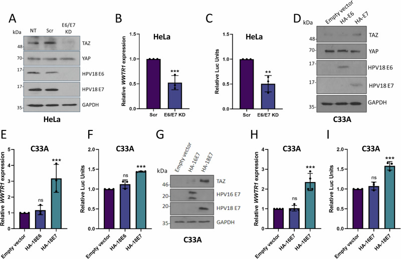
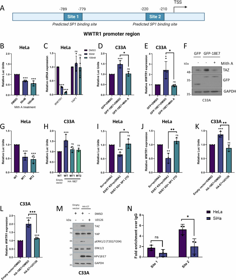
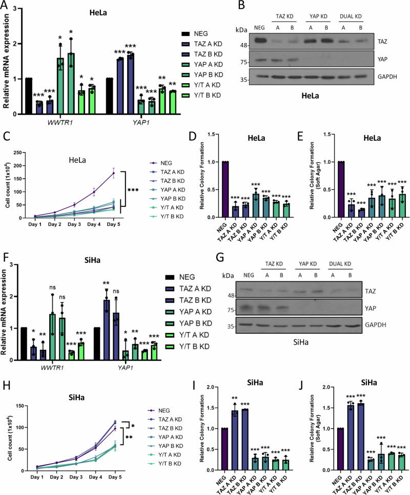
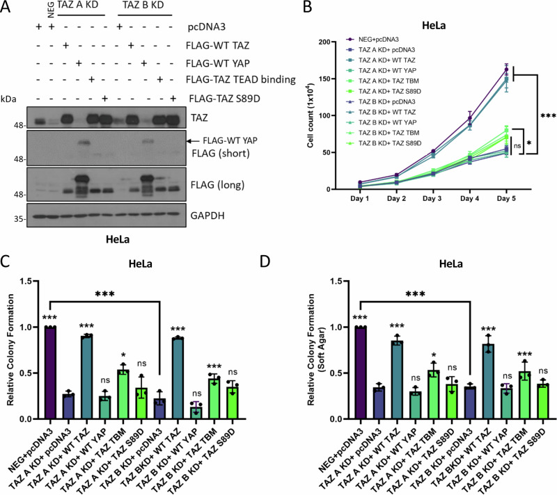
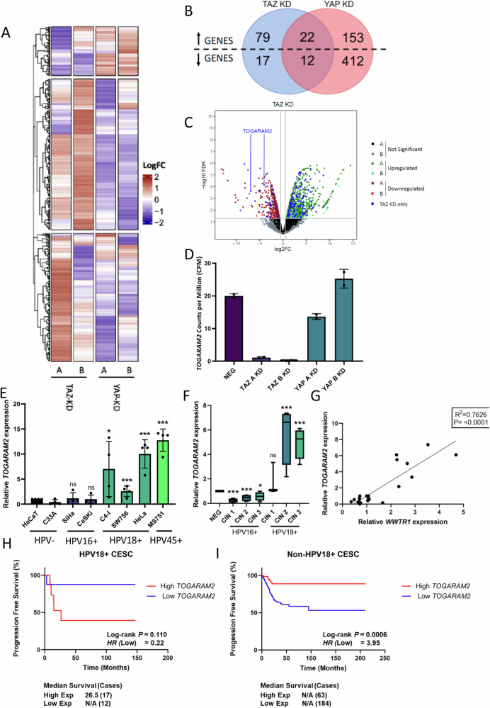
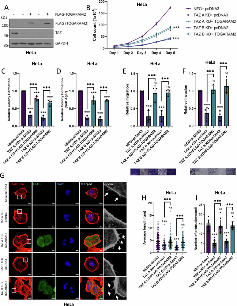
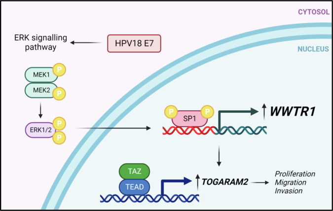
Human papillomaviruses (HPVs) cause most cervical cancers and an increasing number of anogenital and oral carcinomas, with most cases caused by HPV16 or HPV18. HPV hijacks host signalling pathways to promote carcinogenesis. Understanding these interactions could permit identification of much-needed therapeutics for HPV-driven malignancies. The Hippo signalling pathway is important in HPV+ cancers, with the downstream effector YAP playing a pro-oncogenic role. In contrast, the significance of its paralogue TAZ remains largely uncharacterised in these cancers. We demonstrate that TAZ is dysregulated in a HPV-type dependent manner by a distinct mechanism to that of YAP and controls proliferation via alternative cellular targets. Analysis of cervical cancer cell lines and patient biopsies revealed that TAZ expression was only significantly increased in HPV18+ and HPV18-like cells and TAZ knockdown reduced proliferation, migration and invasion only in HPV18+ cells. RNA-sequencing of HPV18+ cervical cells revealed that YAP and TAZ have distinct targets, suggesting they promote carcinogenesis by different mechanisms. Thus, in HPV18+ cancers, YAP and TAZ play non-redundant roles. This analysis identified TOGARAM2 as a previously uncharacterised TAZ target and demonstrates its role as a key effector of TAZ-mediated proliferation, migration and invasion in HPV18+ cancers.


Conflict of interest statement

The authors declare no competing interests.

Figures

Fig. 1
Fig. 1. TAZ is upregulated in HPV18+ cervical cancer.
A Genomic alterations of WWTR1 across human cancers determined by cBioportal analysis of TCGA data (LUSC Lung squamous cell carcinoma (n = 487), USC Uterine serous carcinoma (n = 56), ESCA Eosophageal carcinoma (n = 186), OV Ovarian serous cystadenocarcinoma (n = 579), HNSC Head and Neck squamous cell carcinoma (n = 517), CESC Cervical squamous cell carcinoma (n = 295), BLCA Bladder Urothelial carcinoma (n = 408), PRAD Prostate adenocarcinoma (n = 491), UCEC Uterine corpus endometrial carcinoma (n = 487). B Scatter dot plot analysis of WWTR1 mRNA expression against WWTR1 copy number alterations in cervical cancer determined from TCGA data (n = 278). The correlation coefficient (r) was calculated using Pearson correlation analysis. C Scatter dot plot analysis of WWTR1 mRNA expression against TAZ protein expression in cervical cancer determined from TCGA data (n = 152). The correlation coefficient (r) was calculated using Pearson correlation analysis. D Representative western blot of lysate from HPV-, HPV16+ and HPV18+ cell lines. Lysates were probed for YAP, TAZ and the loading control GAPDH (n = 3). E RT-qPCR analysis of WWTR1 or YAP1 expression in HPV-, HPV16+ or HPV18+ cell lines (n = 3). U6 transcript levels were used as a loading control. F RT-qPCR analysis of WWTR1 expression in negative, HPV16 or HPV18+ patient cervix liquid cytology samples from different CIN grades (n = 4 from each grade). U6 was used as a loading control. Maxima and minima whiskers represent the highest and lowest values, respectively. Bounds of boxes are the 75th and 25th percentiles and the centre represents the median. G Kaplan–Meier curves showing progression free survival in HPV18+ cervical cancer stratified by high or low WWTR1 expression. Survival was compared using the log-rank test. H Kaplan–Meier curves showing progression free survival in non-HPV18+ cervical cancer stratified by high or low WWTR1 expression. Survival was compared using the log-rank test. Error bars represent the mean +/- standard deviation of a minimum of three biological repeats when relevant. *P < 0.05, **P < 0.01, ***P < 0.005 (two-tailed, unpaired Student’s t-test). Source data are provided as a Source Data file.
Fig. 2
Fig. 2. HPV18 E7 upregulates WWTR1 transcription.
A Representative western blots of HeLa cells after transfection with HPV18 E6/E7 targeting siRNA. Cell lysates were probed for TAZ, YAP, HPV18 E6 and HPV18 E7 expression. GAPDH was used as a loading control (n = 4). B RT-qPCR analysis of WWTR1 expression in HeLa cells after transfection with HPV18 E6/E7 targeting siRNA (n = 3). U6 was used as a loading control. C WWTR1-promoter luciferase assay in HeLa cells after transfection with HPV18 E6/E7 targeting siRNA (n = 3). D Representative western blot of C33A cell lysates stably expressing either HA-HPV18 E6 or HA-HPV18 E7 probed for TAZ, YAP, HPV18 E6, HPV18 E7 and the loading control GAPDH (n = 3). E RT-qPCR analysis of WWTR1 expression in C33A cells stably expressing either HA-HPV18 E6 or HA-HPV18 E7 (n = 3). U6 was used as a loading control. F WWTR1-promoter luciferase assay in C33A cells stably expressing either HA-HPV18 E6 or HA-HPV18 E7 (n = 3). G Representative western blot of C33A cell lysate stably expressing either HA-HPV18 E6 or HA-HPV18 E7 probed for TAZ, YAP, HPV16 E7, HPV18 E7 and GAPDH (n = 3). H RT-qPCR analysis of WWTR1 expression in C33A cells stably expressing either HA-HPV16 E7 or HA-HPV18 E7 (n = 4). U6 was used as a loading control. I WWTR1-promoter luciferase assay in C33A cells stably expressing either HA-HPV16 E7 or HA-HPV18 E7 (n = 3). Error bars represent the mean +/- standard deviation of a minimum of three biological repeats. *P < 0.05, **P < 0.01, ***P < 0.005 (two-tailed, unpaired Student’s t-test). Source data are provided as a Source Data file.
Fig. 3
Fig. 3. HPV18 E7-mediated TAZ expression in regulated by the ERK1/2-SP1 signalling axis.
A WWTR1 promoter schematic showing predicted SP1 binding sites. TSS = transcription start site. Made using BioRENDER.com. B WWTR1-promoter luciferase assay in HeLa cells following mithramycin A (Mith A) treatment (n = 3). C RT-qPCR analysis of WWTR1 and YAP1 expression in HeLa cells following Mith A treatment (n = 4). U6 was used as a loading control. D WWTR1-promoter luciferase assay in C33A cells following transfection of GFP-HPV18 E7 plus Mith A treatment (n = 3). E RT-qPCR analysis of WWTR1 expression in C33A cells following transfection of GFP-HPV18 E7 plus Mith A treatment (n = 4). U6 was used as a loading control. F Representative western blots from (E) probed for TAZ, GFP and GAPDH (n = 3). G Luciferase assay in HeLa cells with the WWTR1-promoter SP1 binding site mutants (n = 3). H Luciferase assay in C33A cells stably expressing HA-HPV18 E7 following transfection of the WWTR1-promoter SP1 binding site mutants (n = 3). I WWTR1-promoter luciferase assay in HeLa cells transfected with HPV18 E6/E7 targeting siRNA plus SP1 2TD overexpression (n = 3). J RT-qPCR analysis of WWTR1 expression in HeLa cells transfected with HPV18 E6/E7 targeting siRNA plus SP1 2TD overexpression (n = 3). U6 was used as a loading control. K WWTR1-promoter luciferase assay in C33A cells stably expressing HA-HPV18 E7 following U0126 treatment (n = 3). L RT-qPCR analysis of WWTR1 expression in C33A cells stably expressing HA-HPV18 E7 following U0126 treatment (n = 3). U6 was used as a loading control. M Representative western blot of C33A cells stably expressing HA-HPV18 E7 following U0126 treatment probed for TAZ, pERK1/2 (T202/Y204), ERK1/2, HPV18 E7 and GAPDH (n = 4). N ChIP-qPCR analysis of SP1 binding to the WWTR1 promoter in HeLa and SiHa cells (n = 3). SP1 binding is presented as a fold increase over IgG binding and a gene desert control. Error bars represent the mean +/- standard deviation of a minimum of three biological repeats. *P < 0.05, **P < 0.01, ***P < 0.005 (two-tailed, unpaired Student’s t-test). Source data are provided as a Source Data file. Figure 3A was created with Biorender.com released under a Creative Commons Attribution-NonCommercial 4.0 International License.
Fig. 4
Fig. 4. Inhibition of TAZ reduces HPV18+ cervical cancer cell proliferation.
A Immunofluorescence microscopy analysis of TAZ localisation (green) in HeLa cells treated with 6079510 or Ivermectin. DAPI stained nuclei (blue). Arrows point to nuclear TAZ. Scale bar 10 µm (n = 3). B Immunofluorescence microscopy analysis of nuclear YAP (green) in HeLa cells treated with 6079510 or Ivermectin. DAPI stained nuclei (blue). Arrows point to nuclear YAP. Scale bar 10 µm (n = 3). C Growth curve analysis of HeLa cells treated with 6079510 or Ivermectin (n = 3). D Colony formation assay of HeLa cells treated with 6079510 or Ivermectin (n = 4). E Soft agar assay of HeLa cells treated with 6079510 or Ivermectin (n = 3). F Growth curve analysis of SiHa cells treated with 6079510 or Ivermectin (n = 3). G Colony formation assay of SiHa cells treated with 6079510 or Ivermectin (n = 4). H Soft agar assay of SiHa cells treated with 6079510 or Ivermectin (n = 3). I RT-qPCR analysis of WWTR1 or YAP1 expression in TAZ KD HeLa cells (n = 3). U6 was used as a loading control. J Representative western blot of TAZ KD HeLa cells probed for TAZ, HPV18 E6, HPV18 E7 and GAPDH (n = 3). K Growth curve analysis of TAZ KD HeLa cells (n = 3). L Colony formation assay in TAZ KD HeLa cells (n = 4). M Soft agar assay in TAZ KD HeLa cells (n = 4). N Colony formation assay in C33A cells stably expressing HA-HPV18 E7 plus 6079510 treatment (n = 4). O Colony formation assay in HaCaT cells stably expressing HA-HPV18 E7 plus 6079510 treatment (n = 3). Error bars represent the mean +/- standard deviation of a minimum of three biological repeats. *P < 0.05, **P < 0.01, ***P < 0.005 (two-tailed, unpaired Student’s t-test). Source data are provided as a Source Data file.
Fig. 5
Fig. 5. YAP and TAZ are oncogenic in HPV18 + , but not HPV16+ cervical cancer cell lines.
A RT-qPCR analysis of WWTR1 and YAP1 expression in TAZ, YAP or YAP/TAZ (Y/T) KD HeLa cells (n = 3). U6 was used as a loading control. B Representative western blots of TAZ, YAP and loading control GAPDH from TAZ, YAP or YAP/TAZ (Y/T) KD HeLa cell lysates (n = 3). C Growth curve analysis of TAZ, YAP or YAP/TAZ (Y/T) KD HeLa cells (n = 3). D Colony formation assay of TAZ, YAP or YAP/TAZ (Y/T) KD HeLa cells (n = 3). E Soft agar assay of TAZ, YAP or YAP/TAZ (Y/T) KD HeLa cells (n = 3). F RT-qPCR analysis of WWTR1 or YAP1 expression in TAZ, YAP or YAP/TAZ (Y/T) KD SiHa cells (n = 3). U6 was used as a loading control. G Representative western blot of YAP, TAZ and GAPDH from TAZ, YAP or YAP/ TAZ (Y/T) KD SiHa cell lysates (n = 3). H Growth curve analysis of TAZ, YAP or YAP/TAZ (Y/T) KD SiHa cells (n = 3). I Colony formation assay of TAZ, YAP or YAP/TAZ (Y/T) KD SiHa cells (n = 3). J Soft agar assay of TAZ, YAP or YAP/TAZ (Y/T) KD SiHa cells (n = 3). Error bars represent the mean +/- standard deviation of a minimum of three biological repeats. *P < 0.05, **P < 0.01, ***P < 0.005 (two-tailed, unpaired Student’s t-test). Source data are provided as a Source Data file.
Fig. 6
Fig. 6. YAP or a TAZ mutant defective in TEAD binding cannot rescue the loss of proliferation in TAZ KD HeLa cells.
A Representative western blots from TAZ KD HeLa cell lysates transfected with pcDNA3, FLAG-TAZ, FLAG-YAP, FLAG-TAZ TEAD binding mutant and FLAG-TAZ S89D, probed for TAZ, FLAG and the loading control GAPDH (n = 3). B Growth curve analysis of TAZ KD HeLa cell lysates transfected with either pcDNA3, FLAG-TAZ, FLAG-YAP, FLAG-TAZ TEAD binding mutant and FLAG-TAZ S89D (n = 3). Colony formation assay (C) or soft agar assay (D) of TAZ KD HeLa cells transfected with pcDNA3, FLAG-TAZ, FLAG-YAP, FLAG-TAZ TEAD binding mutant or FLAG-TAZ S89D (n = 3). Statistical analysis of each condition is compared to each TAZ KD cell line. Error bars represent the mean +/- standard deviation of a minimum of three biological repeats. *P < 0.05, **P < 0.01, ***P < 0.005 (two-tailed, unpaired Student’s t-test). Source data are provided as a Source Data file.
Fig. 7
Fig. 7. RNA-seq analysis demonstrates distinct transcriptional profiles controlled by YAP and TAZ and identify TOGARAM2 as a TAZ-dependent gene.
A LogFC Heatmap of differentially expressed genes in TAZ KD (A and B KD cell lines) YAP KD (A and B KD cell lines) HeLa cell lines (n = 2). LogFC are scaled row-wise, where red and blue indicate a LogFC higher and lower than the row-wise statistical mean, respectively. B Venn diagram of DEGs upregulated and downregulated in TAZ KD (A and B) compared with NEG and YAP KD (A and B) compared with NEG, including overlapping DEGs in each comparison contrast. C Volcano plot of TAZ KD RNA-sequencing in HeLa cells defined significant at adjusted P-value < 0.05 and logFC threshold at 0.5. Non-significantly altered genes are highlighted black and grey in A1 and A2 contrasts, respectively. Significantly upregulated genes are highlighted dark green and light green in A1 and A2 contrasts, respectively. Significantly downregulated genes are highlighted dark red and light red in A1 and A2 contrasts, respectively. DEGs significantly altered in TAZ KD only are highlighted blue. D Counts per million of TOGARAM2 expression in RNA-sequencing of NEG, TAZ KD (A and B KD cell lines) and YAP KD (A and B KD cell lines) (n = 2). E RT-qPCR analysis of TOGARAM2 expression in HPV-, HPV16+ or HPV18+ cell lines (n = 4). U6 transcript levels were used as a loading control. F RT-qPCR analysis of TOGARAM2 expression in negative, HPV16 or HPV18+ patient cervix liquid cytology samples from CIN grades (n = 4 from each grade). U6 was used as a loading control. Maxima and minima whiskers represent the highest and lowest values, respectively. Bounds of boxes are the 75th and 25th percentiles and the centre represents the median. G Graph showing correlation between TOGARAM2 and WWTR1 expression from C. The correlation coefficient (r) was calculated using Pearson correlation analysis. H, I Kaplan–Meier curves showing overall survival in cervical cancer stratified by HPV18+ or non-HPV18+. Survival was compared using the log-rank test. Errors represent the mean +/- standard deviation of a minimum of three biological repeats. *P < 0.05, **P < 0.01, ***P < 0.005 (two-tailed, unpaired Student’s t-test). Source data are provided as a Source Data file.
Fig. 8
Fig. 8. TOGARAM2 is pro-oncogenic in HPV18+ cervical cancer cells.
A RT-qPCR analysis of TOGARAM2 expression in TOGARAM2 KD HeLa cells (n = 3). U6 was used as a loading control. B Growth curve analysis of TOGARAM2 KD HeLa cells (n = 3). C Colony formation assays in TOGARAM2 KD HeLa cells (n = 4). D Soft agar assays in TOGARAM2 KD HeLa cells (n = 4). E Immunofluorescence microscopy analysis of Rhodamine-Phalloidin stained (red) TOGARAM2 KD HeLa cells. DAPI stained nuclei (blue). Scale bar 10 µm. F Measurement of filopodia length in TOGARAM2 KD HeLa cells (n = 10). G Average number of filopodia per cell in TOGARAM2 KD HeLa cells (n = 10). Error bars represent the mean +/- standard deviation of a minimum of three biological repeats. *P < 0.05, **P < 0.01, ***P < 0.005 (two-tailed, unpaired Student’s t-test). Source data are provided as a Source Data file.
Fig. 9
Fig. 9. TOGARAM2 contributes towards TAZ-driven oncogenicity.
A Representative western blots of TAZ KD HeLa cell lysates transfected with pcDNA3 or FLAG-TOGARAM2 probed for TAZ, FLAG and GAPDH loading control (n = 3). B Growth curve analysis of TAZ KD HeLa cell lysates transfected with pcDNA3 or FLAG-TOGARAM2 (n = 3). C Colony formation assay of TAZ KD HeLa cell lysates transfected with pcDNA3 or FLAG-TOGARAM2 (n = 4). D Soft agar assay of TAZ KD HeLa cell lysates transfected with pcDNA3 or FLAG-TOGARAM2 (n = 4). E Transwell migration assay of TAZ KD HeLa cells transfected with pcDNA3 or FLAG-TOGARAM2 (n = 3). F Cell Invasion assay of TAZ KD HeLa cells transfected with pcDNA3 or FLAG-TOGARAM2 (n = 4). G Immunofluorescence microscopy analysis of Rhodamine-Phalloidin stained (red) TAZ KD HeLa cells transfected with pcDNA3 or FLAG-TOGARAM2. DAPI stained nuclei (blue). Scale bar 10 µm. H Measurement of filopodia length in TAZ KD HeLa cells transfected with pcDNA3 or FLAG-TOGARAM2 (n = 10). I Average number of filopodia per cell in TAZ KD HeLa cells transfected with pcDNA3 or FLAG-TOGARAM2 (n = 10). Error bars represent the mean +/- standard deviation of a minimum of three biological repeats. *P < 0.05, **P < 0.01, ***P < 0.005 (two-tailed, unpaired Student’s t-test). Source data are provided as a Source Data file.
Fig. 10
Fig. 10. HPV18 E7-mediated upregulation of TAZ expression and activity in cervical cancer.
The HPV18 E7 oncoprotein activates the ERK1/2 MAPK pathway resulting in activation of the SP1 transcription factor, which drives the increased expression of TAZ. Subsequently TAZ is responsible for a transcriptional programme necessary for the increased proliferation, migration and invasion observed in HPV18-positive cancer cells and this is partially dependent on the actions of the TAZ target TOGARAM2. Figure 10 was created with Biorender.com released under a Creative Commons Attribution-NonCommercial 4.0 International License.

References

    1. Sung H, et al. Global cancer statistics 2020: GLOBOCAN estimates of incidence and mortality worldwide for 36 cancers in 185 countries. Cancer J Clin. 2021;71:209–259. doi: 10.3322/caac.21660. - DOI - PubMed
    1. Scarth JA, Patterson MR, Morgan EL, Macdonald A. The human papillomavirus oncoproteins: a review of the host pathways targeted on the road to transformation. J. Gen. Virol. 2021;102:001540. doi: 10.1099/jgv.0.001540. - DOI - PMC - PubMed
    1. Medda A, Duca D, Chiocca S. Human papillomavirus and cellular pathways: Hits and targets. Pathogens. 2021;10:262. doi: 10.3390/pathogens10030262. - DOI - PMC - PubMed
    1. Graham SV. The human papillomavirus replication cycle, and its links to cancer progression: a comprehensive review. Clin Sci. 2017;131:2201–2221. doi: 10.1042/CS20160786. - DOI - PubMed
    1. Moody CA, Laimins LA. Human papillomavirus oncoproteins: pathways to transformation. Nat Rev Cancer. 2010;10:550–560. doi: 10.1038/nrc2886. - DOI - PubMed

MeSH terms

Substances