. 2024 Jul 10;26(1):23.
doi: 10.1186/s12575-024-00250-5.

High-sensitivity whole-mount in situ Hybridization of Mouse Oocytes and Embryos Visualizes the Super-resolution Structures and Distributions of mRNA Molecules

Affiliations

High-sensitivity whole-mount in situ Hybridization of Mouse Oocytes and Embryos Visualizes the Super-resolution Structures and Distributions of mRNA Molecules

Takahiro Sanada et al. Biol Proced Online. .

Abstract

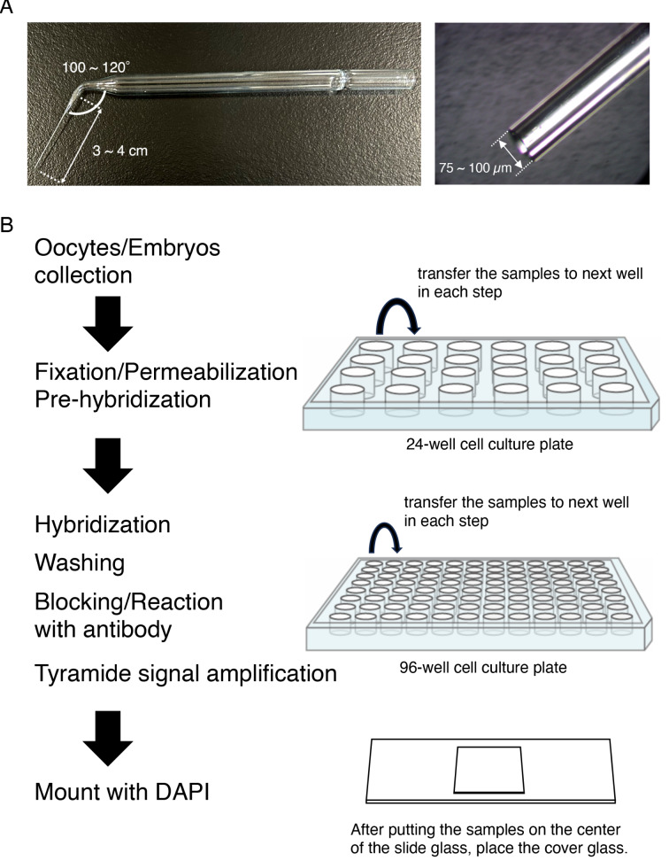
Mammalian oocytes accumulate more than ten thousand mRNAs, of which three to four thousand mRNAs are translationally repressed. The timings and sites of translational activation of these dormant mRNAs are crucial for promoting oocyte maturation and embryonic development. How these mRNAs are accumulated and distributed in oocytes is therefore a fundamental issue to be explored. A method that enables visualization of mRNA molecules with high resolution in a simple manner would be valuable for understanding how oocytes accumulate and regulate the dormant mRNAs. We have developed a highly sensitive whole-mount in situ hybridization method using in vitro-synthesized RNA probes and the tyramide signal amplification (TSA) system optimized for mouse oocytes and embryos. By using this method, Pou5f1/Oct4, Emi2, and cyclin B1 mRNAs were detected in immature oocytes and 2-cell stage embryos. Confocal microscopy showed that these mRNAs formed granular structures in the oocyte cytoplasm. The structures of Pou5f1/Oct4 and cyclin B1 mRNAs persisted in 2-cell stage embryos. Pou5f1/Oct4 RNA granules exhibited a solid-like property in immature oocytes and became liquid-like droplets in 2-cell stage embryos. Double-staining of cyclin B1 mRNA with Emi2 or Pou5f1/Oct4 mRNA revealed that these mRNAs were distributed as different RNA granules without overlapping each other and that the size of cyclin B1 RNA granules tended to be larger than that of Emi2 RNA granules. The structures and distribution patterns of these mRNAs were further analyzed by N-SIM super-resolution microscopy. This analysis revealed that the large-sized RNA granules consist of many small-sized granules, suggesting the accumulation and regulation of dormant mRNAs as basal-sized RNA granules. The method established in this study can easily visualize the structure and distribution of mRNAs accumulated in mammalian oocytes and embryos with high sensitivity and super-resolution. This method is useful for investigating the cellular and molecular mechanisms of translational control of mRNAs by which maturation and early developmental processes are promoted.

Keywords: Embryo; Mammal; Maternal mRNA; Oocyte; Super-resolution microscopy; in situ hybridization.


Conflict of interest statement

The authors declare no competing interests.

Figures

Fig. 1
Fig. 1
Schematic view of the procedure for whole-mount in situ hybridization optimized for mouse oocytes and embryos. A The structure of glass pipettes used in the procedure. B After the oocytes/embryos had been collected in a 24-well cell culture plate, the samples were fixed with 4% PFA/PBS, permeabilized with 2% Triton X-100, and incubated with 66% formamide/10% SSC (pre-hybridization). A 24-well cell culture plate was used in these steps. Then the oocytes/embryos were transferred to a 96-well cell culture plate and hybridized with probes of target RNAs at 45˚C overnight. The samples were washed with a series of SSC and incubated with blocking buffer. Then anti-DIG or Fluorescein antibody conjugated with horseradish peroxidase (HRP) was added, and signals were amplified with the TSA system. A 96-well cell culture plate was used in these steps. Finally, the samples were mounted on a slide glass with mounting medium with DAPI
Fig. 2
Fig. 2
in situ hybridization of Pou5f1/Oct4, Emi2 and cyclin B1 mRNAs in mouse oocytes and embryos. A-C Detection of Pou5f1/Oct4 mRNA in immature oocytes (A-B) and 2-cell stage embryos (C) hybridized with the sense (A) or antisense (B-C) RNA probe. DNA is shown in blue. D The numbers of Pou5f3/Oct4 RNA granules per 100 µm2 in individual oocytes and 2-cell stage embryos were counted (means ± standard deviations). Results from three independent experiments were summarized. E-G Detection of Emi2 mRNA in immature oocytes (E-F) and 2-cell stage embryos (G) hybridized with the sense (E) or antisense (F-G) RNA probe. DNA is shown in blue. H The numbers of Emi2 RNA granules per 100 µm2 in individual oocytes and 2-cell stage embryos were counted. Results from three independent experiments were summarized. I-K Detection of cyclin B1 mRNA in immature oocytes (I-J) and 2-cell stage embryos (K) hybridized with the sense (I) or antisense (J-K) RNA probe. DNA is shown in blue. L The numbers of cyclin B1 RNA granules per 100 µm2 in individual oocytes and 2-cell stage embryos were counted. Results from three independent experiments were summarized. The numbers in parentheses indicate the total numbers of oocytes and embryos analyzed. Statistical significance was analyzed by Student’s t-test. ***p < 0.001. Similar results were obtained from five independent experiments. GC, granulosa cell; GV, germinal vesicle. Bars, 50 μm
Fig. 3
Fig. 3
in situ hybridization of Pou5f1/Oct4 mRNA in mouse oocytes and embryos treated with hexanediol or hexanetriol. A Detection of Pou5f1/Oct4 mRNA (green) in immature oocytes (GV) and 2-cell stage embryos (2-cell) without (Control) and with hexanediol or hexanetriol. The oocytes and embryos are outlined by broken lines. DNA is shown in blue. Similar results were obtained from two independent experiments. GV, germinal vesicle. Bars, 50 μm. B The number of RNA granules per 100 µm2 in immature oocytes was counted (means ± standard deviations). C The number of RNA granules per 100 µm2 in 2-cell stage embryos was counted. The numbers in parentheses indicate the total numbers of oocytes and embryos analyzed. Statistical significance was analyzed by the Tukey-Kramer test. *p < 0.05, **p < 0.01. Cont, without treatment; HD, treated with hexanediol; HT, treated with hexanetriol
Fig. 4
Fig. 4
Double fluorescence in situ hybridization of Emi2 (green) and cyclin B1 (red) mRNA in mouse oocytes. A Detection of Emi2 (left) and cyclin B1 (middle) mRNAs by section in situ hybridization. A merged image is shown (Merge). DNA is shown in blue. Note that the signals in granulosa cells would be background of the TSA system in the section in situ hybridization method, which was caused by strong signal amplification, because similar signals were also observed in control sections hybridized with the Emi2 sense RNA probe. B Detection of Emi2 (left) and cyclin B1 (middle) mRNAs by whole-mount in situ hybridization. A merged image is shown (Merge). DNA is shown in blue. C Enlarged views of the boxed region in (B). Similar results were obtained from two independent experiments. GV, germinal vesicle. Bars, 50 μm in A and B, 1 μm in C
Fig. 5
Fig. 5
Double fluorescence in situ hybridization of Pou5f1/Oct4 (green) and cyclin B1 (red) mRNAs in mouse oocytes. A Detection of Pou5f1/Oct4 (left) and cyclin B1 (middle) mRNAs in an immature oocyte by whole-mount in situ hybridization. A merged image is shown (Merge). DNA is shown in blue. B Enlarged views of the boxed regions in (A). C Views of Pou5f1/Oct4 and cyclin B1 mRNAs in an immature oocyte by super-resolution microscopy. A representative top view (X-Y view) is shown on the left. A 3D view (X-Y-Z view) is shown on the right. D An enlarged view of the 25 µm2 square region in (C). E Views of 3D reconstruction of the 25 µm2 square region in (C). Insets are a top view (upper) and a 3D view (lower) of the x-y axis of the 1-µm-thick z-stacks indicated. Similar results were obtained from two independent experiments. Bars, 50 μm in A and C, 1 μm in B, D and E
Fig. 6
Fig. 6
Reconstruction views of the x-y axis of 1-µm-thick z-stacks of a super-resolution microscope image. A Acquiring a reconstruction image of the x-y axis in a whole oocyte. One µm of z-stacks was trimmed from the whole oocyte image and analyzed. B Top view of the reconstructed image in (A) (upper) and enlarged view of the 100 µm2 square region in the upper image (lower). Bars, 50 μm in A and B (upper), 5 μm in B (lower)
Fig. 7
Fig. 7
Reconstruction views of x-z and y-z axes of super-resolution microscope images. A Acquiring a reconstruction image of x-z and y-z axes in the whole oocyte. One µm y- or x-stacks was trimmed from the whole oocyte images. B X-Z view of the reconstructed image in (A, upper). C Y-Z view of the reconstructed image in (A, lower). Bars, 50 μm

Similar articles

Cited by

References

    1. Hamatani T, Carter MG, Sharov AA, Ko MSH. Dynamics of global gene expression changes during mouse preimplantation development. Dev Cell. 2004;6:117–31. doi: 10.1016/S1534-5807(03)00373-3. - DOI - PubMed
    1. Braude P, Bolton V, Moore S. Human gene expression first occurs between the four- and eight-cell stages of preimplantation development. Nature. 1988;332:459–61. doi: 10.1038/332459a0. - DOI - PubMed
    1. Luong XG, Daldello EM, Rajkovic G, Yang C-R, Conti M. Genome-wide analysis reveals a switch in the translational program upon oocyte meiotic resumption. Nucleic Acids Res. 2020;48:3257–76. doi: 10.1093/nar/gkaa010. - DOI - PMC - PubMed
    1. Hu W, Zeng H, Shi Y, Zhou C, Huang J, Jia L, et al. Single-cell transcriptome and translatome dual-omics reveals potential mechanisms of human oocyte maturation. Nat Commun. 2022;13:5114. doi: 10.1038/s41467-022-32791-2. - DOI - PMC - PubMed
    1. Xiong Z, Xu K, Lin Z, Kong F, Wang Q, Quan Y, et al. Ultrasensitive Ribo-Seq reveals translational landscapes during mammalian oocyte-to-embryo transition and pre-implantation development. Nat Cell Biol. 2022;24:968–80. doi: 10.1038/s41556-022-00928-6. - DOI - PubMed

LinkOut - more resources