

A proof of concept for microcirculation monitoring using machine learning based hyperspectral imaging in critically ill patients: a monocentric observational study
- PMID: 38987802
- PMCID: PMC11238485
- DOI: 10.1186/s13054-024-05023-w
A proof of concept for microcirculation monitoring using machine learning based hyperspectral imaging in critically ill patients: a monocentric observational study
Abstract
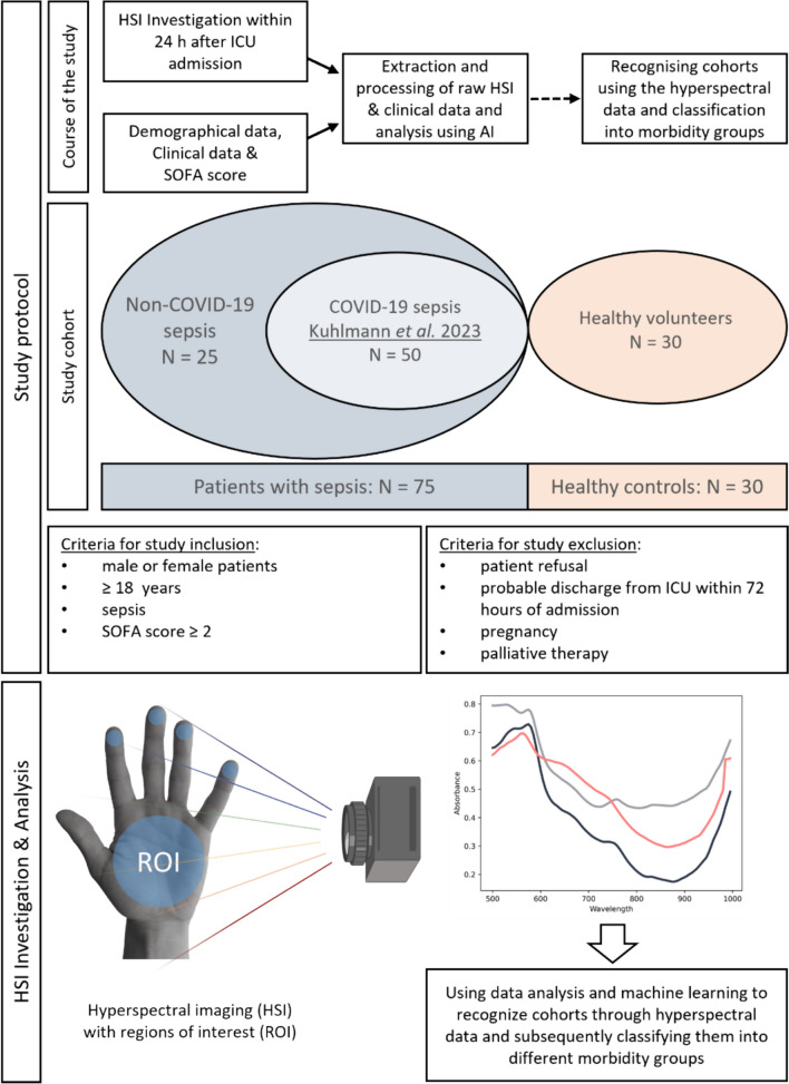
Background: Impaired microcirculation is a cornerstone of sepsis development and leads to reduced tissue oxygenation, influenced by fluid and catecholamine administration during treatment. Hyperspectral imaging (HSI) is a non-invasive bedside technology for visualizing physicochemical tissue characteristics. Machine learning (ML) for skin HSI might offer an automated approach for bedside microcirculation assessment, providing an individualized tissue fingerprint of critically ill patients in intensive care. The study aimed to determine if machine learning could be utilized to automatically identify regions of interest (ROIs) in the hand, thereby distinguishing between healthy individuals and critically ill patients with sepsis using HSI.
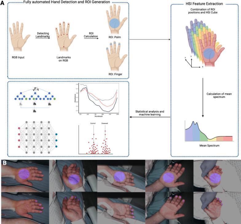
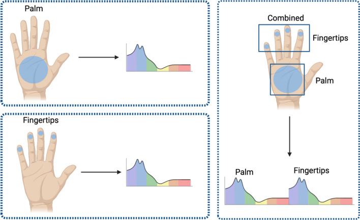
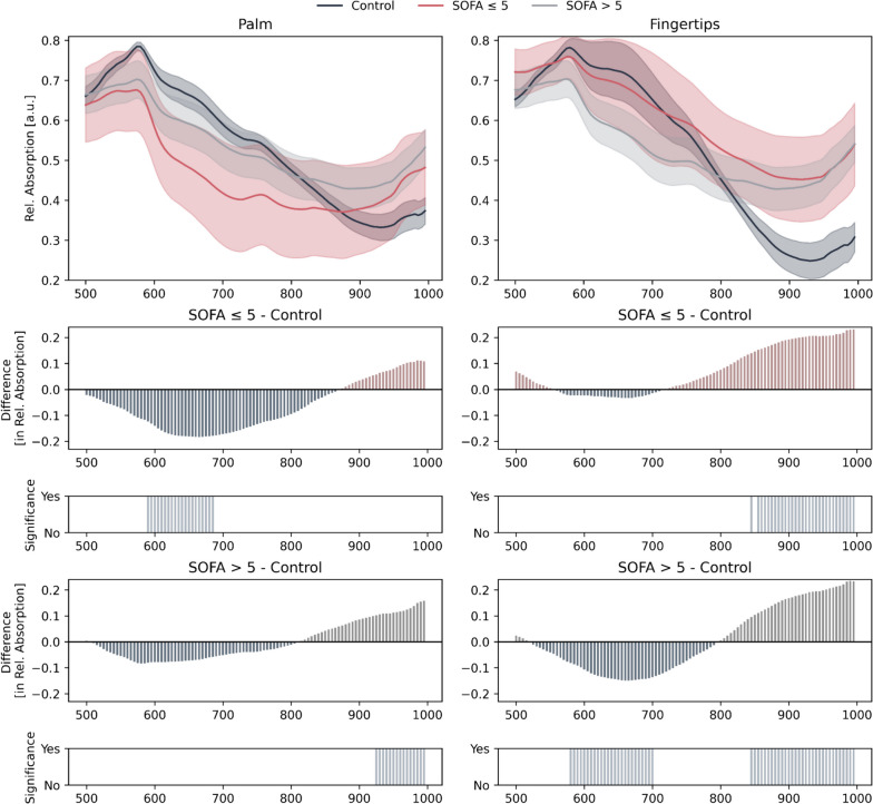
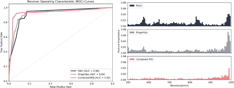
Methods: HSI raw data from 75 critically ill sepsis patients and from 30 healthy controls were recorded using TIVITA® Tissue System and analyzed using an automated ML approach. Additionally, patients were divided into two groups based on their SOFA scores for further subanalysis: less severely ill (SOFA ≤ 5) and severely ill (SOFA > 5). The analysis of the HSI raw data was fully-automated using MediaPipe for ROI detection (palm and fingertips) and feature extraction. HSI Features were statistically analyzed to highlight relevant wavelength combinations using Mann-Whitney-U test and Benjamini, Krieger, and Yekutieli (BKY) correction. In addition, Random Forest models were trained using bootstrapping, and feature importances were determined to gain insights regarding the wavelength importance for a model decision.
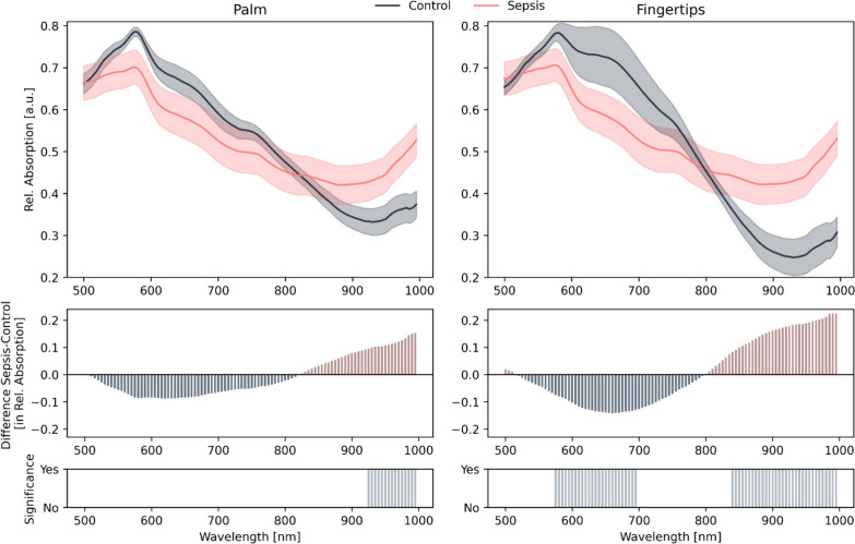
Results: An automated pipeline for generating ROIs and HSI feature extraction was successfully established. HSI raw data analysis accurately distinguished healthy controls from sepsis patients. Wavelengths at the fingertips differed in the ranges of 575-695 nm and 840-1000 nm. For the palm, significant differences were observed in the range of 925-1000 nm. Feature importance plots indicated relevant information in the same wavelength ranges. Combining palm and fingertip analysis provided the highest reliability, with an AUC of 0.92 to distinguish between sepsis patients and healthy controls.
Conclusion: Based on this proof of concept, the integration of automated and standardized ROIs along with automated skin HSI analyzes, was able to differentiate between healthy individuals and patients with sepsis. This approach offers a reliable and objective assessment of skin microcirculation, facilitating the rapid identification of critically ill patients.
© 2024. The Author(s).
Conflict of interest statement
Thorsten Brenner received honoraria for invited academic conference lectures from CSL Behring GmbH, Schöchl medical education GmbH, Biotest AG, Baxter Deutschland GmbH, Boehringer Ingelheim Pharma GmbH, Astellas Pharma GmbH, B. Braun Melsungen AG, MSD Sharp & Dohme GmbH, Akademie für Infektionsmedizin e.V. and Sedana Medical Germany GmbH. Thorsten Brenner is a consultant for Baxter Deutschland GmbH. Thorsten Brenner received re-search funding from German Research Foundation (DFG), Dietmar Hopp Foundation, University of Essen Medical Foundation, Heidelberg Surgery Foundation and Innovation Fund of the Joint Federal Committee (G-BA). Karsten Schmidt received honoraria for invited academic conferences lecture from Dr. Franz Köhler Chemie GmbH, Germany. Karsten Schmidt received research fun-ding from Stiftung Universitätsmedizin Essen and Heidelberger Stiftung Chirurgie. The funders had no role in the design of the study; in the collection, analyses, or interpretation of data; in the writing of the manuscript; or in the decision to publish the results.
Figures
References
-
- Evans L, Rhodes A, Alhazzani W, Antonelli M, Coopersmith CM, French C, Machado FR, McIntyre L, Ostermann M, Prescott HC, et al. Surviving sepsis campaign: international guidelines for management of sepsis and septic shock 2021. Intensive Care Med. 2021;47(11):1181–1247. doi: 10.1007/s00134-021-06506-y. - DOI - PMC - PubMed
Publication types
MeSH terms
Grants and funding
LinkOut - more resources
Full Text Sources
