. 2024 Jul 10;22(1):357.
doi: 10.1186/s12964-024-01708-5.

Macrophage-derived exosomes promote telomere fragility and senescence in tubular epithelial cells by delivering miR-155

Affiliations

Macrophage-derived exosomes promote telomere fragility and senescence in tubular epithelial cells by delivering miR-155

Qing Yin et al. Cell Commun Signal. .

Abstract

Background: Chronic kidney disease (CKD) is highly prevalent worldwide, and its global burden is substantial and growing. CKD displays a number of features of accelerated senescence. Tubular cell senescence is a common biological process that contributes to CKD progression. Tubulointerstitial inflammation is a driver of tubular cell senescence and a common characteristic of CKD. However, the mechanism by which the interstitial inflammation drives tubular cell senescence remains unclear. This paper aims to explore the role of exosomal miRNAs derived from macrophages in the development of tubular cell senescence.

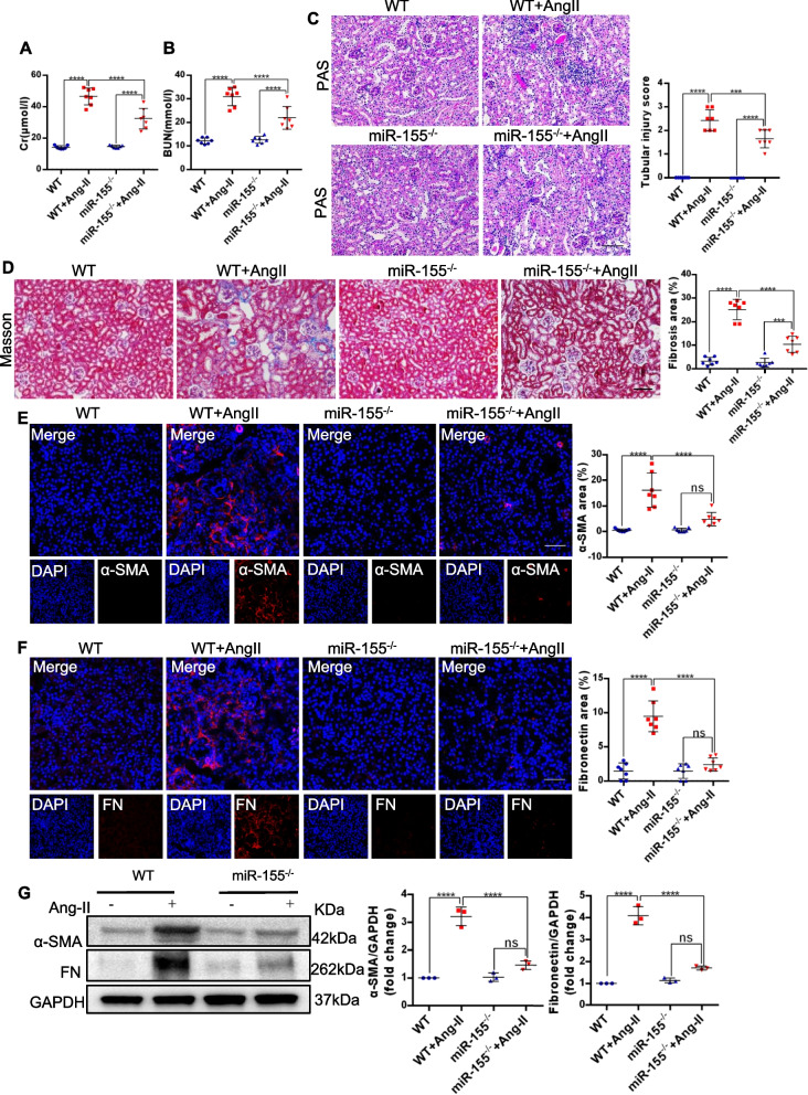
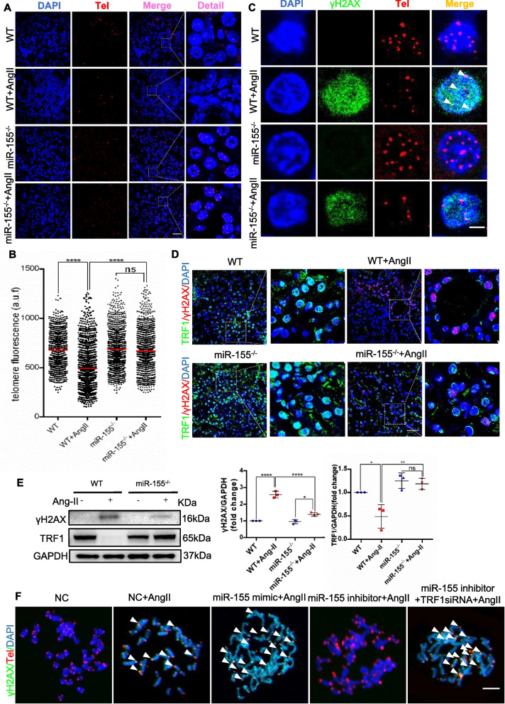
Methods: Among the identified inflammation-related miRNAs, miR-155 is considered to be one of the most important miRNAs involved in the inflammatory response. Macrophages, the primary immune cells that mediate inflammatory processes, contain a high abundance of miR-155 in their released exosomes. We assessed the potential role of miR-155 in tubular cell senescence and renal fibrosis. We subjected miR-155-/- mice and wild-type controls, as well as tubular epithelial cells (TECs), to angiotensin II (AngII)-induced kidney injury. We assessed kidney function and injury using standard techniques. TECs were evaluated for cell senescence and telomere dysfunction in vivo and in vitro. Telomeres were measured by the fluorescence in situ hybridization.

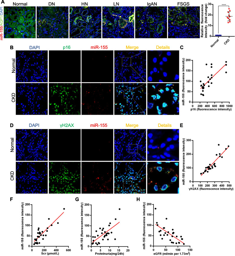
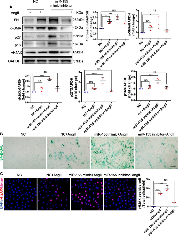
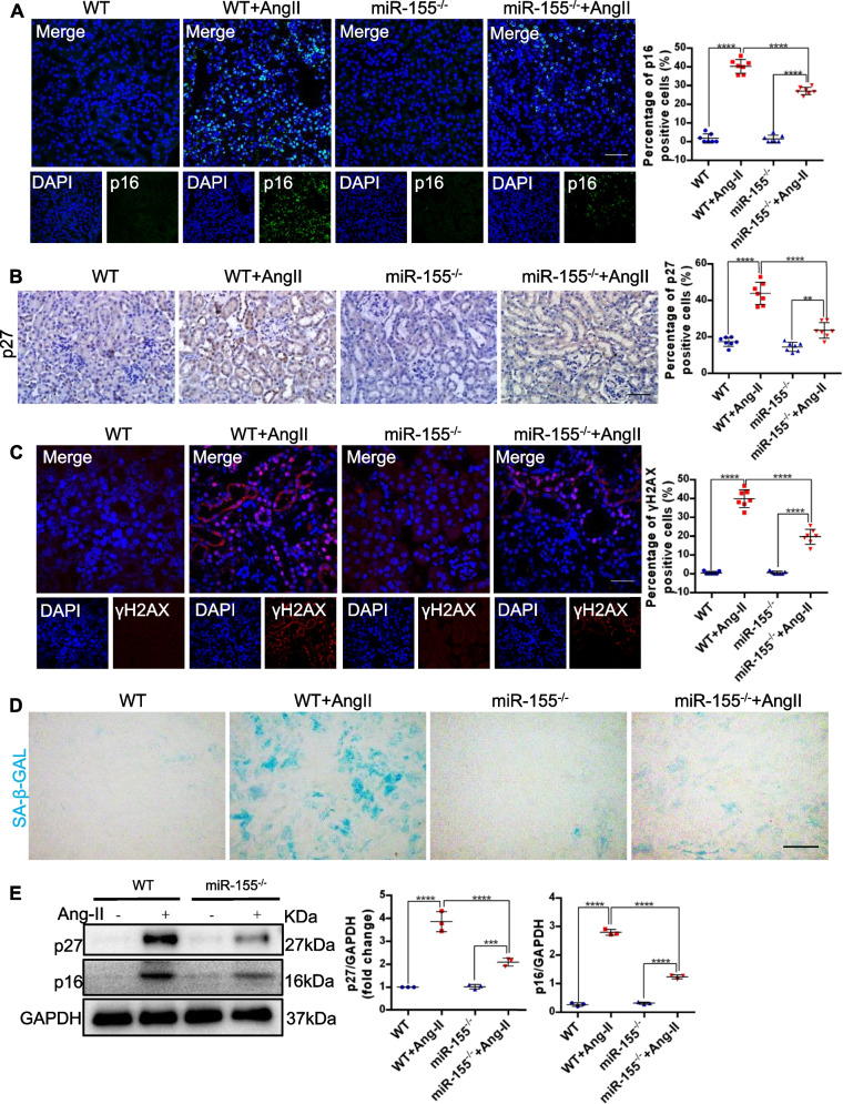
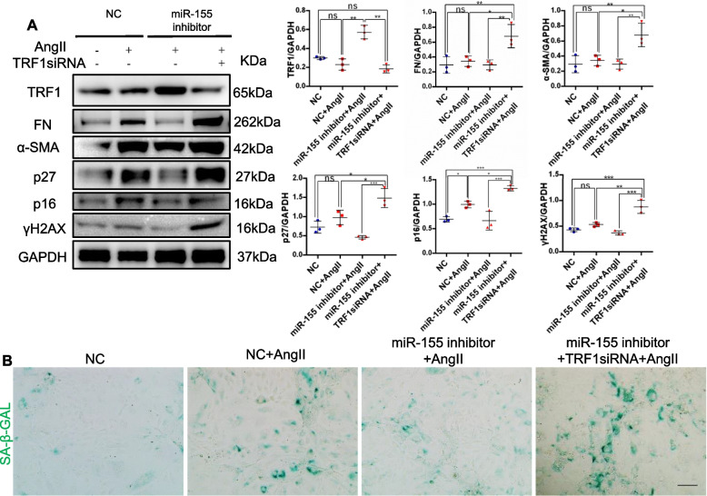
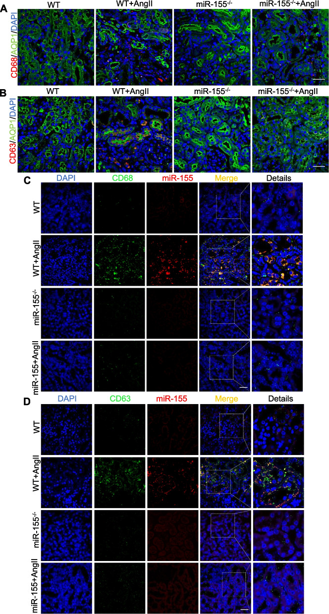
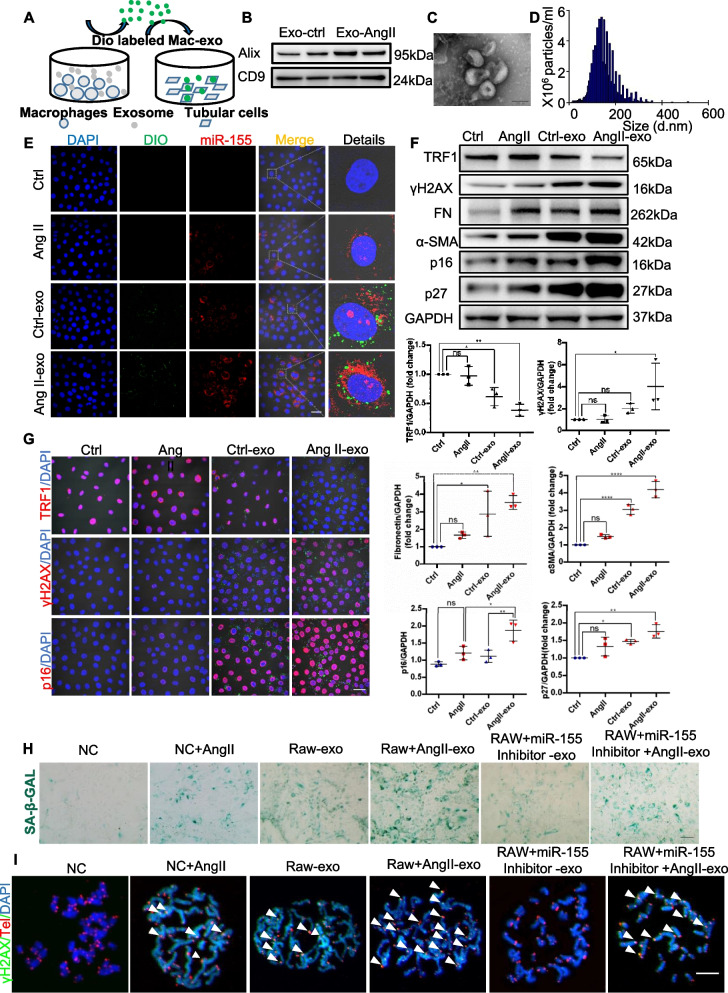
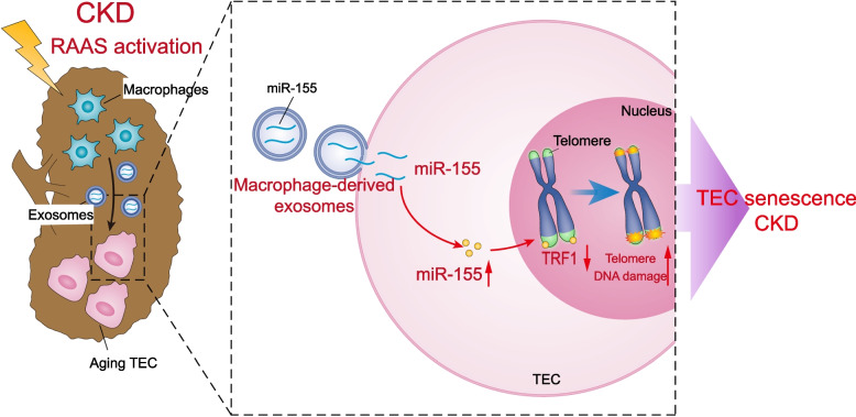
Results: Compared with normal controls, miR-155 was up-regulated in proximal renal tubule cells in CKD patients and mouse models of CKD. Moreover, the expression of miR-155 was positively correlated with the extent of renal fibrosis, eGFR decline and p16INK4A expression. The overexpression of miR-155 exacerbated tubular senescence, evidenced by increased detection of p16INK4A/p21expression and senescence-associated β-galactosidase activity. Notably, miR-155 knockout attenuates renal fibrosis and tubule cell senescence in vivo. Interestingly, once released, macrophages-derived exosomal miR-155 was internalized by TECs, leading to telomere shortening and dysfunction through targeting TRF1. A dual-luciferase reporter assay confirmed that TRF1 was the direct target of miR-155. Thus, our study clearly demonstrates that exosomal miR-155 may mediate communication between macrophages and TECs, subsequently inducing telomere dysfunction and senescence in TECs.

Conclusions: Our work suggests a new mechanism by which macrophage exosomes are involved in the development of tubule senescence and renal fibrosis, in part by delivering miR-155 to target TRF1 to promote telomere dysfunction. Our study may provide novel strategies for the treatment of AngII-induced kidney injury.

Keywords: CKD; Cell senescence; Exosomes; Macrophages; MiR-155; TRF1.


Conflict of interest statement

The authors declare no competing interests.

Figures

Fig. 1
Fig. 1
miR-155 is increased in multiple types of clinical nephropathy and is associated with tubular senescence. A Representative images show the expression and localization of miR-155 in various types of human CKD. miR-155 (Red), AQP1(Green). Scale bar, 50 μm. B Co-staining of p16INK4A (green) and miR-155 (red) in various types of human CKD. C Scatter plots with linear regression show significant correlation between miR-155 expression levels and p16INK4A. r = 0.7115, P < 0.05, n = 30, Scale bar, 50 μm. D Co-staining of γH2AX (green) and miR-155 (red) in various types of human CKD. E Scatter plots with linear regression show significant correlation between miR-155 expression levels and γH2AX. r = 0.9105, P < 0.05, n = 30, Scale bar, 50 μm. F Linear regression shows an positive correlation between miR-155 expression level and serum creatinine levels (r = 0.7794, P < 0.05, n = 30), G proteinuria (r = 0.6191, P < 0.05, n = 30) and H an inverse correlation between miR-155 expression level and eGFR (r = -0.7130, P < 0.05, n = 30). DN: diabetic nephropathy; HN: hypertensive nephropathy; LN: lupus nephritis; IgAN: IgA nephropathy; FSGS: focal segmental glomerulosclerosis; CKD: chronic kidney disease
Fig. 2
Fig. 2
Transfection of miR-155 into cultured mouse tubular cells (mTECs) exacerbates cellular senescence in vitro. A Representative western blotting and summarized data of fibronectin, ɑ-SMA, p27, p16INK4A and γH2AX in the mTECs (n = 3). B Representative images of SA-β-gal activity staining of mTECs. Scale bars, 50 µm. C Representative images of γH2AX stained sections of mTECs. Data are presented as mean ± SD, * p < 0.05, ** p < 0.01, *** p < 0.001, **** p < 0.0001
Fig. 3
Fig. 3
MiR-155 knockout attenuates renal fibrosis in AngII-induced kidney injury. A Serum creatinine levels and B BUN over time 4 weeks after vehicle or AngII infusion (n = 7). C Representative kidney histology as shown by PAS staining. Dot plot shows the corresponding quantification of tubular injury score (n = 7). Scale bar: 100 μm. D Representative images of masson staining of the kidney. Scale bar: 100 μm. The dot plot showed the corresponding quantification of fibrosis area (n = 7). E Representative images of ɑ-SMA-stained kidney sections from wild type and miR-155−/− mice. Scale bars: 50 μm. The quantification of ɑ-SMA fluorescence intensity in the kidneys (n = 7). F Representative images of fibronectin-stained kidney sections from wild type and miR-155−/− mice. Scale bars: 50 μm. The quantification of fibronectin fluorescence intensity in the kidneys (n = 7). G Representative western blotting gel documents and summarized data showing the protein levels of ɑ-SMA and fibronectin in the kidneys of mice. Data are presented as mean ± SD, * p < 0.05, ** p < 0.01, *** p < 0.001, **** p < 0.0001
Fig. 4
Fig. 4
MiR-155 knockout attenuates renal TECs senescence in AngII-induced kidney injury. A Representative images of p16INK4A staining in kidney tissues and quantification of the p16INK4A positive cells (n = 7). Scale bars, 50 µm. B Representative images of p27 staining in kidney tissues and quantification of the p27 positive cells (n = 7). Scale bars, 50 µm. C Representative images of γH2AX-stained kidney sections. Scale bars: 50 μm. D SA-β-gal activity staining of frozen kidney sections, which appeared as bright blue granular staining in the cytoplasm of renal TECs. Scale bars, 50 µm. E Representative western blotting gel documents and summarized data showing the protein levels of p16INK4A and p27 in the kidneys of mice. Data are presented as mean ± SD, * p < 0.05, ** p < 0.01, *** p < 0.001, **** p < 0.0001
Fig. 5
Fig. 5
miR-155 inhibition protects telomere function by binding to TRF1 in renal epithelial cells. A Representative images of telomere-stained kidney sections from wild type and AngII mice 4 weeks after vector or AngII infusion. Scale bars: 50 μm. B Telomeric length measurements by quantitative telomeric DNA FISH in the kidney sections; a.f.u, arbitrary fluorescence units. C Telomeric DNA damage was detected by the co-localization of γH2AX (green) and telomeres (red) in the immunofluorescence of the kidney sections. D Representative images of TRF1- (green) and γH2AX- (red) stained sections of kidney sections from mice. Scale bars: 50 μm. E Representative western blotting and summarized data of TRF1 and γH2AX in kidney tissues of mice (n = 3). F Telomere DNA damage in mTECs treated with AngII, as demonstrated by the co-localization of γH2AX (green) and telomeres (red) in the immunofluorescence. White arrows indicate TIFs. Scale bars: 10 μm. TIFs, telomere dysfunction induced foci. Data are presented as mean ± SD, * p < 0.05, ** p < 0.01, *** p < 0.001, **** p < 0.0001
Fig. 6
Fig. 6
miR-155 inhibition protects cell senescence and renal fibrosis in mTECs by binding to TRF1. A Representative western blotting and summarized data of TRF1, fibronectin, ɑ-SMA, p27, p16INK4A and γH2AX in the mTECs (n = 3). B Representative images of SA-β-gal activity staining of mTECs. Scale bars, 50 µm. Data are presented as mean ± SD, * p < 0.05, ** p < 0.01, *** p < 0.001, **** p < 0.0001
Fig. 7
Fig. 7
Macrophage-derived exosomes transfers miR-155 into tubules in AngII-induced kidney. A Representative images of AQP1- (green) and CD68 (red) stained sections of the kidney sections. Scale bars: 50 μm. B Representative images of AQP1- (green) and CD63 (red) stained sections of the kidney sections. Scale bars: 50 μm. C Representative images of miR-155 FISH (red) co-stained with CD68 (green) immunofluorescence. Scale bars: 50 μm. D Representative images of miR-155 FISH (red) co-stained with CD63 (green) immunofluorescence. Scale bars: 50 μm
Fig. 8
Fig. 8
Macrophage-derived exosomes transfers miR-155 into tubules in AngII-induced kidney. A Exosomes harvested from the medium of RAW 264.7 were labeled with DIO and added to primary cultured mouse renal tubular cells for 48 h. B Western blotting for Alix and CD9 exosome markers. C and D Macrophage original exosome was verified by transmission electron microscopy, ranging in size from 80 to 130 nm. E DIO-labeled exosomes were introduced into mTECs for 48 h, and miR-155FISH was performed. Scale bars: 20 μm. F Western blotting analysis of TRF1, γH2AX, Fibronectin, α-SMA, p16INK4A and p27 in primary cultured mouse renal tubular cells. (n = 3). G Representative confocal images of TRF1, γH2AX and p16INK4A staining in kidney tissues. H Representative images of SA-β-gal activity staining of primary cultured mouse renal tubular cells. Scale bars, 50 µm. I Telomere DNA damage in mTECs treated with AngII, as demonstrated by the co-localization of γH2AX (green) and telomeres (red) in the immunofluorescence. White arrows indicate TIFs. Scale bars: 10 μm. TIFs, telomere dysfunction induced foci. Data are presented as mean ± SD, * p < 0.05, ** p < 0.01, *** p < 0.001, **** p < 0.0001
Fig. 9
Fig. 9
Schematic illustration of the mechanism by which macrophage-derived exosomes deliver miR-155 to promote telomere fragility, chromosomal instability, and senescence in TECs. MiR-155 was synthesized and loaded into the exosomes in increased infiltration of macrophages in AngII-infused mice. The released exosomal fusion with the plasma membrane leads to the release of miR-155 into the TECs and the translational repression of TRF1 in the TECs. Finally, macrophage-derived miR-155-containing exosomes were shown to promoted TEC tubular senescence and renal fibrosis by directly targeting TRF1 in AngII-infused mice

References

    1. Carney E. The impact of chronic kidney disease on global health. Nat Rev Nephrol. 2020;16:251. doi: 10.1038/s41581-020-0268-7. - DOI - PubMed
    1. Sturmlechner I, Durik M, Sieben C, Baker D, van Deursen J. Cellular senescence in renal ageing and disease. Nat Rev Nephrol. 2017;13:77–89. doi: 10.1038/nrneph.2016.183. - DOI - PubMed
    1. Liu B, Tang T, Lv L, Lan H. Renal tubule injury: a driving force toward chronic kidney disease. Kidney Int. 2018;93:568–579. doi: 10.1016/j.kint.2017.09.033. - DOI - PubMed
    1. Huang W, Hickson L, Eirin A, Kirkland J, Lerman L. Cellular senescence: the good, the bad and the unknown. Nat Rev Nephrol. 2022;18:611–627. doi: 10.1038/s41581-022-00601-z. - DOI - PMC - PubMed
    1. Xu L, Wang J, Yu H, Mei H, He P, Wang M, et al. GLIS1 alleviates cell senescence and renal fibrosis through PGC1-α mediated mitochondrial quality control in kidney aging. Free Radic Biol Med. 2023;209:171–184. doi: 10.1016/j.freeradbiomed.2023.09.037. - DOI - PubMed

Publication types

MeSH terms

LinkOut - more resources