. 2024 Dec 1;109(12):3941-3951.
doi: 10.3324/haematol.2024.285101.

Umbilical cord blood T cells can be isolated and enriched by CD62L selection for use in 'off the shelf' chimeric antigen receptor T-cell therapies to widen transplant options

Affiliations

Umbilical cord blood T cells can be isolated and enriched by CD62L selection for use in 'off the shelf' chimeric antigen receptor T-cell therapies to widen transplant options

Christos Georgiadis et al. Haematologica. .

Abstract

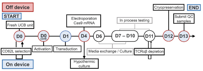
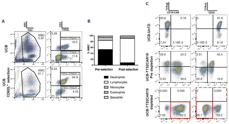
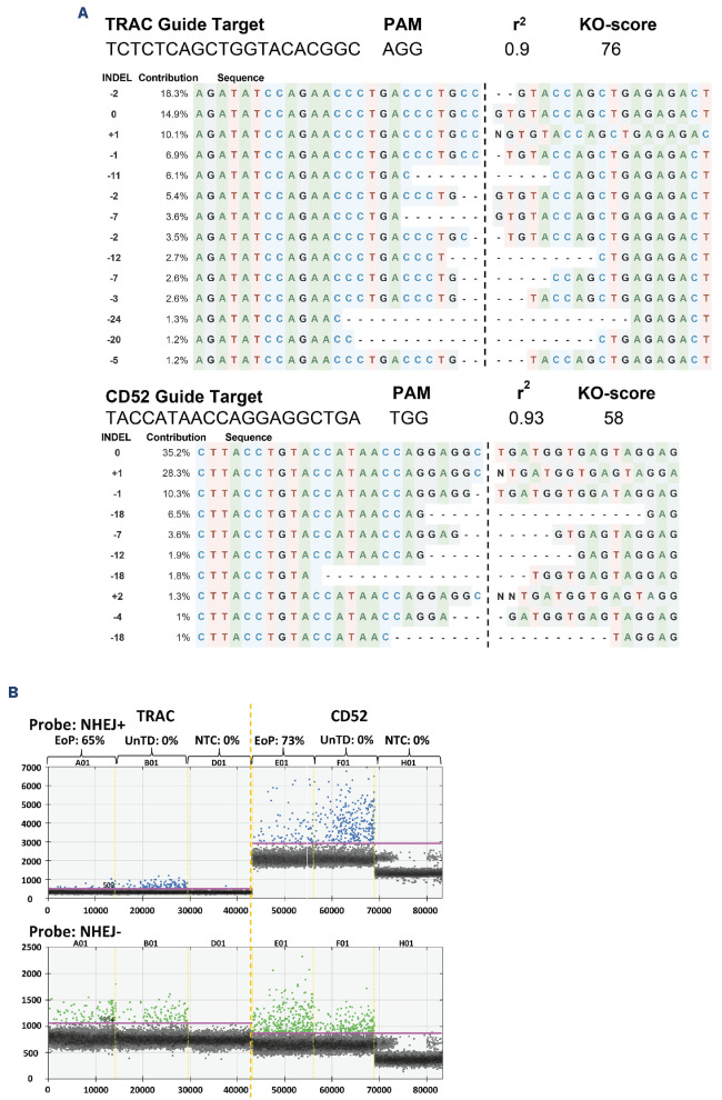
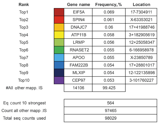
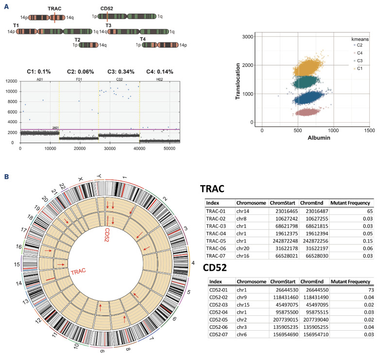
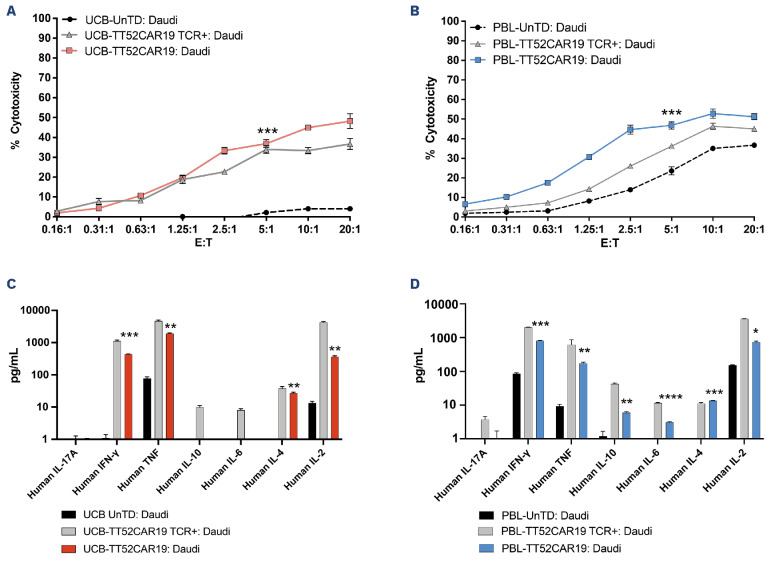
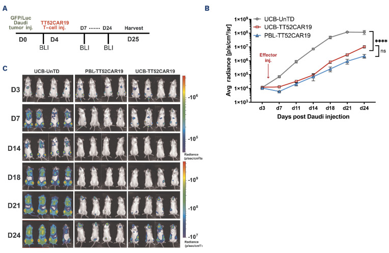
Umbilical cord blood (UCB) T cells exhibit distinct naïve ontogenetic profiles and may be an attractive source of starting cells for the production of chimeric antigen receptor (CAR) T cells. Pre-selection of UCB-T cells on the basis of CD62L expression was investigated as part of a machine-based manufacturing process, incorporating lentiviral transduction, CRISPR- Cas9 editing, T-cell expansion, and depletion of residual TCRαβ T cells. This provided stringent mitigation against the risk of graft-versus-host disease (GvHD), and was combined with simultaneous knockout of CD52 to enable persistence of edited T cells in combination with preparative lymphodepletion using alemtuzumab. Under compliant manufacturing conditions, two cell banks were generated with high levels of CAR19 expression and minimal carriage of TCRαβ T cells. Sufficient cells were cryopreserved in dose-banded aliquots at the end of each campaign to treat dozens of potential recipients. Molecular characterization captured vector integration sites and CRISPR editing signatures, and functional studies, including in vivo potency studies in humanized mice, confirmed anti-leukemic activity comparable to peripheral blood-derived universal CAR19 T cells. Machine manufactured UCB-derived T-cell banks offer an alternative to autologous cell therapies and could help widen access to CAR T cells.


Figures

Figure 1.
Figure 1.
Schematic timeline for Good Manufacturing Practice manufacturing of TT52CAR19 cell banks from umbilical cord blood starting material. Fresh umbilical cord blood (UCB) units were enriched for lymphocytes by CD62L+ magnetic bead positive selection and activated on day 0 (D0). T cells were transduced 24 hours later (D1) with TT52CAR19 vector and electroporated on day 4 (D4) with SpCas9 mRNA. On day 11 (D11), cells underwent automated TCRαβ T-cell depletion. After this step, the final cell product was harvested and cryopreserved in therapeutic doses. Steps itemised below the timeline were performed on a Miltenyi Prodigy and are shown as ‘On device’,, with the remaining items (above the timeline) undertaken ‘Off device’. cont.: continued; QC: quality control.
Figure 2.
Figure 2.
Whole umbilical cord blood CD62L selection using the CliniMACS Prodigy and TT52CAR19 batch manufacture. (A) Umbilical cord blood (UCB) cells pre- and post-CD62L+ selection using the CliniMACS Prodigy demonstrates enrichment of CD3+ T cells. (B) Cord blood cells from UCB donors (N=4) were analyzed by Sysmex pre- and post-CD62L selection using the CliniMACS Prodigy. (C) Flow cytometric T-cell transduction and knockout analysis of UCB-TT52CAR19 GMP1 (UCB1) cell bank. UCB-TT52CAR19 cells were stained before and after up to 2 rounds of magnetic TCRαβ depletion alongside untransduced (UnTD) cells or non-edited UCB-TT52CAR19 TCR+ cells. Transduction efficiency was measured by quantifying transgene expression using F(ab’)2, and CRISPR-Cas9-mediated protein knockout was determined through staining for TCRαβ and CD52. FSC: forward scatter; WBC: white blood cells; CAR: chimeric antigen receptor.
Figure 3.
Figure 3.
Molecular characterization of ‘on-target’ CRISPR-Cas9-mediated cleavage. (A) ICE analysis of Sanger sequence traces identified indels as signatures of NHEJ at the TRAC locus and at the CD52 target site. (B) Quantification of indels by ddPCR at both the TRAC and CD52 sites using separate probes, one specific to the predicted NHEJ region (NHEJ+) and a second outside the NHEJ region (NHEJ). EoP: end of production; UnTD: non-transduced controls; NTC: non-treated control.
Figure 4.
Figure 4.
Integration site analysis. Ligation-mediated polymerase chain reaction (LMPCR) detection and quantification of vector integration sites (IS) where the top 10 most frequent sites comprised <0.1% of integrants.
Figure 5.
Figure 5.
Detection for translocation and ‘off-target’ CRISPR-Cas9-mediated cleavage events. (A) (Top left) Droplet Digital polymerase chain reaction (ddPCR) was used for the detection and quantification of possible translocations after ‘on-target’ DNA scission. Four predicted recombination events (C1-C4) are presented in the schematic with TRAC (Chr14q) (red), CD52 (Chr1p) (green), and SpCas9 cleavage site (red line). (Right) Colours (yellow, green, blue, magenta) discriminate 4 possible translocations. (Bottom left) Low frequency translocation events (blue dots) C1-C4 arising between the edited TRAC and CD52 loci. Cumulative events for all 4 possible events were <1%. (B) (Left) Circos plot with verification of quantification using targeted NGS across the 6 highest scoring predicted ‘off-target’ sites. TRAC-01 (solid red line marking locus in outer yellow circle), CD52-01 (solid red line marking inner yellow circle), and at predicted ‘off-target’ sites TRAC-02-TRAC-07 (red arrows marking outer yellow circle) and CD52-02-CD52-07 (red arrows marking inner yellow circle). (Right) Table shows that negligible ‘off-target’ events were detected for both the TRAC and CD52 guides.
Figure 6.
Figure 6.
In vitro killing potential and cytokine production of umbilical cord blood-TT52CAR19 cells against CD19+ targets. In vitro cytotoxicity of umbilical cord blood (UCB)-TT52CAR19 GMP1 (UCB1) cell bank compared to respective UCB-TT52CAR19 transduced but not edited (UCB-TT52CAR19 TCR+) or non-transduced (UnTD) controls (A) or PBL-TT52CAR19 effectors and respective controls (B) when measured by 51Cr chromium release of labeled CD19+ Daudi cells following four hours of co-culture at incremental effector (E) : target (T) ratio. Cell co-cultures ranged from 1x10⁵ : 5x10⁴ (at E:T of 20:1) to 8x10² : 5x10⁴ (at E:T of 0.16:1). Effector responses were considered successful if ≥50% lysis was detected compared to UnTD controls at E:T cell ratios between 1.25:1 and 5:1. Error bars represent Standard Error of Mean (SEM), N=3 replicate/wells. In vitro target specific cytokine secretion of UCB-TT52CAR19 GMP1 cell bank (UCB1), and respective UCB-TT52CAR19 TCR+ or non-transduced (UnTD) controls after co-culture with CD19+ Daudi cells overnight (C). Cytokine release also quantified for PBL-TT52CAR19 effectors and respective PBL-TT52CAR19 TCR+ and UnTD control at 1:1 E:T (1.25x10⁵ of each effector and target cells) co-cultures with CD19+ Daudi cells (D). The presence of cytokines in the co-culture supernatant was measured by cytokine bead array and levels >50 pg/mL were considered positive responses. Significance was calculated between UCB-TT52CAR19 GMP1 (UCB1) or PBL-TT52CAR19 banks and respective UnTD controls. *P<0.05, **P<0.01, ***P<0.001, ****P<0.0001 by t test. Error bars represent SEM, N=3 replicates.
Figure 7.
Figure 7.
In vivo tumor clearance in human : murine xenograft model of CD19+ disease. Timeline of in vivo human:murine xenograft modeling indicating target and effector intravenous injection (inj.) days and bioluminescent imaging (BLI) time-points (A). Serial measurement of bioluminescence of Daudi CD19+ B-cell disease in immunodeficient NOD/SCID/yc (NSG) mice (N=12) infused with 5x10⁵ GFP/luciferase expressing Daudi CD19+ B cells were treated on day 4 with 5x10⁶ of either umbilical cord blood (UCB)-TT52CAR19 GMP1 (UCB1) (N=4) or peripheral blood lymphocyte (PBL) PBL-TT52CAR19 (N=4) and were monitored over a 4-week period (B, C). Non-transduced (UnTD) (N=4) T cells were used as controls. Error bars represent Standard Error of Mean (SEM). Significance compared by area under the curve using one-way ANOVA (F-value 35.86) with Tukey multiple comparison post-hoc; not significant (ns) P≥0.05, ****P<0.0001.

References

    1. Maude SL, Laetsch TW, Buechner J, et al. . Tisagenlecleucel in children and young adults with B-cell lymphoblastic leukemia. N Engl J Med. 2018;378(5):439-448. - PMC - PubMed
    1. Qayed M, McGuirk J, Myers G, et al. . Leukapheresis guidance and best practices for optimal chimeric antigen receptor T-cell manufacturing. Cytotherapy. 2022;24(9):869-878. - PubMed
    1. Abecassis A, Roders N, Fayon M, et al. . CAR-T cells derived from multiple myeloma patients at diagnosis have improved cytotoxic functions compared to those produced at relapse or following daratumumab treatment. EJHaem. 2022;3(3):970-974. - PMC - PubMed
    1. Brudno JN, Somerville RP, Shi V, et al. . Allogeneic T cells that express an anti-CD19 chimeric antigen receptor induce remissions of B-cell malignancies that progress after allogeneic hematopoietic stem-cell transplantation without causing graft-versus-host disease. J Clin Oncol. 2016;34(10):1112-1121. - PMC - PubMed
    1. Zhang C, Wang XQ, Zhang RL, et al. . Donor-derived CD19 CAR-T cell therapy of relapse of CD19-positive B-ALL post allotransplant. Leukemia. 2021;35(6):1563-1570. - PMC - PubMed

Substances

LinkOut - more resources